Category Archives: Outreach Project

Too Much Protein? Let Me Get Rid of That

Imagine if we could change the levels of proteins in our body the same way we make changes on our iPhones, that is, quickly, efficiently, reliably, and reversibly (“reset button”). This may seem highly unlikely. However, Wu Yang Jin and his lab succeeded in developing a new approach to alter the levels of specific proteins in rats. Proteins are crucial for humans as they perform a wide variety of functions such as building, maintaining and replacing tissues in our bodies.

Alzheimer’s disease is characterized by too much proteins in the brain called amyloid-beta plaques. Source: Flickr Commons. Credit to: alle banane.

Alzheimer’s disease is characterized by too much proteins in the brain called amyloid-beta plaques. Source: Flickr Commons. Credit to: alle banane

Proteins are indeed critical for our survival. However, inappropriate levels of proteins can also be harmful to humans, as is the case in Parkinson’s disease and Alzheimer’s disease. The method by Jin and his colleagues could be of importance when considering new research avenues to overcome shortcomings of protein manipulations in current clinical research. His research may lead to promising treatment options for treating human diseases such as Parkinson’s disease. Below is a video description of the plaque formation that causes Alzheimer’s disease.

Credit: Danton O’Day

As opposed to current techniques that regulate levels of proteins, Jin’s proposed method allows to remove proteins quickly and reversibly, such that proteins can regain their initial states, if need be. Specifically, his method consists of injecting a small protein, known as a peptide, into an organism. The peptide has the ability to track down specific proteins and eliminate them in order to bring the levels of proteins back to normal. An additional feature of this new method is that the peptide can remove targeted proteins in the brain. Due to its small size, the peptide can cross the blood brain barrier (BBB) and can enter the brain from the circulatory system. The BBB is a barrier that protects the brain from harmful compounds that could present in the circulatory system.

https://www.youtube.com/watch?v=Un_x59nQiIg

Credit: Daniel Hsiao, Harrimal Cheema, Sara Lariviere, and Amirhossein Tashakor

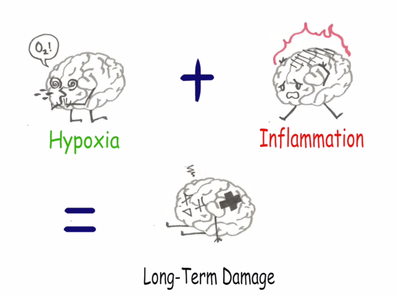
Jin has demonstrated the efficiency of the method by reducing the levels of various proteins. One such example targeted a protein called death-associated protein kinase 1 (DAPK1), which plays an important role in triggering cell death after a stroke. The implication of this protein is that it kills cells in the brain after a stroke, thereby increasing irreversible damage in the brain. When Jin and his colleagues injected the peptides into the circulatory system of stroke induced experimental rats, they found that the peptides successfully targeted and eliminated DAPK1 in areas of the brain affected by the stroke. As a result, damage done by the stroke were immensely reduced.


Parkinson’s disease is associated with symptoms such as resting tremor, postural instability, and slow walking. Source and Credit to: Wikipedia Commons.

In the podcast, we explained the paper in more detail.

https://www.youtube.com/watch?v=WtC5HhFQNHA

Credit to: Daniel Hsiao, Harrimal Cheema, Sara Lariviere, and Amirhossein Tashakor

Daniel Hsiao, Harrimal Cheema, Sara Lariviere, and Amirhossein Tashakor

Caught in the Act: Partners in Crime behind Brain Diseases

The first step to treating a disease is to catch the culprit behind it. Researcher Jingfei and her team set out to do just that and discovered that hypoxia (a lack of oxygen) and inflammation act as partners in crime to cause damage to the brain.

hypoxia plus inflammation

The main result of Jingfei’s research. (Image: credits to Sean)


Jingfei’s recent publication from the University of British Columbia (UBC), highlighted the key role that hypoxia plays in brain damaging diseases such as Alzheimer’s, something that has been overlooked in past studies. They found that hypoxia and inflammation combined is what causes long term damage to the brain.

“This is a never-before-seen mechanism among three key players in the brain that interact together in neurodegenerative disorders,” says Jingfei’s supervisor, Brian MacVicar, from the Djavad Mowafaghian Centre for Brain Health at UBC. The three key players that he is referring to are hypoxia, inflammation, and microglia (the immune cells of the brain).

By manipulating the brain slices of rodents using various techniques, the researchers tracked the movement of microglia and found that the two factors, hypoxia and inflammation, work together to permanently weaken the connections between brain cells. Furthermore, the damaging effects of the two factors may worsen people’s memory, which is one of the early symptoms of brain diseases like Alzheimer’s.

Pictures (or rather videos) are worth a thousand words so watch below to see exactly how they went about conducting this exciting research and what they found.

YouTube Preview Image

 (Video: Credits to Ian, Siana, Shikha, and Sean)

So, who will benefit from this research? Jingfei said, “Well I think right now it still will be researchers, because this paper is more like a new finding of something people didn’t know before, so hopefully […] they can build more realistic model on top of it.”

Moreover, some researchers have already built upon her research in the short  time that has passed since her paper was published. Researchers at Cambridge University have broadened our understanding of microglia in a recent study. They found that microglia are not just important after injury to the brain, but also for daily functioning.

In another study, researchers looked at human brain diseases like Alzheimer’s and Parkinson’s to see if they’re related to inflammatory conditions in multiple sclerosis (MS). They found that the type of inflammation found in brain diseases is completely different from the inflammatory conditions in MS.

At the rate that research around the world is being conducted, we can only hope to see clinical applications in the near future that hopefully bring us a step closer to finding a cure for brain diseases.

We can do our part in helping researchers gain more support in their studies by raising awareness of brain diseases. It’s important for people to be aware of these diseases and realize how devastating they can be to the patient, and their  loved ones.

Participants of Walk to End Alzheimer’s 2014

Participants of Walk to End Alzheimer’s 2014. (Flickr Image: Yooperann)

Below is our “Myth or Fact” podcast that aims to raise awareness for Alzheimer’s, which affects over 15% of Canadians over the age of 65. Listen to find out just how much some of the brightest students at UBC know about Alzheimer’s.


https://soundcloud.com/e-thereal-1/so-project-podcast

(Podcast: Credits to Ian, Siana, Shikha, and Sean.
Music: Credits to House Theme Song from YouTube User: Damaster00777)
No copyright infringement intended

– Written by Ian, Siana, Shikha and Sean

Think you know your spit? Think again.

How would you feel if you couldn’t taste or swallow your food? How about if speaking was uncomfortable? Having a dry mouth, or not having enough saliva, can cause these unpleasant symptoms.

Most people don’t know all the uses of saliva or the importance of it. Take a listen to our podcast below to see for yourself.

Audio source: Own project group

As was mentioned in the podcast, saliva is necessary not only for digestion of food, but also for tasting, oral health, prevention of bad breath, chewing, fighting germs, preventing tooth decay and communication. Researcher Hal Clark and his team looked into saliva loss resulting in a condition called xerostomia, more commonly known  as dry mouth.

Xerostomia is known to cause a decrease in patient’s quality of life, such as discomfort in speaking and swallowing, pain and possibly anxiety and sleep disturbance. So what causes xerostomia? One of the main causes of xerostomia is linked to radiation therapy for patients with head-and-neck cancers. Radiation therapy consists of targeting X-rays to the area of the tumor (external) or inserting a device near the tumor that emits radiation. Hal Clark and his team investigate loss of saliva due to dose of radiation therapy, or amount of X-rays, for head-and-neck cancer patients.

Source: Own project interview

Source: Own project interview

In this recent study, patients underwent radiotherapy treatment for head-and-neck cancers at the BC Cancer Agency. The researchers collected saliva output from the patients 3 months and one year after radiation therapy. Hal then compared this output with that of baseline, or the saliva output before radiation therapy. Clark found that the average loss of saliva after 3 months was 72% of baseline and the average loss after 1 year was 56% of baseline. To conclude his study, Hal suggested a minimum radiation dose to the main salivary gland to greatly reduce the chances of xerostomia.

In the following video, Hal and his supervisor, Dr. Steven Thomas explain saliva output measurements and radiation therapy treatment.

YouTube Preview Image

Video source: Own project interview

The machine seen in the following video of the patient’s point of view when receiving radiation treatment is the same technology that Hal Clark and his team used for radiation treatment in their study.

Video source: from Vimeo

The level of saliva output affects the patients’ quality of life. Therefore, to reduce the side effect of radiation treatment, researchers are working hard to find the right balance between killing the tumor and maintaining the saliva output of patients. Tasting, swallowing and even speaking would be uncomfortable and painful if you had xerostomia. To put it simply, the fact that efforts are being made to reduce dry mouth shows that our saliva is important. So…don’t forget about your spit!

Surekha Gangar, Seungwon (David) Lee, Jay Wong, Uttara Kumar

Do you have control over your weight loss?


Screen Shot 2015-04-10 at 2.05.16 AM

There’s always a new dieting trend around the corner. Photo Source: Flickr Commons By: Alan Cleaver

Dieting pills, the latest workout trend, juice and tea cleanses, there’s a lot of advertising claiming a certain method is a surefire way for you to lose weight. Countless people fall for these claims, joining the latest bandwagon in the hopes of achieving their dream beach body.

Then there are those who feel like they are losing the never-ending battle against their genes. Yes, their genes – and not their jeans. They don’t even bother trying the latest weight loss trend because they feel like their weight is at the mercy of their genes.

Benjamin Cheung and the members of his research team sought to answer the question “Can merely learning about obesity genes affect eating behaviour”, which is the title of their upcoming research paper. Our video highlights the main points of his research.

YouTube Preview Image

Video Source: UBC SCIE 300 212 Scientific Outreach Project Group 4

Although they mainly studied the implications of beliefs about obesity, Cheung also connects his research to weight loss. Take a look at what he has to say:

YouTube Preview Image

Video Source: UBC SCIE 300 212 Scientific Outreach Project Group 4

The weight loss industry and the media are hugely influential when it comes to physical activity, weight loss, and the genetic vs. environmental debate regarding obesity. Since the media is part of why people have been led to believe their weight is controlled by their genes, our podcast covers the media’s influence.

Podcast Source: UBC SCIE 300 212 Scientific Outreach Project Group 4    Podcast Photo Source: Flickr Commons By: Yutaka Tsutano

Some have indicated there is a strong evidence for specific genes causing obesity. Knowing that certain mutations can be responsible for a lack of fulness after eating a meal and craving of fatty foods, there is good support for a genetic source of weight loss struggles.

Burger

Craving fatty foods can come along with those who have the so called ‘fat gene’. Photo Source: Flickr Commons By: reynermedia

However, there is a huge environmental impact on your ability to lose weight. In fact, it has been proposed that very little of our weight can be blamed on our genes. The main question is: do we really believe we can lose weight?

While there are influences on both sides suggesting weight loss is genetic or environmental, from Cheung’s work we realize it isn’t that simple. We simply don’t know how much genes and the environment  control your weight loss. So for those out there seeking to shed some pounds, don’t loose hope!

– SCIE 300 212 Group 4                                                                                                 Selamawit Joseph, Samantha Mee, Manpreet Takhi, Kevin Nand

SNAPSHOT: A Clearer Picture of the Brain

Cutting, staining and imaging brain slices has been a vital technique to study the brain and its intricate structures. Lasse Dissing-Olesen, a researcher at the University of British Columbia, has produced a simple and effective approach that will allow researchers to view brain slices like never before. SNAPSHOT, as he termed it, literally gives you a picture of a brain slice at that moment in time, preserving its structure.

During an interview, Lasse described his unwavering interest of the brain’s immune system. Lasse talked about the immune cells of the brain called microglial cells and their multiple functions.  Not only are these microglial cells responsible for defending the brain against virus, bacteria and injury, they play an important role in the maintenance of the brain’s neural connections. For Lasse, the prospect of studying the most complex immune system in the human body was just motivation in itself. And for this, he needed a way to image the brain such that he could preserve its morphology.

Here is an image of a microglial cell made possible with the SNAPSHOT method

Here is an image of a microglial cell made possible with the SNAPSHOT method Source: Lasse Dissing-Olesen

Previous preserving methods forced researchers to freeze the brain slices which produced several problems. Firstly, as Lasse alluded to in the interview, freezing brain slices kills the tissue and so live tissue cannot be observed. In addition, freezing the brain slice distorts the structure of the brain because as you freeze it, the water molecules expand. SNAPSHOT provides a solution to this problem. In fact, Lasse does not freeze the brain slice at any point, allowing live, undistorted tissue to be observed.

 Lasse uses this two-photon microscope in the lab to view the brain slices he has prepared with SNAPSHOT

Lasse uses this two-photon microscope in the lab to view the brain slices he has prepared with SNAPSHOT Source: Lasse Dissing-Olesen

The reason why Lasse’s method provides a clearer image is because of better antibody penetration. These antibodies are special proteins that attach to certain cells in the brain slice, for example microglial cells. Given that they have fluorescent markers attached to them, researchers can see these structures underneath a microscope. Since SNAPSHOT provide researchers with better antibody penetration, they will have a clearer picture of the microglial cells as well as other structures in the brain slice. Finally, as compared to other techniques, SNAPSHOT’s simplicity allows it to be completed in an afternoon at a very cheap price.

Since microglial cells are implicated in diseases such as Alzheimer’s, SNAPSHOT may allow researchers to further study how the microglial cells respond to the progression of this mysterious disease. In addition, Lasse talked about how he can mimic injuries such as strokes and then observe how the brain responds; this type of live imaging can help researchers learn much more about what goes on at a microscopic level during such injuries. To conclude, it’s important to note that SNAPSHOT is just one tool that will undoubtedly further the research in the field of neuroscience.

Here is a video illustrating how the SNAPSHOT method can be used to study different types of strokes:

YouTube Preview Image

Below is a podcast talking more about SNAPSHOT’s ability to study neurodegenerative disease:


– Gagandeep, Elice, Anne and Gurtaj

Can Vegetarians Say Goodbye to Some Cancers?

Cut the meat, cut the crap? It appears this is the case when considering colorectal cancer, a cancer of the large intestine and rectum, and its reduced risk in vegetarians. This cancer is the second leading cause of cancer death in the United States. A new study found that a vegetarian diet may cut ones risk of colorectal cancer by 20%! In a society that’s obsessed with weight loss, a vegetarian diet seems to be increasing in popularity. What is it about the vegetarian diet that may be causing this association?

Vegetarian diet: Flickr Commons by Ano Lobb

Vegetarian diet: Flickr Commons by Ano Lobb

Vegetarian diet: Flickr Commons by bangdoll

Vegetarian diet: Flickr Commons by bangdoll


The biggest reason for the association between a vegetarian diet and reduced risk of colorectal cancer is little to no consumption of red and processed meat. A nutrition report addressed the relationship between consumption of red or processed meat and risk of colorectal cancer. Examples of red meat include, beef, goat, lamb and pork. On the other hand, examples of processed meats are ham, bacon, sausage and hot dogs. It was stated, in this nutrition report as well as a review, that studies from 2007 to 2011 continued to support and strengthen the finding that as consumption of red or processed meat increases, the risk of colorectal cancer does as well. However, this has not been concluded as a cause-and-effect relationship. It is this association that seems to be the biggest reason why vegetarians appear to have a reduced risk of colorectal cancer.

Red meat: Flickr Commons by Jeff Attaway

Red meat. Source: Flickr Commons by Jeff Attaway

Processed meat: Flickr Commons by Steven Depolo

Processed meat. Source: Flickr Commons by Steven Depolo


It is important to remember that a vegetarian diet is a lifestyle choice and there may be other reasons why it is associated with reduced colorectal cancer risk. Not only does this diet include a decreased meat intake, it also includes decreased consumption of unhealthy foods, increased consumption of many healthy foods and healthier activities. For example, vegetarian diets show a reduced sugar intake especially through decreasing caloric beverages. Increased fruit and vegetable consumption is also a key aspect of vegetarian diets. This diet has also been demonstrated to include healthy activities such as, not smoking and avoiding/moderate consumption of alcohol, both of which are linked to reduced cancer risk. Therefore, as was mentioned in the previously talked about nutrition report, it is necessary to clearly understand how different aspects of a vegetarian diet may be influencing cancer risk.

You may be wondering, don’t these diets also result in decreased consumption of other essential nutrients and protein, which could increase cancer risk? As Professor Marion Nestle addresses in the following video posted by Big Think, vegetarians don’t lose out on much.

YouTube Preview Image

Towards the end, Professor Nestle talks about how being a vegetarian plays a positive role in the environment. Therefore, not only is vegetarianism thought to be benefiting the individual, by appearing to reduce risk of colorectal cancer, it has broader implications on the world around us.

Source: Flickr Commons by QuotesEverlasting

Source: Flickr Commons by QuotesEverlasting

It seems Einstein believed it too, in many ways, cutting the meat is cutting the crap.

– Surekha Gangar