
PRIMARY AND SECONDARY ROOTS
In this section we will examine the primary root, the formation of lateral roots, and the development of secondary roots.
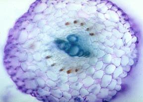
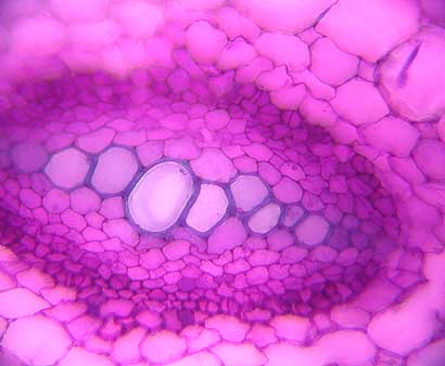
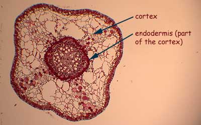
Primary roots are also called herbaceous roots. Here is a cross-section through a primary root of Ambrosia chamissonis grown in tissue culture. This preparation has been stained with toluidine bule. The primary xylem tracheary elements are stained dark blue. In the cortex you can see intercellular spaces which are quite common in roots. One thing that occurs in a small number of plants is the presence of canals associated with the endodermis. You can see the red contents of these canals which almost surround the vascular cylinder. They probably have a defensive function.
Ranunculus (buttercup)
Some plants, such as Ranunculus, do not exhibit any secondary growth and so have no secondary roots. This is not to be confused with lateral root formation which are root “branches”.
This is a cross-section through Ranunculus root.
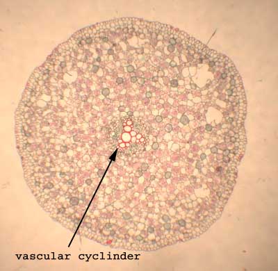
Here is a closer look at the vascular cyclinder.
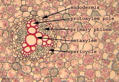
Examine the image of a Ranunculus root cross section and note the following:
The 4-armed arrangement (can have 3) of primary xylem, with the smaller, older (firstformed) protoxylem of the cross towards the arms and the younger metaxylem in the centre. Both protoxylem and metaxylem are primary xylem. The tissue between the arms of this primary xylem is primary phloem, consisting of thinwalled sieve tubes and companion cells. The layer outside the primary xylem and primary phloem is called the pericycle; it consists of somewhat larger parenchyma cells, and is the layer from which lateral roots develop. The endodermis, which lies just outside the pericycle, is the innermost layer of the cortex. Endodermal cells have bands of waxy material called Casparian strips which may be important in controlling what goes into the xylem of the root. Ranunculus roots may undergo a very limited amount of secondary growth. Most of what you see in these slides is basically primary growth. (See Raven Raven 7th, p. 535, Fig. 24-10; 8th, p. 566, Fig. 24-11).
Raphanus (radish)
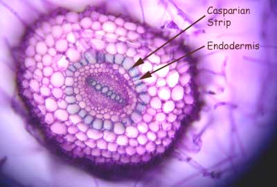
This hand-section through a the vascular bundle of a young radish root shows you a different pattern of primary xylem. There are two protoxylem poles with the metaxylem between. This slide has been stained with toluidine blue.
Here you see the entire section through a root, again stained with toluidine blue. The endodermis and Casparian strips are indicated.
Helianthus (sunflower)
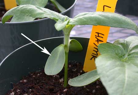
Here is a sunflower plant. The arrow indicates the cotyledon (first embryonic leaf).
A plant has been uprooted to show the three organs: stem, leaves, and roots.
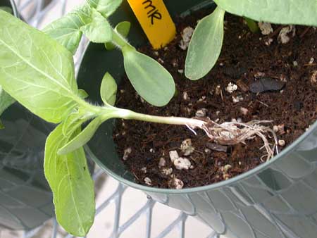
A closer look a the roots. Note the abundance of lateral roots. Each one has an apical meristem and root hairs.
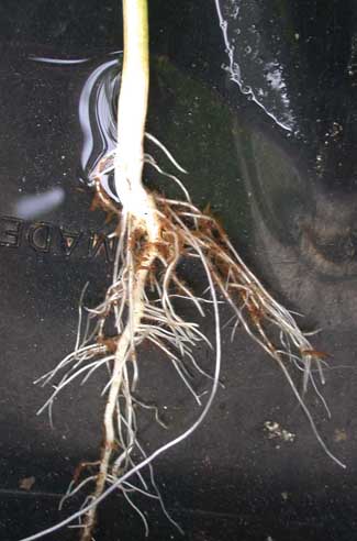
This is the prize-winning root cross-section. You can see that there are three lateral roots arising from this part of the primary root.
A close-up shows you that the vascular tissue of the new lateral root is continuous with the taproot.
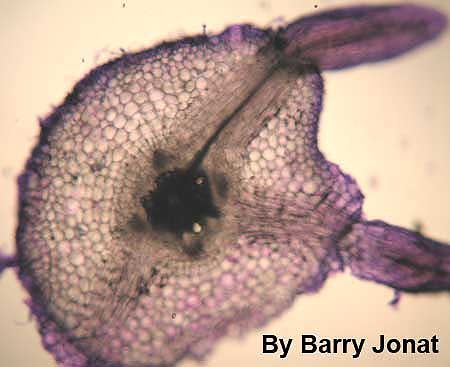
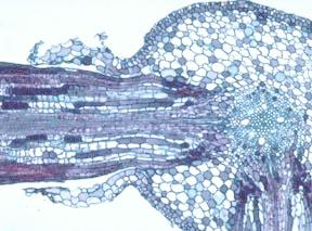
Salix (willow)
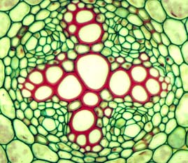
This is a cross-section through the young Salix root through a region where a lateral branch is being produced. You can see four protoxylem arms of the primary xylem. Look for the same tissues as you saw in the Ranunculus root, and note the origin of branch roots. A lateral root is generated opposite a protoxylem pole. See Raven Raven 7th, p. 541, Fig. 24-17; 8th, p. 572, Fig. 24-17.
The lateral roots are generated in the primary root. Secondary growth occurs once primary (elongational) growth is complete. The vascular cambium develops first (residual procambium and pericycle begin to divide). Cork cambium is intitiated in the outer layer of the pericycle. The increase in girth due to the development of the secondary tissues (secondary xylem, secondary phloem, cork cells, and phellogen) cause the cortex and epidermis to break apart and get sloughed off (as you can see in the picture). See Raven 7th, p. 541; 8th, p.572.
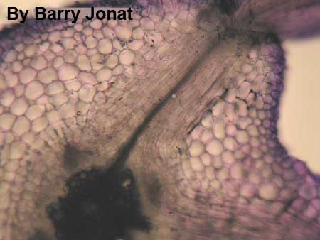
Below is a close-up of the vascular cylinder of the section above. Can you see the four-armed primary xylem? If not click here to see the same picture, but with the primary xylem enhanced.
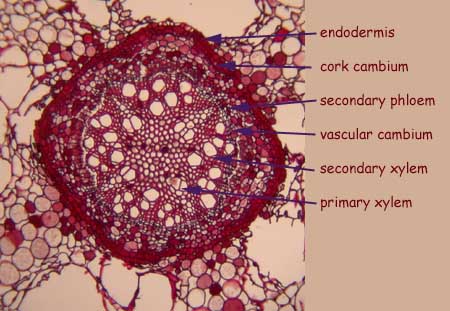
Below is a close-up of edge of the vascular bundle of the root. You can see the endodermis (the innermost layer of the cortex) and the beginning of the periderm (remember the periderm is made up of the cork cambium, cork and phelloderm). The cork cambium is usually initiated in the outer pericycle.

Be sure you understand the changes brought about by secondary growth which take place. Use Raven 7th, p.545, Fig. 24-23; 8th, p.578, Fig. 24-14 to aid your understanding.
ROOT APEX
HERBACEOUS AND WOODY ROOTS
ROOT MODIFICATIONS
BACK TO ROOT FRONTPAGE