
3. PRIMARY TO SECONDARY GROWTH
Primary Eudicot Stem
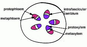
The primary eudicot stem has a vascular system arranged in a eustele (in a ring). The ring of vascular bundles is derived from procambium. The xylem differentiates in the bundle from “pith-side” outward. The protoxylem thus differentiates first and then the metaxylem. The phloem differentiates from the outerpart inward. It is important to keep in mind that the stem is elongating. Once the differentiating tracheary elements (eg. vessels) and phloem elements are functional they cannot withstand too much stretching and therefore must be replaced. That is why the gradual maturation of the vascular bundles is important in growing organs. The metaxylem and metaphloem mature after elongation has ceased. In some plants where there is only primary growth (like the buttercup) the metaxylem is the functioning part of the vascular tissue for the life of the plant.
Ranunculus (buttercup)
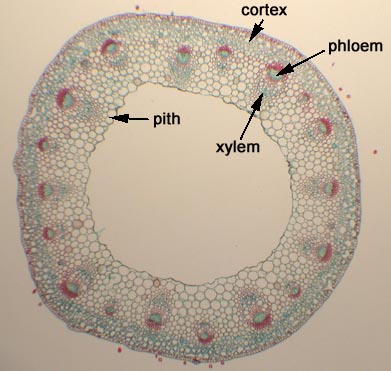
This is a picture of the prepared slide of Ranunculus stem. You can see the cortex and pith. These are two regions of the ground tissue system.
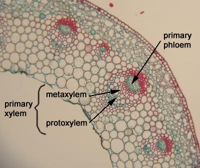
A closer look gives us a better view of the vascular bundles. The protoxylem are both part of the primary xylem. The protoxylem differentiates during stem elongation whereas the metaxylem differentiates once elongation has finished. There is no clear line between the two. In this plant all of the procambium is used up in the development of the primary tissues. No vascular cambium is ever initiated so no secondary tissues are produced. Therefore this is called a closed vascular bundle.
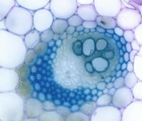
Here is a section through a vascular bundle of buttercup stem. By now you should be able to discern the different parts of a vascular bundle.
Secondary Growth in a Eudicot Stem
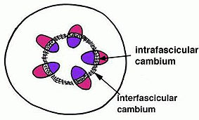
In many plants after primary growth is complete there is often a layer of residual procambium between the primary vascular tissues which will become vascular cambium. This is referred to as intrafascicular (or fascicular) cambium. Parenchyma cells in between the bundles (interfascicular) differentiates to become vascular cambium as well. You can thus see how a complete circle of vascular cambium is formed to start the production of secondary vascular tissues. Primary vascular bundles that will give rise to vascular cambium are said to be “open”.
Ricinus (castorbean)
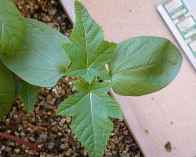
Here is a picture of the seedling of castorbean. You can see the round contyledons and the lobed foliage leaves.
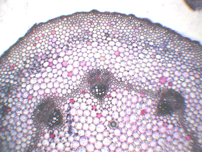
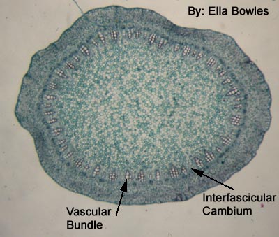
This cross-section through a young part of the castorbean stem shows the distribution of the vascular bundles.
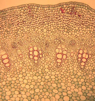
Here is a picture through a vascular bundle. Can you determine the location of the primary xylem and the primary phloem?
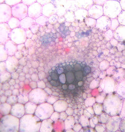
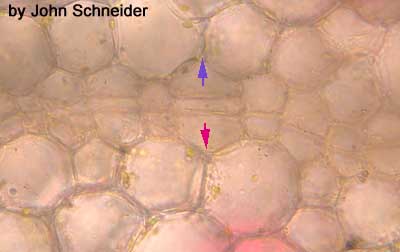
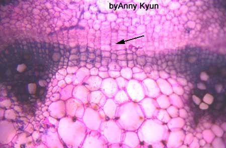
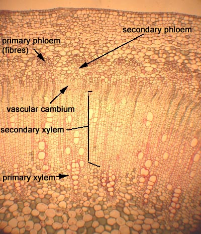
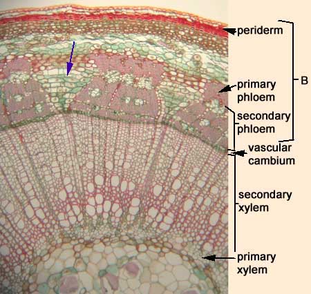
The residual procambium within the bundles will give rise to the vascular cambium by starting to divide in two directions. Secondary xylem is generated toward the inside and secondary phloem toward the outside. Cells of the ground tissue system between the bundles (interfascicular) also start to divide. Here you can see the vascular cambium has been initiated and cells produced to the outside (purple arrow) will become secondary phloem and cells produced to the inside (pink arrow) will become secondary xylem.
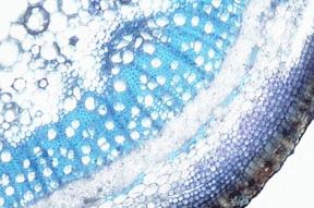
The vascular cambia generated in the vascular bundle and in between the bundles form a continuous layer of vascular cambium. You can also see that there is already some secondary vascular tissue. This slide has been stained with toluidine blue.
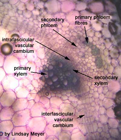
In this cross-section (stained with toluidine blue) you can see the stained elements of the secondary xylem in the interfascicular rregion.
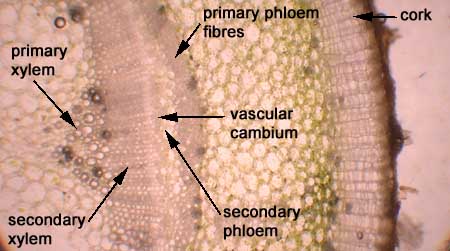
You can see the primary xylem toward the inside of the bundle. Can you see the primary phloem fibres to the outside of the bundle? This is all that remains of the primary phloem.
A closer look at the interfascicular region.
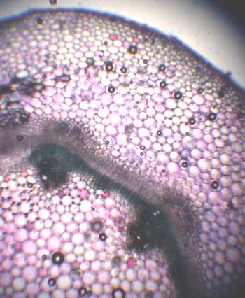
The castorbean stem of the prepared slide has vascular bundles much closer together than our “fresh” plant. Interfascicular cambium is indicated.
Hmm…. a closer look….still can’t see that interfascicular vascular cambium????
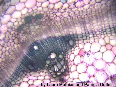
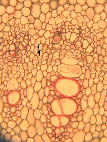
The interfascicular vascular cambium is just starting to divide. You can see it indicated with the black arrow.
This is a picture of a prepared slide of an older castorbean stem. Note the secondary vascular tissues.
Sambucus (elderberry)
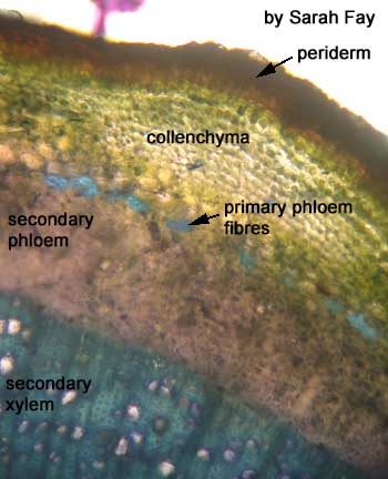
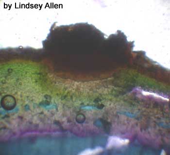
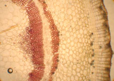
This is a cross-section through the stem of Sambucus (stained with toluidine blue). Note the central pith region which is made up of parenchyma cells outside of this is: the xylem (stained bright blue); the vascular cambium; the secondary phloem; the islands of primary phloem; the lamellar collenchyma; and the thick brown cork on the outside.
Here is another cross-section though the woody stem. It has also been stained with toluidine blue.
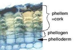
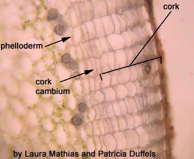
Here is a close-up of the periderm. The cork cambium gives rise to cork cells outward and phelloderm cells inward. The presence of (also called files) of cells is a characteristic feature of tissues derived from cambium.
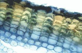
Here is another picture of a section through the periderm. Note the meristematic cells which are called cork cambium (phellogen). The cells to the outside (cork or phellem) have their walls thickened with a cork-like substance called suberin, and give protection to the inner tissues of the stem. The cells inside the cork cambium are called phelloderm, and are living parenchyma cells with dense cytoplasm.
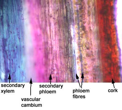
This longitudinal section has been stained with toluidine blue….you can see the same layers as you did in the cross-section…just a different perspective.
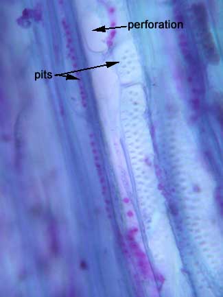
This is a longitudinal view through the secondary xylem. Here you can see a vessel element. The perforation plate is a key characteristic.
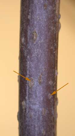
You may have noticed that the cells of the cortex of Sambucus are green. Once a cork layer is produced it makes gas exchange a little difficult. Many plants have lenticels (indicated with arrows) on their bark. These are areas where the cork is spongy, allowing for gases to pass to the photosynthetic tissue.
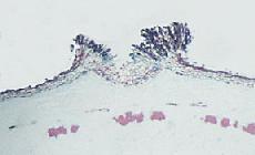
Here is a section through a lenticel.
This is a prepared slide of a section through the lenticel of Sambucus
This is a section through the stem of Ricinus showing a young lenticel. It is the activity of cork cambum which precludes the formation of the lenticel. Lenticels are often the first site of cork cambium formation.
Pelargonium (geranium)
Here is an unstained section through the stem.
This section has been stained with phloroglucinol. Can you identify the different tissue types a little more easily?
To the left you can see a close-up of the periderm of this plant.
Tilia (basswood)
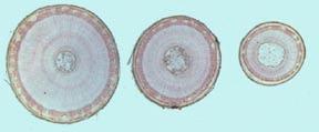
Examine the cross-sections of Tilia. They range in age from one to three years old. Note the thick zone of ringed secondary xylem (wood) and the vascular rays, flaring through the secondary phloem into the cortex. Fibres develop from primary and secondary phloem. The bark in trees consists of all the tissues outside the vascular cambium.
Here you can see the rays extend into the xylem and the phloem. In the phloem they become dilated (indicated with an arrow). Why would such dilation occur? What functions does it serve?
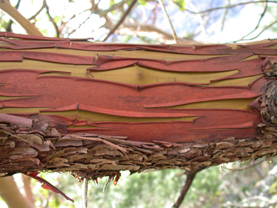
You probably have noticed that bark varies from tree to tree. This is a branch of an Arbutus tree which has paper-like bark.
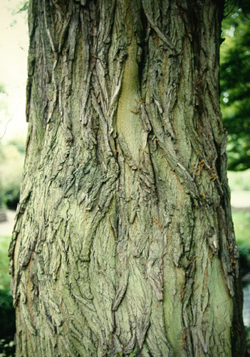
Cedars have very fibrous bark.
3. PRIMARY TO SECONDARY GROWTH