MESOPHYTE LEAVES
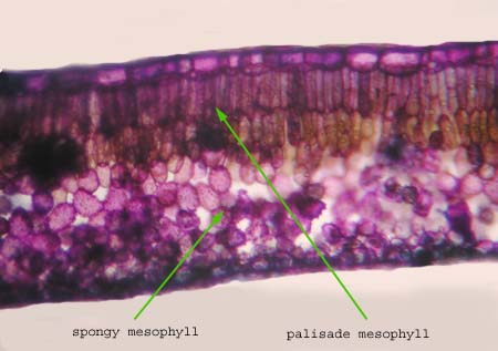
In mesophytes there is usually a cuticle over the epidermis and stomata are more common on the lower (abaxial) than the upper leaf surface. The mesophyll is often divided into adaxial palisade and abaxial spongy layers.
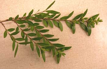
Lonicera leaf
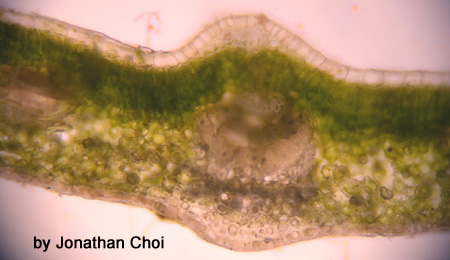
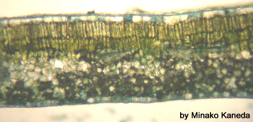
This is a hand section of Lonicera leaf. It has not been stained. Note the tissue types, particluarly the photosynthetic tissues.
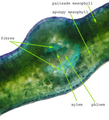
This section is stained with toluidine blue.
This is a section through the midrib stained with toluidine blue. Note the vascular tissue as well as the support cells.
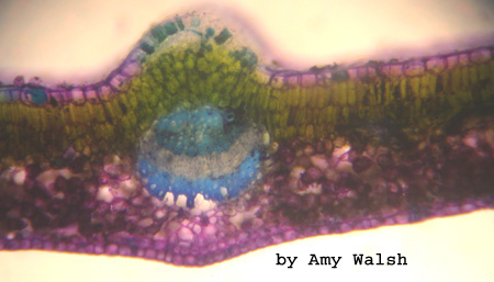
….and another great section.
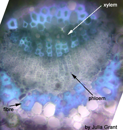
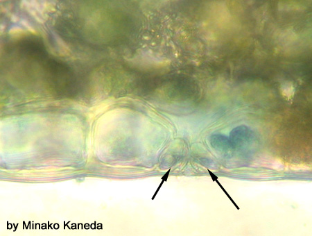
This section is stained with toluidine blue. You can see the guard cells.
Zea (corn) leaf
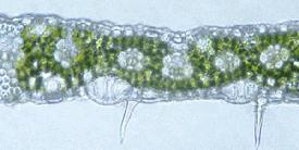
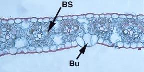
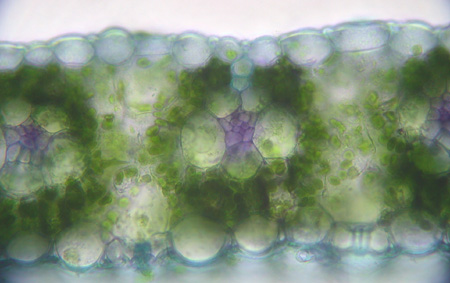
These two sections through the leaf of corn demonstrate that there is no differentiation of the mesophyll into spongy and palisade layers. This is common in corn and other grasses. See Raven 7th, p. 133, Fig. 7-23; 8th, p. 143, Fig. 7-23.
The bundle sheath (BS) surrounds the smaller vascular bundles in the leaf. The bulliform cells (Bu) are believed to be involved in the rolling of the leaves of grasses.
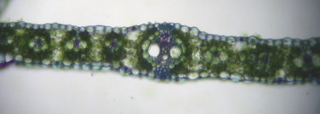
This is a section through a corn leaf. You can see that some of the veins are larger than others.
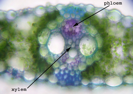
Here is a picture through a large vein. You can see the same type of bundle you saw in the stem cross-section….kind of looks like a clown’s face. It is difficult to tell the top from the bottom of the leaf because there is no palisade mesophyll. Remember that the phloem is toward the lower side of the leaf and the xylem toward the top so this slide is actually upside down.
When you see sections through parts of the leaves with smaller veins being able to determine top from bottom is even more difficult.
MESOPHYTES (there is no shortage of available water)
XEROPHYTES (dry habitat)
HYDROPHYTES (lots of water)
TRICHOMES AND LEAF APPENDAGES
LEAF MODIFICATIONS
BACK TO LEAF FRONTPAGE

