FERN LEAVES = FRONDS
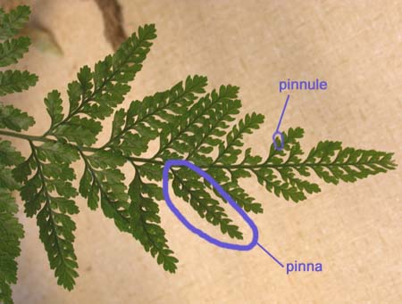
You are probably familiar with the typical leaf (frond) of a fern. The following structures are found: the pinnae (the primary divisions of the leaf), pinnules (the ultimate divisions of the leaf), the petiole (the bare stalk of the frond) and the rachis (the extension of the petiole to which the pinnae are attached).
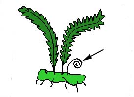
You have probably seen (or eaten) the curled up young leaves called fiddleheads. This characteristic feature of young fern leaves is called circinnate vernation.
Pinna cross-section
This is a section through the midrib of the pinna. You can see two vascular bundles which make up the midrib.
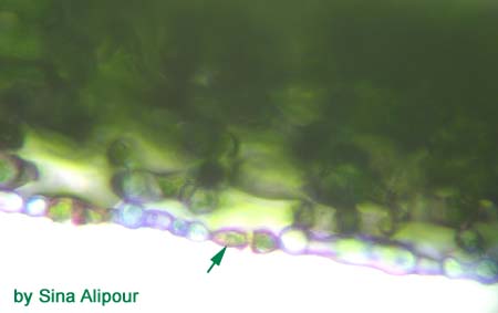
Here you can see the guard cells of the lower epidermis. They are green. Remember that the guard cells are the only photosynthetic cells of the epidermis. they are also found on the lower side of the leaf. They are one way that you can determine which side is up!
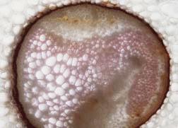
Petiole – cross-section, stained with phloroglucinol
The outer part of the cortex is made up of lignified cells (sclerenchyma). You can see the vascular bundles which make up a ring. On the right is a close-up of a vascular bundle of the petiole. The lignified cells of the xylem have stained red. The endodermal cell walls are thickened.
SPOROPHYTE INTRO
FRONDS
RHIZOME
SPORANGIA


