
Leaf Morphology and Anatomy
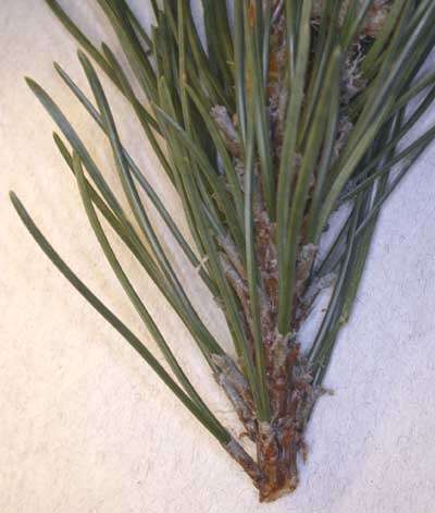
The leaves of the conifers are often needle-like like the pine seen here.
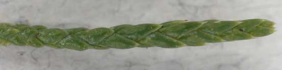
The leaves can also be scale like. Juniperus (juniper), pictured below, for instance has scale-like leaves.
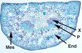
Pinus needle cross section
The mesophyll (Mes) is made up of only spongy parenchyma. There is an endodermis (End) surrounding “transfusion tissue” in the leaves of many conifers. You can see the two vascular bundles with the xylem (X) and phloem (P). The number of vascular bundles is species specific and has given rise to a theory that some conifer leaves are the result of the fusion of two leaves.
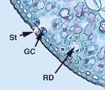
Here is handmade section through the needle of pine.
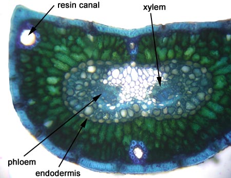
The epidermis is covered with a thick cuticle. The stomata (St) are sunken; you can see the guard cells (GC) below the surface of the leaf. You can see the hypodermis which is one cell thick (just underneath the epidermis). From this information what type of habitat do these organisms live in (or have evolved in)? Resin ducts (RD) are found in the leaves. Where else have you seen them in pine?
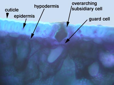
This section shows the overarching subsidiary cells and a whole lot more!
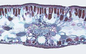
Sequoia sempivirens (coastal redwood) needle cross-section
Some conifers, such as Sequoia do have palisade mesophyll in their leaves as this section through the needle demonstrates.
LEAF ANATOMY