Collenchyma Tissue
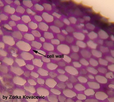
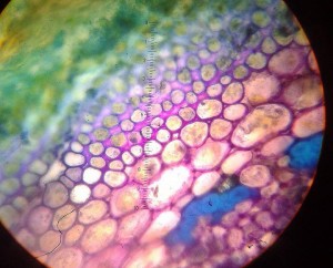
Collenchyma cells, like parenchyma, are living cells, but are usually elongated with unevenly thickened, unlignified primary walls. There are many types of distribution of thickenings. For instance the corners between cells may be thickened as in the example here to the right. This is called angular collenchyma.
The thickened walls provide support, but can still stretch to accommodate growth. For this reason collenchyma functions as support in plant organs which are still elongating. It is most commonly found just beneath the epidermis.
Celery petiole
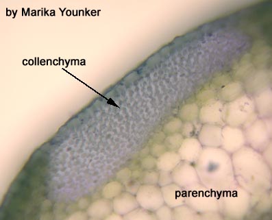
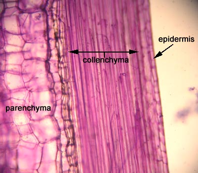
The collenchyma cells are the thicker walled cells found in bundles just beneath the epidermis of the petiole. Larger thin walled parenchyma cells can be seen making up the bulk of the petiole tissue, so this is a good opportunity to compare the two cell types. This is an unstained section through the celery petiole.
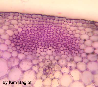
Here is a stained section. The cell walls are primary and therefore stain a purple-pink colour with toluidine blue.
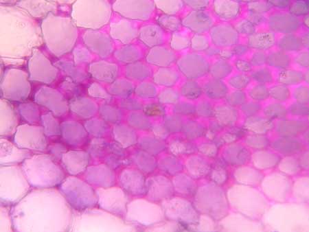
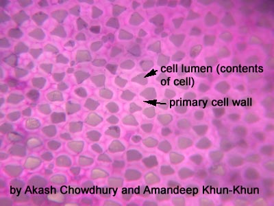
Here is a close-up. Can you see the thickened primary cell walls?
The collenchyma cell walls in celery are like those illustrated in figure 2-2A of your lab manual, i.e. with walls thickened at the corners.
another great section…….
Another characteristic of collenchyma cells is that they are elongate. This longitudinal section demonstrates this characteristic.
Sambucus (elderberry) stem
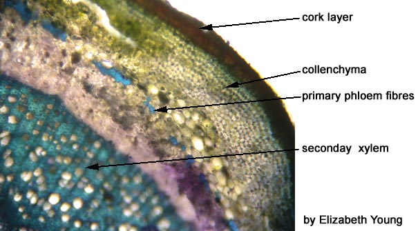
Below is a cross-section through a Sambucus. You should find a layer of collenchyma just inside the brownish periderm (cork).
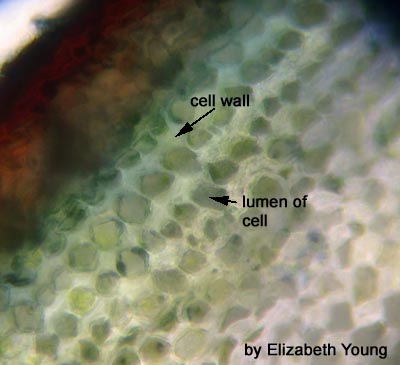
Collenchyma in Sambucus exhibits wall thickenings on the inner and outer cell faces, like those in Fig. 2-2B of your lab manual. (Refer to Raven 7th p. 513, Fig. 23-5; 8th p. 542, Fig. 23-5)
Stained with toluidine blue – Section through Sambucus (elderberry) showing some lovely collenchyma as well as primary phloem fibres
Other tissue types:
PARENCHYMA
SCLERENCHYMA
BACK TO GROUND TISSUE SYSTEM FRONTPAGE


