VESSELS
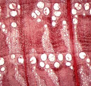
Quercus (oak) Wood
In cross-sections of wood of most angiosperms the vessels appear as large holes. The wood is thus said to be porous. This is a cross-section through oak wood. As you can see it is very heterogeneous tissue made up of many different cell types. The large pores you see are vessels. This wood is said to be ring-porous because the larger vessels are found at the beginning of the growth ring (earlywood). Vessels offer little in the way of structural support. This function is performed by fibres which look darker in this preparation. The lighter coloured areas you see have parenchyma, tracheids and smaller vessels. You may see lines running perpendicular to the growth rings. These are rays which are made up of parenchyma.
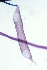
This is a picture of a vessel. You can see the perforation (hole) on both ends of this cell. The presence of the perforation is the most important characteristic that sets the vessel apart from the tracheid.
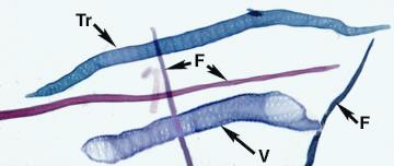
This is a preparation of macerated tissue (the cells have been chemically separated). The different cell types are labelled (V=vessel, Tr=tracheid, F=fibre). Parenchyma is also found in the xylem tissue.
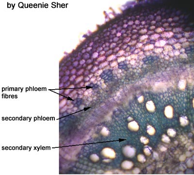
Coleus
Here is a section through the stem of Coleus. You can see the large vessels very clearly.
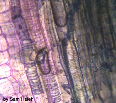
This longitudinal section through Coleus stem shows vessel elements with a couple of different types of secondary wall thickenings? Can you see them? If not keep reading and then come back.
Here you can see a vessel element with annular wall thickenings. The cell has been stretched so the thickenings are far apart.
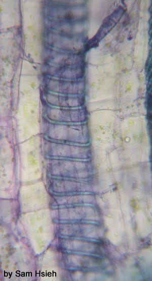
Here is a good example of helical (or spiral) wall thickenings.
Here is a vessel with scalariform wall thickenings.
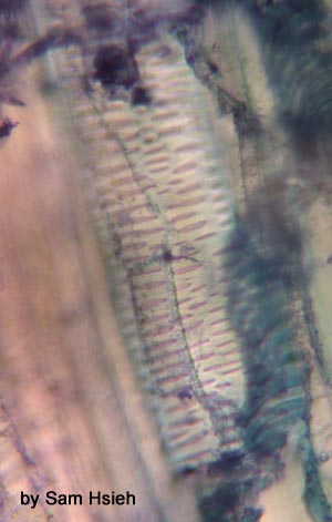
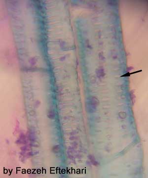
Celery Petiole
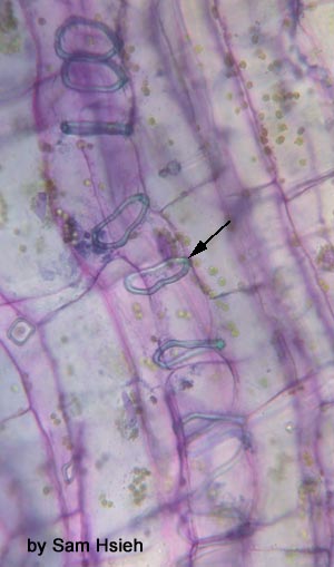
This is a longitudinal section through the petiole of celery. Here you can see that the secondary wall thickenings can vary even within the same plant. Can you name the types of wall thickenings pictured here? The arrow is pointing at a pit.
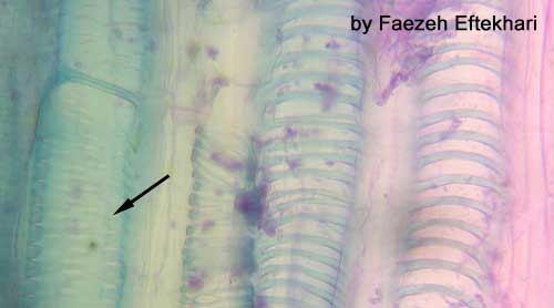
Here is a close-up of vessels of the same plant. Why would you expect vessels such as these in tissues that have completed primary growth?