
Sclerenchyma Tissue
Sclerenchyma cells may be living (at least at the initial stage) but lose their protoplasts at maturity. In contrast to collenchyma, sclerenchyma has secondary walls, and lignin is present. Two types of sclerenchyma cells are usually recognized: 1. Fibres and 2. Sclereids.
1. Fibres
Fibres are very elongate cells which have have thickened secondary cell walls. They function in support for the plant and are generally not living at functional maturity.
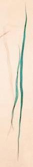
Sansevieria (Mother-in-law’s tongue) Leaf
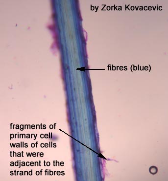
Here is a picture of fibres which have been pulled from a plant leaf. They have been stained with toluidine blue. You can see that each strand that you pulled from the plant is actually made up of a bunch of fibre cells.
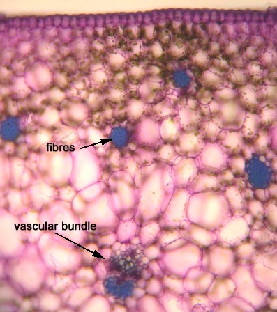
This is a cross-section through a leaf.
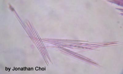
Here is a picture of some needle-like structures that can be found within some of the cells of this plant. Pretty nasty for some unsuspecting herbivour!!!
Other examples of fibres:
CYMBIDIUM (an orchid).
LINUM (linen)
2. Sclereids
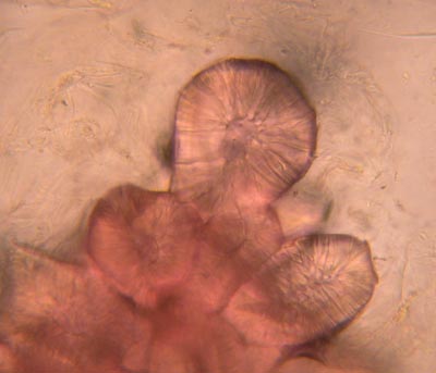
Pyrus communis (pear) Pulp
This is a slide of pear pulp which has been stained with phloroglucinol. The walls of the sclereids have stained pink because of the lignin. Observe the clusters of sclereids (refer to Raven 7th p.516, Fig. 23-10; 8th p. 544, Fig. 23-10). The center of the cell (lumen) is very small.
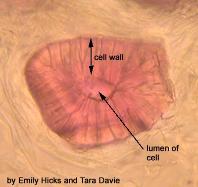
The shape of these sclereids is isodiametric. This close-up of one sclereid shows the deep pits running through the thick cell wall. The lumen is visible in the center of the sclereid.
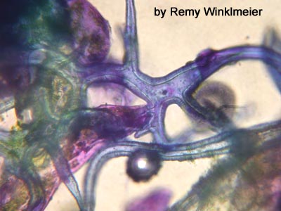
Sciadopitys (Japanese Umbrella Pine) Leaf
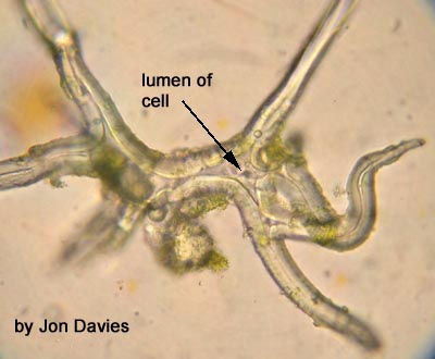
This cross section of the needle of Sciadopitys (an exotic conifer) demonstrates the branched sclereids. They stain light blue with toluidine blue. Can you think of possible functions of these sclerenchyma cells? (Note the thick cuticle on the outer surface of the leaf).
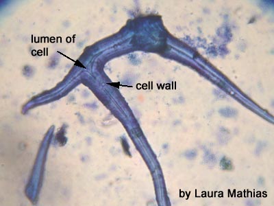
Here is a close-up of the sclereid. You can see it branches and that it has a very thick cell wall.
This is a sclereid which wasn’t stained.
Malus (apple) Endocarp
This is what the core of your apple looks like under the microscope. It is very difficult to separate the sclereids, even after the sample is boiled in potassium hydroxide (KOH) for two hours to partially macerate the tissue. You are looking at many sclereids laid down in sheets.
Other tissue types:
COLLENCHYMA
PARENCHYMA
BACK TO GROUND TISSUE SYSTEM FRONTPAGE