Author Archives: cdrohan

Final Project Outline

One of the intermediate stages of my final project.

Topic chosen: Cancer and DNA methylation

Specific question: Does loss of DNMT3A lead to changes in HOXA9 methylation that contribute to leukemogenesis?

How is this question novel and original? DNMT3A null mutations in acute myeloid leukemia were only discovered in 2010. To date, the majority of studies have only examined the presence of DNMT3A mutations in clinical leukemia samples, with few performing “add something” or “remove something” experiments. Those that did examine DNMT3A knockout’s effect in AML did not delve into the mechanisms through which this increased propensity to cancer might be occurring. This study will investigate a potential avenue through which DNMT3A exerts its leukemogenic function.

Hypothesis: I hypothesize that knockout of DNMT3A will lead to hypomethylation of the HOXA9 gene, which will in turn increase the cells’ propensity to develop into AML.

Evidence on which the hypothesis is based:

  • DNMT3A is frequently mutated in acute myeloid leukemia (Yang et al., 2015)
  • DNMT3A mutation is one of the first mutations to occur in DNMT3A-/- AML and increases likelihood of developing into cancer (Shlush et al., 2014)
  • Homeobox-containing (HOX) genes were found to be hypomethylated in DNMT3A-/- cancers (Qu et al., 2014)
  • HOXA9 overexpression has been implicated in AML development and poor prognosis (Collins and Hess, 2016)

Predictions: DNMT3A knockout will result in hypomethylation of the HOXA9 promoter, which will in turn lead to increased gene expression from the HOXA9 locus. This upregulation of HOXA9 will render cells predisposed towards AML.

Experimental approach to test prediction:

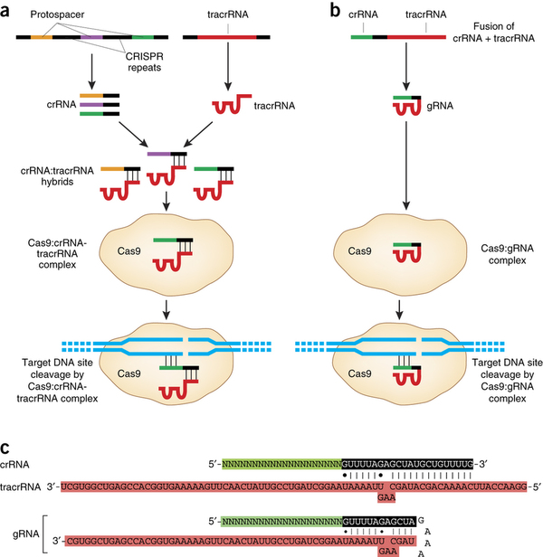
  • Knockout of DNMT3A gene – catalytic domain, likely using CRISPR/Cas9 system or siRNA
  • Methylation status of HOXA9 – measure via bisulfite conversions and pyrosequencing
  • Protein expression, mRNA expression of HOXA9 – Western blot, RT-qPCR
  • Xenograft or tumourigenesis assay in vitro to assess leukogenic capacity of cells

List of relevant primary and review articles read:

Yang, L., Rau, R., Goodell, M.A. (2015). DNMT3A in hematological malignancies. Nature Reviews Cancer. 15:152-165.

This review summarizes the research connecting mutations in the DNA methyltransferase 3A (DNMT3A) enzyme to blood malignancies such as acute myeloid leukemia (AML). This paper provided crucial information surrounding the structure of the DNMT3A enzyme, its catalytic activity, and the role it plays during development. The authors also examine the research implicating DNMT3A mutations in various blood malignancies of the myeloid and leukoid lineage, and propose a model for leukemia development in which DNMT3A acts to transform cells into a “pre-leukemic” state, and subsequent mutations in key proteins such as NPM1 result in different types of blood malignancies. This model greatly informed my own hypothesis and predictions for my research project.

Qu, Y. et al. (2014). Differential methylation in CN-AML preferentially targets non-CGI regions and is dictated by DNMT3A mutational status and associated with predominant hypomethylation of HOX genes. Epigenetics. 9(8):1108-1119.

This primary research study characterized the genome-wide methylation differences between cytogenically normal AML (CN-AML) cells and healthy control bone marrow cells. A key finding from this study was that homeodomain-containing (HOX) genes experienced the most significant changes in methylation, with hypomethylation occurring in genes including HOXA5 and HOXA9. While this study showed only a correlation between DNMT3A mutation and hypomethylation of homeobox genes, it provides an intriguing potential mechanism through which DNMT3A may initiate cancer.

Collins, C.T., Hess, J.L. (2016). Role of HOXA9 in leukemia: dysregulation, cofactors, and essential targets. Oncogene. 35(9):1090-8.

This review summarizes the role of the homeobox family protein HOXA9 in leukemia. HOXA9 is a transcription factor that plays several important roles in development, specifically in the expansion of hematopoietic stem cells (HSCs). The molecular mechanisms through which HOXA9 can induce leukemia are not yet understood, and it is hypothesized that several additional upstream genetic alterations leading to overexpression of HOXA9 have yet to be identified. Given that HOXA9 has also been found to be hypomethylated in DNMT3A-null cancers, this paper brought up the question of whether loss of DNMT3A and HOXA9 hypomethylation are connected to the gene’s overexpression and leukemogenic activity.

Potential ways to make your question known to the public at large:

  • Biggest thing that people need to understand with cancer therapy and research is the heterogeneity of cancers – no two tumours are identical, which makes treatment especially challenging
  • Need to explain concept of DNA methylation – enzyme (small molecular “machine”) adds a tag to DNA that turns it on or off
  • Also emphasize the prevalence of AML – most common in older adults (65+) but still does affect younger people, has a poor prognosis (1 in 4 5-year survival rate)


Learning Journal 5

Name of the classmate whose portfolio you will be viewing: Leon Lai

What would you say the biggest strengths of this person are and why?

Leon is great at reflecting upon his experiences. When I was going through his posts, I was impressed with the depth of reflection on projects and assignments, and the effort that he had put into identifying successes and challenges. I think that this is a great skill that will serve Leon well in his future in the sciences.

What is the most valuable thing you learned from doing your final project?

The most valuable thing I learned from doing this project is how challenging it can be to come up with an original research question that a) fills a gap in the present scientific knowledge, and b) is in logical accordance with the existing data in the literature. I found myself constantly having ideas of things to investigate, but I would quickly realize that my line of reasoning was disproven or called into question by other studies. I must have gone through 10 different research questions and hypotheses before finally settling on one. I have a newfound respect for grad students and scientific researchers, who must constantly be looking for new areas to investigate. It requires far more delving into the existing literature than I had previously imagined, and requires you to synthesize these dozens of papers and research findings into a model that makes sense to investigate. I will definitely be bringing my PI a coffee next time I hear that he is working on grant proposals, since I now understand what a struggle it can be!

What do you think your classmate may have learned that you have not, or vice versa?

Leon learned a lot about clinical research by investigating his final project. His project was examining the effect of alcohol consumption on the immune system of children, which has the potential to be ethically contentious – both because it involves a substance that can be damaging for health, and because it involves children, which requires additional ethical safeguards. Through working on his project, it is clear that Leon learned how to ethically conduct a clinical study, ensuring the safety and comfort of all participants.

Contrary to Leon, my final project was focused on model systems – specifically, cell culture and mouse models of disease. Therefore, I learned how to choose a model system that works well for my purposes and that best recapitulates the physiological conditions in humans.

The genome revolution and molecular biology

Thinking about the general field of molecular biology and gene regulation, what has been the most impactful discovery or invention over the past 50 years? Why do you think it was the most impactful? How did it impact the field?

The Human Genome Project has been getting quite a bit of flak recently. In a 2010 article in Scientific American, Stephen S. Hall stated that the Human Genome Project (HGP) had to date “failed to produce the medical miracles that scientists promised”. The HGP, which was completed in 2003, was a decade-and-a-half-long international research effort to sequence and map the entirety of the human genome. Upon the project’s completion, the scientific and medical communities were excited about the potential of this information to personalize medicine and discover the genes that were responsible for every human disease. However, things turned out to be a bit more complicated than anticipated. Many of the conditions that are most prevalent around the world, such as diabetes, do not have a single gene to blame. Instead, genome-wide association studies (GWAS) have demonstrated that a multitude of potential genes interact in complex and mysterious ways to produce the metabolic condition. When it comes to investigating the causes of disease, it seems that every new discovery leads to another ten questions. As a result, the personalized medicine revolution does not seem likely any time soon.

However, while it is true that the sequencing of the human genome has not delivered all of the exceptional results that were initially promised, the ability to sequence an entire genome rapidly and efficiently has undoubtably revolutionized the way in which research in molecular biology is done. The DNA sequence of a protein one wishes to investigate can be found with a quick Google search. Sequencing an entire genome can take place in a matter of hours. Mutations can be quickly and easily identified, and compared to a reference genome. Many new technologies, including next generation sequencing techniques, have been developed as well, undoubtably inspired by the quest to sequence the entire genome. These changes have reverberated across all fields of research within the life sciences, and have the potential to lead to discoveries more efficiently. Therefore, although the impact of the Human Genome Project may not be seen in hospitals and clinics, it can be easily observed in the research labs located around the world.

Final Project – The role of DNMT3A and HOXA9 hypomethylation in AML

Final Project

My final project is a proposal for a research project investigating the connection between DNA methyltransferase 3A (DNMT3A), HOX9A, and the development of acute myeloid leukemia (AML).

Lay Person Summary

Small molecules called enzymes carry out a wide variety of important processes in the body. One such molecule is DNMT3A, which adds modifications to DNA regulatory regions to prevent genes from being expressed as proteins. This process is called DNA methylation, and it is used to regulate the expression of different genes in the cells of the body.

Studies have shown that DNA methylation patterns are altered in many types of cancer, including acute myeloid leukemia (AML). This is a type of blood cancer in which blood stem cells fail to grow into mature blood cells, leaving immature cells called “blasts” crowding the bone marrow and impairing its function. It is a fast-developing cancer with a five-year survival rate of less than 1 in 4. Mutations in the DNMT3A gene that prevent it from functioning properly has been observed in up to 22% of AML cases, as well as in other types of cancer. DNMT3A is one of the earliest mutations that occurs in AML, which suggests that its dysfunction might predispose cells to leukemia development.

But how does this happen? Few studies have investigated the mechanisms through which defective DNMT3A can affect AML. However, a recent study showed that there may be a link between the DNMT3A mutation and decreased methylation of a group of genes called the homeobox-containing (HOX) genes. One member of this family is HOX9A, . Since decreased methylation of a gene can cause increased expression of the gene’s protein product, it is possible that the dysfunctional DNMT3A protein is unable to methylate the HOXA9 gene, leading to overexpression and AML. This study will investigate this possibility by destoying the DNMT3A gene in blood cells, assessing the methylation levels of the HOXA9 gene, and transplanting the cells into mice to determine their ability to cause leukemia. By developing a better understanding of DNMT3A’s role in leukemia development, it will be possible to identify new targets for therapies to help treat those with AML.

Reflection

The learning objectives that I believe I demonstrated through this assignment are:

  • Approach questions, concepts, and facts with curiosity
  • Deal positively with frustration, setbacks, and failure – there was a lot of frustration in the process of developing a research question!
  • Search the literature and effectively extract relevant information from it
  • Integrate new factual and conceptual information into your models/thoughts/ideas
  • Communicate information, ideas, and findings clearly and succinctly at a variety of levels – demonstrated through the write-up and the lay person summary
  • Develop new questions that can be investigated experimentally
  • Construct testable hypotheses based on evidence
  • Predict the outcomes of observations, experiments, and correlational studies based on a given hypothesis or model
  • Select the most suitable techniques to test a hypothesis
  • Evaluate what can and what can’t be concluded from given or predicted data/results

Appreciating the negative in scientific research

Granqvist, E. (2015). Why Science Needs to Publish Negative Results. https://www.elsevier.com/reviewers-update/story/innovation-in-publishing/why-science-needs-to-publish-negative-results.

“Publish or perish” is a commonly touted phrase within academic research. The main way in which scientists are able to gain funding, publicity, and prestige is through publishing impactful and important results in a journal. But this can be a very stressful path.

For a post-doctorate fellow, especially in the biological sciences, attempting to get a professorial position without a long list of impressive published papers to your name is virtually impossible, as I have seen firsthand. In one of my co-op positions at an academic research lab, I worked under an absolutely brilliant post-doctoral fellow. After several years of research in this particular lab, she was hoping to obtain a professor position and run her own lab at another university. However, this proved incredibly challenging, and I could tell that she constantly felt the pressure to publish novel research findings in high-impact journals. And novel findings usually do not come from experiments with negative results.

Failing to publish negative results is not only damaging for the individual, but for the scientific community at large. When a researcher simply keeps negative results to themselves, there is a possibility that other scientists who are working on a similar problem may attempt the same experiment. While duplication of experiments is also important (and frequently overlooked), this can ultimately be a waste of resources, as the question has already been answered. Instead of pursuing more promising avenues, researchers find themselves toiling over a hypothesis that has already been disproven, which does nothing to advance knowledge within the field. Furthermore, this high-stress culture can in turn make it more tempting to falsify results in order to make your research seem more “exciting”. Retraction Watch, a site that documents retracted papers (frequently retracted due to fraudulent results), is filled with stories of scientists that have doctored images or tinkered with raw data in order to make their research publishable. While the vast majority of scientists will not go to such extremes in order to obtain positive results, this phenomenon can be extremely problematic for the scientific community at large, fostering a community of mistrust and cutthroat competition rather than open collaboration.

However, the picture is not entirely bleak. In recent years, new journals have begun to spring up that specialize in publishing negative results, such as the Journal of Negative Results, New Negatives in Plant Science, and the Journal of Negative Results in Biomedicine. While these journals may not have an impact factor approaching that of Science or Nature, they still provide an outlet for this much-needed research, and are slowly working to dismantle the stigma surrounding “failed” experiments. This can be extremely beneficial for research as a whole, since it promotes a more open culture of science and prevents unnecessary experiments from being repeated. Furthermore, becoming more accepting of negative results could also help younger people who are considering a career in research. Instead of teaching trainees that negative results do not matter, we could adopt the mindset that any results are “good” results, as they . As Thomas Edison said, “I have not failed. I’ve just found 10,000 ways that won’t work.”

Honeybee Epigenetics Assignment

Paper: Shi et al. (2011). Diet and cell size affect queen-worker differentiation through DNA methylation in honeybees (Apis mellifera, Apidae). PLoS One.

What are some other “types” of honeybees, other than queens and workers?

Drones are male bees that result from a haploid unfertilized egg. They do not have stingers, nor do they gather nectar or pollen. Intercaste is another type of bee that has an intermediate phenotype between that of a queen or worker.

What prompted the authors to investigate the effects of DNA methylation on honey bee development?

Recent studies had suggested that DNA methylation was a factor involved in the caste differentiation of honeybees. Honey bees had been found to have DNA methyltransferases that are orthologs of the vertebrate Dnmt1 and Dnmt3 enzymes, and silencing of the Dnmt3 gene was found to result in a higher proportion of queens. The authors were interested in examining whether royal jelly exerts its action through changes in methylation, mediated by Dnmt3. The authors were also interested in examining whether different cell sizes could also contribute to the caste differentiation process, as there had not been much attention previously devoted to this factor.

Considering Table 1, what are the main question(s), experiment, results, and possible conclusions? How do conclusions support the article’s title claim?

Question: What effect(s) do(es) food and cell type have on the queen-worker differentiation process?

Experiment: Examined larval development when they were fed royal jelly versus worker jelly. Also examined development when larvae were grown in different sizes of cells.

Results: There were changes in methylation at 4 sites as the duration of royal jelly feeding increased, indicating that royal jelly duration is correlated with methylation at CpG locations.

Conclusion: This experiment demonstrates a correlation between the duration of royal jelly consumption or growth within a queen cell and development into queens, which provides some support for the article’s title claim. However, another experiment must be done in order to demonstrate convincingly that the methylation pattern changes are what cause the larvae to develop into either queens or workers. Until then, the effects of diet and cell size cannot be said to function “through DNA methylation” in order to affect differentiation.

Consider Figure 2. What is/are the experiment, results, conclusions, and possible inferences? How do the conclusions support the title claim?

Experiment: Larvae were fed with royal jelly for different durations (3, 4, or 5 days). The levels of Dnmt3 enzyme activity, Dnmt3 expression, and methylation of the dynactin p62 gene were measured, and the differentiated state of the honeybees were observed at day 6.

Results: A longer duration of royal jelly feeding was found to be correlated with an increase in the percentage of adults that differentiated into queens. Dnmt3 activity and expression, as well as methylation of dynactin p62, were shown to decrease as the duration of royal jelly feeding increased.

Conclusions: While this experiment does not explicitly establish causation between methylation activity and differentiation into queens, the fact that 5 days of royal jelly feeding was sufficient to make all of the larvae differentiate into queens provides strong support that an element of the royal jelly is responsible for the differentiation into a queen phenotype. 

Consider Figure 3. What does cell size refer to? What was the experimental question, the experiment, results, and conclusions? Do the conclusions support the title?

Cell size refers to the type of cell in which the larva develops – queen cell or worker cell.

Question: What is the effect of cell size on the methylation patterns of honeybees? Does a change in methylation patterns correlate with a change in the percentage of adults that develop into queens?

Experiment: Reared larvae in either worker or queen cells, and assayed them at 3 and 5 days of age. Measured the levels of Dnmt3 activity, Dnmt3 RNA expression levels, and percent methylation of the dynactin p62 gene.

Results: Larvae that were reared in the queen cells had lower levels of Dnmt3 activity, mRNA expression, and overall methylation in the dynactin p62 gene relative to larvae raised in worker cells. However, no queens resulted from larvae reared in queen cells, with 19% differentiating into intercastes.

Conclusions: Rearing larvae in queen cells is not sufficient to differentiate honeybees into queens. However, queen cell size is correlated with decreased levels of Dnmt3 activity, expression, and methylation. These data may suggest that the cell size may be another component of the differentiation process, as rearing larvae in non-worker cells (i.e. queen cells) resulted in a lower proportion of workers overall.

Propose a model, incorporating the conclusions made from the data presented in the article and the information presented in class, that explains how the difference in diet between queen and worker larvae could result in their phenotypic differences.


The KCNQ1OT1 imprinting control region and non-coding RNA.

Chiesa et al. (2012). The KCNQ1OT1 imprinting control region and non-coding DNA: new properties derived from the study of Beckwith-Wiedemann syndrome and Silver-Russell syndrome cases. Hum Mol Genet. 21(1): 10-25.

Introduction

  • Imprinting control regions (ICRs) are cis-acting regulatory elements of the imprinted loci
  • Chromosome 11p15.5 has a large cluster of imprinted genes
  • Divided into 2 separate domains that each have their own ICRs
  • ICR1 is telomeric, ICR2 is centromeric, have different mechanisms of action
  • ICR2 is the promoter of KCNQ1OT1 gene (non-coding, imprinted)
    • KCNQ1OT1 is antisense, contained within the protein-coding KCNQ1 gene
    • Non-coding KCNQ1OT1 transcript silences imprinted genes of the centromeric domain on the paternal chromosome
    • Methylation of ICR2 on maternal chromosome, not transcribed and imprinted genes are expressed
  • Changes in methylation patterns on the imprinting control regions (ICRs)
  • Loss of methylation on the maternal allele of ICR2 results in BWS
    • Most common defect in BWS
    • Leads to bi-allelic activation of KCNQ1OT1 and silencing of the imprinted genes
    • This includes the cell growth inhibitor CDKN1C
  • Inverse defects in DNA methylation at ICR1 causes BWS or SRS
    • With associated changes in the expression of IGF2/H19 transcripts
  • Mutation of the cell growth inhibitor CDKN1C (5% of BWS)
  • Loss-of-function mutation in a trans-acting factor demonstrated in familial case of BWS
  • Uniparental disomy frequently observed in BWS
  • Chromosomal abnormalities – rarer, usually involve paternal duplications, maternal deletions or balanced maternal translocations in BWS
    • Maternal duplication usually observed in SRS
  • 2 cases in this paper:
    • 2 Mb long inverted duplication of entire imprinted gene cluster à SRS phenotype
    • 160 kb duplication including ICR2 and 5’ 20 kb of KCNQ1OT1 co-segregating with BWS phenotype in 3 generations
      • Expression of truncated KCNQ1OT1 transcript, silencing of CDKN1C results

Results

  1. SRS family
  • SRS patient born in family with no signs of SRS
    • Intra-uterine growth restriction, low birth weight and length
    • Height and weight below the 3rd centile
  • Slight ICR1 hypomethyation, ICR2 hypermethylation observed in patient
    • Parents were both normal at both loci (50%)
  • Used microsatellite and SNP analysis to find de novo maternal duplication in the patient
  • 2 Mb duplication encompassing the entire imprinted gene cluster, only in cis (confirmed via FISH)
  • Hypermethylation of CpGs throughout SRS patient 1, consistent with duplication of methylated maternal allele
    • Shows duplicated chromosome acquired imprinted methylation of ICR2
  1. BWS family
  • Female patient born to unrelated parents
    • Mother born with elevated birth weight, father normal
    • Maternal uncle and aunt also had elevated birth weight
    • Umbilical hernia also runs in the family on the maternal side
  • MS-MLPA – hypomethylation at ICR2 and normal methylation at ICR1 in III-6, II-4, II-3, I-4
    • Normal methylation in ICR2 and ICR2 at II-5, III-5
    • Increased copy number of KCNQ1 exons 12-15 and ICR2 in all family members with hypomethylation
    • Maternal transmission of 11p15.5 duplication from I-4 to II-3 and II-4, from II-4 to III-6
  • Identified in cis duplication of a portion of the KCNQ1 gene (exon 12-15), ICR2 and most 5’ 20 kb of KCNQ1OT1
    • Excluded mutations of CDKN1C gene in patient 1 via exon sequencing
  • Extensive hypomethylation at ICR2 in all individuals with the 160 kb duplication
    • Loss of imprinted methylation of maternal allele evident in BWS patients 1 and 2
    • 160 kb duplication leads to imprinting alteration and BWS phenotype only with maternal transmission
  • By cloning the patient cells, determined that hypomethylation was likely due to lack of methylation of one of the two ICR2 copies present on the maternal chromosome
  • Found KCNQ1OT1 gene expressed on the maternal chromosome
    • Bi-allelic expression of KCNQ1OT1 in duplication region but normal in non-duplicated region
  • Level of CDKN1C RNA was lower in BWS patient 2 vs. age-matched controls
    • Indicates silencing of CDKN1C by expressed KCNQ1OT1
    • Confirmed that KCNQ1OT1 interacts with chromatin
    • Interaction exerted at least partially by 5’ 20 kb sequence
    • The silencing of CDKN1C likely occurs via interaction with chromatin

Chiesa et al. Assignment

1. Based on the article, what are the known causes of SRS and BWS? Which of these causes are genetic, and which are epigenetic?

The known causes of SRS and BWS all involve defects in the 11p15.5 imprinted gene cluster, which contains two independent domains that are each under control of a different imprinting control region (ICR):

The genetic causes:

  • Mutation of the DNA sequence of the cell growth inhibitor CDKN1C (5% of BWS cases)
  • Loss-of-function mutation in a trans-acting factor
  • Uniparental disomy
  • Chromosomal abnormalities – maternal deletions, paternal duplications, balanced maternal translocations (BWS), maternal duplications (SRS)

The epigenetic causes are:

  • Loss of DNA methylation on the maternal allele of ICR2
  • DNA methylation defects at ICR1, associated with changes in IGF2-H19 expression

2. What do the data in Figure 5B show, and what do they tell us about the methylation state of the ICR2 region in individuals I-4, II-4 and III-6?

Figure 5B shows the methylation status of the ICR2 region, which is located within the imprinted gene cluster at 11p15.5, in three individuals in the BWS family. Two of these individuals carry the BWS phenotype (II-4 and III-6), while I-4 is normal phenotype. Individuals II-4 and III-6 have the maternal 160 kb duplication that was identified in the paper. The data show that I-4, II-4, and III-6 have hypomethylated ICR2 alleles relative to an unrelated control. The overall methylation pattern is quite similar across the three cases, but I-4 has the maternal allele almost entirely methylated, while the duplicated maternal allele is only partially methylated in II-4 and III-6.

3. Notice how I-4, II-4 and III-6 all have the same number and methylation pattern of ICR2 ‘loci’. How can their difference in terms of having vs. not having BWS be explained?

Individuals II-4 and III-6 have the BWS phenotype, while I-4 does not. This difference can be explained based on the origin of the 160 kb duplication in ICR2. I-4 has a duplicated paternal allele, while II-4 and III-6 have duplicated maternal alleles. In both of the BWS patients, the maternal allele is only partially methylated, which indicates disruption of the imprinted methylation process in these individuals. In I-4, however, the maternal allele is still almost entirely methylated and the paternal allele is unmethylated. This difference suggests that the 160 kb duplication alters the imprinted methylation of ICR2 only when maternally transmitted, and that this is associated with the presence of the BWS phenotype.

4. Explain what Figure 7B shows and how you interpret the data.

This figure shows the interaction between the chromatin and the KCNQ1OT1 RNA transcript in four different cases: an unaffected control, two individuals with BWS from the analyzed pedigree (II-4 and III-6), and an individual with BWS that has ICR2 hypomethylation but no 160 kb microduplication. The data comes from chromatin RNA immunoprecipitation (ChRIP), a technique that precipitates RNA that is bound to specific regions of the chromatin. By synthesizing cDNA from the precipitated RNA transcripts and subsequently performing PCR, the authors were able to determine whether KCN1OT1 transcripts interact directly with the chromatin or exert their silencing through some other mechanism.

The data shows that the KCNQ1OT1 transcript is significantly enriched in the BWS patients relative to control (Fig 7A). The individual alleles were then analysed in Figure 7B, and the data show that the BWS maternal alleles’ interaction with chromatin is significantly higher relative to the control. There does not appear to be a significant difference in chromatin interaction with the paternal allele. Finally, in panel C the authors analyzed the sequence of the cDNA obtained from the ChRIP experiments. In the BWS samples, both parental alleles (A/G) were present, while only G was present in the control.

I interpret these data as indicating that the KCNQ1OT1 transcript interacts with chromatin, and that the maternal duplication of this region observed in the BWS cases correlates with increased interaction. This suggests that the duplicated region of the KCNQ1OT1 transcript is at least partly responsible for the interaction with the chromatin. Taken together with the other data presented in the paper, it also suggests that this chromatin interaction with the duplicated region may be the mechanism of action through which the transcript silences CDKN1C.

5. Propose two possible mechanisms that would explain how the duplication of ICR2 in these patients causes a reduction in the expression of CDKN1C. Based on what you know about Airn, Igf2r, and slc22a3, which of the two hypotheses is most likely and why?

Two possible mechanisms of action:

The mechanism that I think is more likely

Airn is also a cis-acting silencer – it controls the imprinting of the genes in Igf2r locus, which contains Igf2r, slc22a3, and Airn.

6. After reading this paper, how do you think clinical papers describing just a few patients can contribute to our understanding of the regulation of developmentally relevant genes?

Studies like this one are very useful when studying genetic conditions that are fairly rare, such as BWS and SRS. Silver-Russell syndrome’s exact incidence is unknown, but is estimated to affect between 1 in 30,000 to 1 in 100,000 people worldwide [1]. Beckwith-Wiedemann syndrome affects approximately 1 in 13,700 newborn infants worldwide, although this estimate is likely conservative since some individuals with mild phenotypes are never formally diagnosed [2]. Both of these conditions are therefore quite rare, and it can be challenging to find a sufficiently large population to perform large clinical studies. Furthermore, both of these conditions are quite heterogeneous in their etiology – see the causes listed under question 1. The authors also note in the paper that microduplications are rarer occurrences in both of these conditions, which further complicates the issue.

Using a few patients as case studies for specific molecular defects can provide interesting information that may be overlooked or unavailable from larger studies. Additionally, the fact that this data is clinical provides “real-world” examples of the disease’s molecular pathogenesis, which can sometimes be a limitation of animal model studies and cell culture experiments. Nevertheless, there are still limitations to this approach. The findings may not be applicable to . Additionally, since specific family pedigrees are usually studied, it is possible that typical familial resemblances in the genetic sequence could be misinterpreted as being correlated with the disease phenotype. Therefore, it is critical to couple these case studies with data from other studies. The authors of this paper attempted to do this, bringing in findings from mouse models regarding the function of the Kcnq1ot1 transcript to help support their own data. This paper demonstrates that there are many different ways to approach a scientific problem, and that examining specific biological questions from a variety of perspectives can be key to obtaining novel and important insights.

Reflection

I chose to showcase this assignment in particular since I enjoyed working through this paper. I do not typically work with rare genetic conditions and case studies of this nature, so it was interesting to see a different way of approaching the study of a molecular biology question. This paper also taught me quite a bit about the ChRIP technique, which I was not previously familiar with. I needed to do a bit of extra research on the technique in order to understand the results that were presented in Figure 7, which was helpful for me. I also looked into the prevalence of the two syndromes discussed in the paper, since I was curious to know how common it was in the general population. Once I realized how rare these conditions are, I understood more clearly how valuable familial case studies like this can be.

Techniques Cafe: CRISPR

CRISPR_Cas9 Writeup – Prepared by Campbell Drohan, Denny Gombalová, Leon Lai, Carl-Johan Claudi Risom, and Ida Vinggaard Kjeldsen

Naturally-ocurring and engineered CRISPR/Cas9 systems. via Sander & Joung (2014). Nature Biotechnology. doi:10.1038/nbt.2842

Reflection

The questions I investigated in this assignment were:

  1. What general biological, chemical, and/or physical principles and concepts is this technique based on?
  2. What does this technique “do”?”

I had previously been somewhat familiar with CRISPR, having studied it briefly in MEDG 420 last year. In that course, we learned about gene therapy and gene editing technologies, gaining a surface-level understanding of the technology and its capabilities. This assignment, however, required me to know the in-depth mechanisms through which CRISPR functions, and allowed me to delve much deeper into the technique than I had done previously. The fact that I would be presenting this technique to my peers and would be expected to answer questions made me reflect on the components of the technique that I was a little confused about.

Over the course of this assignment I was able to learn about some new applications of the CRISPR-Cas9 technology. For instance, I was not previously aware that one could use an inactive Cas9 enzyme to target specific genes and add a marker, such as GFP or a radioactive marker. I always find it amazing how the scientific community can take a technique and tweak it so tat it can serve a with variety of purposes. This project has made me think about CRISPR-Cas9 beyond the classic “gene-editing” or knockout applications. I chose to showcase this assignment because it demonstrates my ability to work collaboratively with my peers, to share information in an engaging and accessible way, and to delve deeper into my own existing knowledge of scientific concepts.

This assignment demonstrates that I was able to achieve the following learning objectives:

  • Assess your own level of knowledge in a field/area of interest and identify “gaps” in your knowledge or skills
  • Integrate new factual and conceptual information into your models/thoughts/ideas
  • Search the literature and effectively extract relevant information from it
  • Communicate information, ideas, and findings clearly

Mammalian Genomic Imprinting

  • Small number of genes are marked on their parental allele
  • Only one parental allele is expressed
  • 1980: pronuclear transplantation experiments demonstrated that mammalian development requires contribution from both parents
    • Maternal uniparental embryos were exclusively embryonic tissues
    • Paternal uniparental embryos developed into extra-embryonic structures only
  • Engineering of offspring dome through combination of nuclei from nongrowing and fully grown oocytes with mutations in 2 different imprinted loci
    • Resulting bimaternal offspring had normal imprinted gene expression
  • Some methods used to identify imprinted genes:
    • Molecular characterization
    • Gene targeting experiments
    • Genome wide studies – more popular once sequencing of the whole genome possible
  • Igf2r: insulin-like growth factor type 2 receptor, first of 3 imprinted genes reported in 1991
  • H19 gene encodes a ncRNA, maternal-specific expression (identified via SNPs)
  • To date, ~100 imprinted genes have been found in mammals
  • ICRs: imprinting control regions, shows parent-of-origin specific epigenetic modifications that are set up in the germline
  • Many imprinted genes are dosage sensitive, with over/under expression resulting in consequences
    • e.g. prenatal growth control, brain function, postnatal energy homeostasis
  • Conditions that result from faulty imprinting:
  1. Prader-Willi syndrome: loss of expression of SNRPN implicated, but contribution unknown
  2. Angelman syndrome: caused by the absence of UBE3A transcript expressed from maternal chromosome
  3. Beckwith-Wiedemann syndrome:
  4. Silver-Russell syndrome:
  • Placenta and the brain are sites of widespread imprinted gene expression
  • Absence of Igf2r causes impaired nutrient transport to the fetus
  • Peg10, Rtl1 are imprinted genes required for placental development, originated from retrotransposons
  • In the brain, imprinted genes are implicated in metabolic axes, behaviour, learning, maternal care
  • G5a: gene expressed from the maternally inherited chromosome in the hypothalamus, controls melanocortin-mediated energy expenditure
  • Peg1, Peg3 paternally expressed imprinted genes, strongly transcribed in brain, KO leads to decreased maternal care

Properties of the Imprinting Mechanism

4 important properties of the genomic imprinting process:

  1. The mark must be able to influence transcription
  2. It must be heritable in somatic lineages
  3. The mark is likely to be placed on the chromosomes when the paternal and maternal chromosomes are located in different nuclei
  4. Must be a mechanism of erasure so that paternally inherited chromosomes in the female germline can establish a new mark and vice versa