Tag Archives: Medicine

Track Your Stress with Sweat

Do you hate having your blood drawn for your lab tests? Could there be a non-invasive way to obtain your lab results?  Perhaps, sweat samples could be used to measure our health status instead.

Sweating is a naturally occurring process, whether it is from exercising or getting nervous on a test. Although sweat can be perceived as wet and smelly, sweat contains various types of biomarkers, such as the stress hormone, cortisol. Since excessive stress can contribute to various health problems, such as high blood pressure, could we use cortisol in our sweats to monitor our stress levels in real-time?

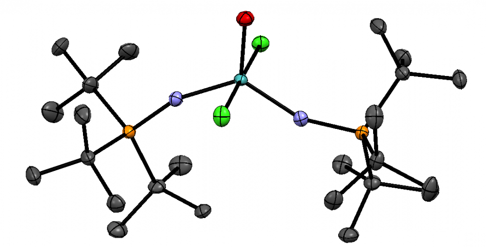
In a recent study, Sekar et al. has developed a wearable electrochemical sensor that can measure cortisol in sweat. The researchers has integrated iron (III) oxide (Fe2O3) in conductive carbon yarn (CCY) to make a semiconductive platform. After that, the platform is coated with antibodies (anti-Cmab) in an electrochemical apparatus, which would make the sensor specific to cortisol. The final product would then become a Fe2O3/CCY immunosensor. The purpose of the study is to see if they can use CCY as a suitable platform for biosensors when monitoring sweats.

Adapted from Figure 1b in the Sekar et al (2019) paper. The black rectangle is the CCY with iron (III) oxide (orange spots). The green cylinder is the electrochemical apparatus. Licensed under a Creative Commons Attribution 4.0 International License

The researchers were able to test the sensor’s detection ability with different concentrations of cortisol. According to Figure 8b below, the line graph shows a linear relationship between the electrical current response from the Fe2O3/CCY immunosensor and the logarithm of cortisol concentration.

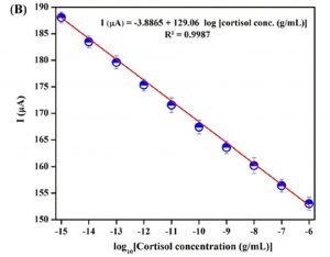
Adapted from Figure 8b in the Sekar et al (2019) paper. Each data point with error bar is the result from three successive experiments. Licensed under a Creative Commons Attribution 4.0 International License

The researchers also tested the sensor with real sweat samples from participants after performing cardio exercise. In the bar graph below, the error bars in the pink bar gives the RSD or relative standard deviation of 3.403%, 3.874%, and 4.064% from sweat sample 1, 2, and 3 respectively. These RSDs show small variations in averaged results from three successive experiments when testing with the Fe2O3/CCY immunosensor. According to the paper, the bar graph below shows a correlation between the two methods: the CLIA (chemiluminescence immunoassay) and their Fe2O3/CCY immunosensor. As a results, using CCY may be a possible choice for designing a biosensor that monitors cortisol in sweats.

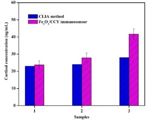
Adapted from Figure 11 in the Sekar et al (2019) paper. Each pink bar with error bar is the result from three successive experiments. Licensed under a Creative Commons Attribution 4.0 International License

In addition, there are other similar studies that focus on wearable sweat sensors, such that they can transmit data to your phone, and diagnose cystic fibrosis. Therefore, sweat sensors are potential non-invasive diagnostic tools, which may lessen the burden on more invasive blood samples to measure our health status.

References

Stress and Heart Health. https://www.heart.org/en/healthy-living/healthy-lifestyle/stress-management/stress-and-heart-health (accessed Mar 26, 2019).

Sekar, M.; Pandiaraj, M.; Bhansali, S.; Ponpandian, N.; Viswanathan, C. Carbon fiber based electrochemical sensor for sweat cortisol measurement. Scientific Reports 2019, 9, 1-14. https://doi.org/10.1038/s41598-018-37243-w.

Stephanie, Relative Standard Deviation: Definition & Formula. https://www.statisticshowto.datasciencecentral.com/relative-standard-deviation/ (accessed Mar 26, 2019).

Geddes, L. Wearable sweat sensor paves way for real-time analysis of body chemistry. http://www.nature.com/news/wearable-sweat-sensor-paves-way-for-real-time-analysis-of-body-chemistry-1.19254 (accessed Mar 26, 2019).

Dusheck, J. Wearable sweat sensor can diagnose cystic fibrosis, study finds. http://med.stanford.edu/news/all-news/2017/04/wearable-sweat-sensor-can-diagnose-cystic-fibrosis.html (accessed Mar 26, 2019).


The Antibiotic Resistance Crisis


“Antimicrobial Resistance – Mutation.” National Institute of Allergy and Infectious Diseases, Feb. 2009, www.niaid.nih.gov/topics/antimicrobialResistance/Understanding/Pages/mutation.aspx.

The development of antibiotic-resistant bacteria is a growing concern that is quickly sweeping up the attention of medicinal chemists and doctors. The growing use of antibiotics as the standard for treatment has led to the increase of drug-resistant bacteria; commonly known as “superbugs.” As the drug is repeatedly introduced to the bacteria, eventually mutations will arise in future generations of bacterium¹ that will allow it to be antibiotic-resistant and allow it to multiply and thrive. This increased number of antibiotic-resistant bacteria has led to an increase in sick patients² with bacterial infections that are antibiotic-resistant. As before due to the trivial nature of the infection simple antibiotics would cure the infection, but as per the nature of these “superbugs”, common treatments won’t work anymore, and new drugs or different treatments must be used to cure the infection.

“Causes of Antibiotic Resistance .” World Health Organization, Nov. 2015, www.who.int/drugresistance.

There are many causes to the antibiotic crisis, but one of the more prevalent causes are the over-prescribing of antibiotics and the over-use of antibiotics in livestock and fish farming. Currently, more than 50% of the antibiotics produced are going directly to the feeds of livestock³to keep them from getting sick due to their poor living conditions. By introducing bacteria to a consistent and high volume of antibiotics, eventually, the bacteria will go through mutations in future generations that will allow it to survive these antibiotics and ultimately result in the discontinued effectiveness of that drug. The same concept applies to the over-prescribing of antibiotics, by always introducing the bacteria to the drug eventually it will not be practical to use anymore4. At that point, different drugs would need to be used until they eventually stop working and so on until we reach a point where there would be no more antibiotics left to use to fight these infections.

What causes antibiotic resistance? – Kevin Wu. https://www.youtube.com/watch?v=znnp-Ivj2ek (accessed Feb 16, 2019).

Potential solutions to combat the problem involve something as trivial as proper hygiene. As making sure to wash your hands often, the chance for infection will go down and as a direct result will cut down on antibiotic use. A more futureproof method would be the development of new antibioticsor potential re-use of old antibiotics6 that could be re-purposed to combat the problem. No matter the method used to combat this problem or a combination of every method available, this a problem that needs to be addressed as soon as possible, or we are looking at a world where trivial infections can run rampant with no good method of treatment.

~ Danial Yazdan

References:

¹Blair, Jessica M. A., et al. “Molecular Mechanisms of Antibiotic Resistance.” Nature Reviews Microbiology, vol. 13, no. 1, 2014, pp. 42–51., doi:10.1038/nrmicro3380.

²Duin, David Van, and David L. Paterson. “Multidrug-Resistant Bacteria in the Community.” Infectious Disease Clinics of North America, vol. 30, no. 2, June 2016, pp. 377–390., doi:10.1016/j.idc.2016.02.004.

³Bush, Karen, et al. “Tackling Antibiotic Resistance.” Nature Reviews Microbiology, vol. 9, 2 Nov. 2011, pp. 894–896., doi:10.1038/nrmicro2693.

4Chellat, Mathieu F., et al. “ChemInform Abstract: Targeting Antibiotic Resistance.” ChemInform, vol. 47, no. 29, 22 Mar. 2016, pp. 6600–6626., doi:10.1002/chin.201629282.

5Nathan, Carl, and Otto Cars. “Antibiotic Resistance — Problems, Progress, and Prospects.” New England Journal of Medicine, vol. 371, no. 19, 2014, pp. 1761–1763., doi:10.1056/nejmp1408040.

6Frieri, Marianne, et al. “Antibiotic Resistance.” Journal of Infection and Public Health, vol. 10, no. 4, 2017, pp. 369–378., doi:10.1016/j.jiph.2016.08.007.

Link

Photoswitchable drugs: the light at the end of the tunnel?

Photoswitchable drugs: the light at the end of the tunnel?

For many developed nations, cancer has become the leading cause of death. Regrettably, the current state of cancer treatment still rests heavily on chemotherapy and its toxic side effects. More than ever, our efforts to further develop targeted cancer therapeutics are of paramount importance.

In more recent years, chemists have begun designing light-activated molecules that can be activated upon contact with its target tumor cell and deactivated following cell death.  That said, photoswitchable drugs are not a novel concept; in fact, scientists have been considering synthetic light-switching molecules as promising treatments for blindness, diabetes, Alzheimer’s disease, and antibiotic resistance, to name a few.

Previously, treatments for skin cancer relied on photodynamic therapy (PDT), a process during which patients receive dyes that convert oxygen molecules into their toxic singlet forms capable of killing diseased cells upon activation by light. Given its requirement of oxygen in the body’s tissues, the applicability and potency of PDT are limited by hypoxic tumor environments in which cancerous cells survive without oxygen.

Comparison of (a) classic chemotherapy and (b,c) photopharmacological chemotherapy DOI:10.1002/chem.201502809

Dr. Phoebe Glazer at the University of Kentucky believes that photoswitchable therapies offer a possible strategy for overcoming this restriction. By deriving energy from photons to induce a chemical reaction, photoswitchable therapies enable molecular changes conducive to the recognition and destruction of diseased cells. This approach, unlike current chemotherapy treatments, is capable of killing tumors and saving healthy tissue with specificity, thereby maximizing possible dosage and minimizing dangerous side effects.

Glazer promotes photoactivated chemotherapy drugs that can function as both PDT sensitizers and one-way photoswitches. Using a ruthenium (II) polypyridyl complex, Glazer irreversibly ejected a methylated ligand, and with light, induced the complex to bind to DNA for ultimate cell damage. By modifying the drugs’ ligands, Glazer tuned the molecules’ solubility and the light absorbance wavelength.

ruthenium(II) polypyridyl complexes DOI: 10.1021/ja3009677

Many chemists, including Dr. Wiktor Szymanski at University Medical Center Groningen, are experimenting with molecules that can be switched on and off by light. Once developed, the resulting drugs can be turned on by contact with a targeted cancer cell and turned off after cell destruction. By adding the photoswitchable group, azobenzene, and using UV light to convert the molecule’s configuration, Szymanski produced photoswitchable molecules. 

Photoswitchable molecule developed by Szymanski, Feringa, et al. DOI: 10.1021/jacs.7b09281

Of course, a handful of concerns must be addressed before such “on and off” drugs can become reality. Scientists need to ensure that their switches can work at tolerable wavelengths, specifically ones that can pass through tissue without causing damage. Dr. Achilefu at the Washington University School of Medicine has developed a method called stimulated intercellular light therapy where light is captured by the molecules that target tumor cells. This light has been designed to reach tumor cells beneath the surface of the body (explained in the video below). 

Because of the extreme difficulties and complications related to the synthesis of small molecular drugs, many chemists are skeptical about the approval process of photoswitchable drugs. However, with more research and development, I believe that photoswitchable drugs offer a viable pathway for the future of cancer treatment.

-Brina Kim