Monthly Archives: March 2020

Life-saving improvements to blood transfusion

An article published by Nature Microbiology in June 2019, studied a new method in converting type A blood to the universal type O blood using bacteria found in the human gut! [1] A team led by Dr. Stephen Withers at the University of British Columbia has developed a method which would eliminate the need for blood-type compatibility, reducing the risks of blood transfusions.  

There are 8 different blood types, and before these findings, these blood types were not all compatible with each other. Each blood type can only receive from other specific types.  When doctors are going to transfuse blood to patients, they need to match the type of blood to one that can safely match with the patient’s blood type. If they don’t, the body will not know how to handle the new type of blood. This can cause blood vessels to rupture, which can be fatal.

This problem has existed since blood transfusions were first scientifically achieved, and scientists have been looking for a solution for just as long. It turns out the solution was hiding right under our noses; inside our stomachs, to be specific!

Inside the human gut are thousands of microscopic bacteria which we use to digest food and convert it into energy. As it turns out, these bacteria are very good at safely interacting with the human body in helpful ways. The researchers extracted these microorganisms through human faeces and found they could be used in exactly the way they were hoping.

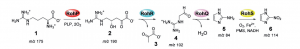
         “Why would they be looking in our stomachs for this solution?” you might ask. The Withers group were on a hunt for a special kind of protein called an enzyme. Enzymes are produced by the body with a very specific task, and that task varies based on what the body wants it to do. Since our gut has the ability to process blood and turn it into energy, Withers and his team decided to see if these enzymes could be harnessed for their research.  As it turns out, they were completely correct.

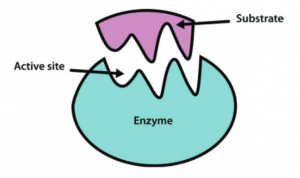
An enzyme interacting with a specific molecule (known as the substrate) in the body.[2]

The group screened more than 20,000 samples to find two enzymes that were particularly good at cleaving the A-type blood. These were extracted and tested on real red blood cells and found that the enzymes could efficiently cleave a specific part of the A-type blood, essentially leaving us with type O red blood cells. These two types of enzymes were 30 times more efficient than previous methods, which means we only need a tiny amount of these enzymes to convert A, B, and AB types of red blood cells to O type red blood cells.

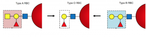
Image depicting the difference between blood types A, O, and B. The image shows that removing the yellow square in A type blood, is the same as O type blood. Modified from [1].

In January 2020, the American Red Cross announced that it has a ‘critical’ shortage of type O blood. In the United States and Canada alone, 4.5 million patients need blood transfusions every year.[3] This high demand means that oftentimes, the supply cannot meet the demand. With this new discovery, incompatible blood types can be made compatible. This would increase the supply of compatible blood, which means more people can be helped.

While the process has been completed in the lab, it has yet to be scaled up to convert massive amounts of blood at a time.  This may take some time to accomplish. However, it is impossible to quantify exactly how many people this new method will help, or even how many lives it will save. One thing is for certain, the world of blood donations will forever feel the impact of these findings.

References
  1. Rahfeld, P., Sim, L., Moon, H., Constantinescu, I., Morgan-Lang, C., Steven, J. H., Kizhakkedathu, J. N., Withers, S. G.; An enzymatic pathway in the human gut microbiome that converts A to universal O type blood. Nature Microbiology. 2019, 1475-1485.
  2. Western Oregon University: Chemistry. CH450 and CH451: Biochemistry – Defining Life at the Molecular Level. Chapter 6: Enzyme Principles and Biotechnological Applications. https://wou.edu/chemistry/courses/online-chemistry-textbooks/ch450-and-ch451-biochemistry-defining-life-at-the-molecular-level/chapter-7-enzyme-kinetics/ (accessed Mar 21, 2020)
  3. Community Blood Bank of Northwest Pennsylvania and Western New York. 56 facts about blood. https://fourhearts.org/facts/ (accessed March 22, 2020)

– Griffin Bare, Eric Ding, Chantell Jansz
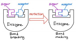
Sugars – The Key to Talking to our Cells

We are one step closer to achieving communication with those 37.2 trillion tiny cells making up our bodies. Our cells communicate using sugars, and different modifications to these sugars can change the way our cells communicate with each other. Just last year, researchers at the University of British Columbia, led by Dr. Stephen Withers, did exactly this! They designed a unique way to tinker with sugars’ chemical structures by using chemicals found in bacteria. The same bacteria living in our gut.

what exactly are sugars?

No ambiguity here! Chemists work with sugars based off molecules in this sugar cube. Credits: The Verge

To be specific, the type of sugars Wither’s team studied were simple sugars; hexagonal-shaped molecules, often joined to other molecules known as “acceptors”. The problem chemists face is like the average love life. Similar to starting a relationship with someone, joining acceptors to sugars is also quite difficult. Luckily for sugars, Mother Nature has come up with some solutions: enzymes, which are helper molecules that speed up the pairing or unlinking of two molecules. 

A SOLUTION UNDER OUR NOSES…

Instead of struggling to find ways of joining sugars and acceptors, Wither’s team thought: Why not hijack Mother Nature and use these enzymes? To make their idea a reality, they extracted sugar-specific enzymes from E. Coli, a type of bacteria that lives inside the human gut. These bacteria manufactured 175 sugar-specific enzymes, and from these they chose eight enzymes that were compatible with the sugars and acceptors they were interested in.

It seemed like Wither’s team now had everything needed to join the desired acceptors to the sugars; however, there was still a problem. The sugar-specific enzymes they got from E. Coli did the exact opposite of what they wanted; instead of forming sugar-acceptor linkages, they were specialized in breaking them. Enzymes that break sugar-acceptor linkages are analogous to using a screwdriver to loosen a screw, while enzymes that form sugar-acceptor linkages are like tightening a screw.

A small modification changes the function of the enzyme. Credits: Blog post authors

To solve this problem, they reverse-engineered the enzymes specialized in breaking linkages into those that form linkages, by changing a small part of the enzymes’ structure, similar to changing the tips on a screwdriver. As a result, Wither’s team now had eight enzymes specialized in forming different sugar-acceptor linkages. 

More than just a bond…

Now being able to freely and efficiently modify sugars, there is a big potential for researchers to join in on the conversations with our cells. Why is this important? Often, there is miscommunication within our cells which can lead to serious trouble. 

One example is cancer; which is partly caused by cancer cells using abnormal sugar molecules as a form of miscommunication, to avoid being cleared up by immune cells. One potential treatment is a sugar-based vaccine, which tells our immune cells to ignore this miscommunication and target tumor cells.

The challenge of designing a sugar-based vaccine isn’t just relevant to cancer, but other diseases as well which occur also as a result of miscommunication. With Wither’s research, designing these sugar-based drugs won’t be as difficult thanks to their novel way of bonding sugars to other molecules. This research brings us one step closer to talking to our cells, helping with the battle against diseases.

Story source

Armstrong, Z.; Liu, F.; Chen, H.-M.; Hallam, S. J.; Withers, S. G. Systematic Screening of Synthetic Gene-Encoded Enzymes for Synthesis of Modified Glycosides. ACS Catalysis 20199 (4), 3219–3227.

– Kenny Lin, Pricia Ouyang, Tom Hou, Aron Engelhard (CO-10)

Magical Materials Revolutionize the Pharmaceutical World

What if there’s more to caffeine than meets the eye? What if we can chemically change the way caffeine works to create a better version of itself. 

If you are a coffee lover, you probably know the naturally occurring substance, caffeine, and its stimulating properties to our nervous system. Why and how does it happen?

Using a special type of reaction, the biological activity of caffeine can be transformed to suit a variety of different applications. Past studies can be improved by changing the materials needed and the methods used. 

Dr. Schafer’s team, at the University of British Columbia, examines an approach to synthesize pseudoalkaloids (Fig. 1), chemicals present in caffeine, with great accuracy.

Figure 1. Examples of pseudoalkaloids. The pseudoalkaloids on the left are produced naturally or synthetically. The pseudoalkaloids on the right are the researchers’ target molecules. (molecules were drawn with ChemDraw 19.0; Credits: Wilson, Young, and Rachel)

Pseudoalkaloids can be artificially made, but time-consuming multiple synthetic steps limit the production of  pseudoalkaloids. Importantly, the challenge lies in the reactivity of the starting materials, and whether it can react to produce the desired pseudoalkaloid without byproducts. 

Similar studies show different products have formed, which proves to be a problem. 

Existing studies experimented with different types of metal catalysts showing potential improvement for the results. A tantalum catalyst, a tool that speeds up the reaction, is tested on a reaction to observe its effectiveness on producing the final product. 

An idea was proposed to add molecules that bind to the tantalum catalyst. This binding molecule improves the reactivity of the catalyst, thereby converting as many initial materials as possible to the final product.

Of many possible starting materials, terpenes (Fig. 2) were used for the synthesis of pseudoalkaloids. In addition to the inactive alkene groups in terpenes, various chemical structures make them attractive starting substrates to explore a new synthetic route. 

Figure 2. Examples of terpenes. Terpenes are naturally occurring organic molecules, produced by plants. The terpenes on the right are starting materials chosen by the researchers. Every terpene has unsaturated functional groups which may react with other molecules.  (molecules were drawn with ChemDraw 19.0; Credits: Wilson, Young, and Rachel)

Another key building block, an amine, can be any, as long as the nitrogen atom of an amine is directly bound to one hydrogen atom. However, a clever choice can even make the final products useful building blocks, allowing further modifications (Fig. 3)

 Figure 3. Amines with varying R groups. After reacting with a terpene, the R group of pseudoalkaloid can be further modified to form a new molecule. (molecules were drawn with ChemDraw 19.0; Credits: Wilson, Young, and Rachel)

The catalytic reaction between amines and terpenes with the tantalum catalyst showed great selectivity. Without the help of the tantalum catalyst, an amine could potentially select any active spot on a terpene and react with it, causing a mixture of pseudoalkaloids at the end of the reaction.

However, the tantalum catalyst results in one dominating product. Although the final pot contains some residual starting materials, the target pseudoalkaloids are the major product that can be easily isolated.

By constructing a pathway to ultimately arrive at the designated point, new and better options can be achieved. Caffeine is one of many that can be innovated upon.


Reference

Dipucchio RC, Rosca SC, Athavan G, Schafer LL. Exploiting Natural Complexity: Synthetic Terpenoid‐Alkaloids by Regioselective and Diastereoselective Hydroaminoalkylation Catalysis. ChemCatChem. 2019;11(16):3871–6.


-Group 9 (Wilson, Young, Rachel)


The Next Step in Antibiotics

Did you know that not all types of bacteria in this world are harmful to humans? Some bacteria can be beneficial such as those used for the production of cheese and yogurt. Furthermore, these tiny creatures could even potentially be used to manufacture antibiotics for human use.

Antibiotics are used to cure bacterial infections by killing bacteria. For example, penicillin saved thousands of people in the 19th century.

Bacteria

As Amazing as they Sound, Antibiotics are not a Permanent Solution

The over-usage of antibiotics results in bacteria that can build resistance and cause the medication to be ineffective. The World Health Organization announced: “A serious threat is no longer a prediction for the future, it is happening right now in every region of the world and has the potential to affect anyone” on the development of drug-resistant bacteria.

Once a type of bacteria develops a resistance to a specific antibiotic, other antibiotics must be used to treat the infection. For example, nitroimidazole is a drug found in antibiotics such as azomycin. Due to its low resistance by bacteria, it has been used extensively to treat bacteria that have developed resistance to certain antibiotics.

Nitroimidazole

Research Looking into Nitroimidazole

Dr. Jason Hedges and Dr. Katherine Ryan of the University of British Columbia took a look into finding new ways to synthesize nitroimidazole. Their recent publication in 2019 showed how they were able to convert an amino acid (a building block of proteins) into azomycin. 

The researchers used bioinformatics, a data analysis tool, to find all previous work that has been done on this topic. Bioinformatic searches are performed on databases such as NCBI.

They found that back in 1953, azomycin was extracted from a strain of bacteria called Streptomyces eurocidicus. Furthermore, they found a specific gene in a separate strain of yeast called Streptomyces cattleya, which had similarities to S. eurocidicus

The two researchers aimed to find new ways to synthesize nitroimidazole, and proposed to synthesize nitroimidazole from L-arginine. Once a pathway had been developed, their end goal was to synthesize and extract azomycin in S. cattleya.

Hedges and Ryan were able to develop a multi-step pathway for the conversion of L-arginine to nitroimidazole through experimentation. However, they were unable to detect azomycin in S. cattleya. They noted that the gene of interest was unable to synthesize azomycin, and a separate drug may have synthesized in its place.

Synthesize of nitroimidazole from L-Arginine

A Bright Future

The significance of this research transcends the synthesis of azomycin. Although they failed to detect azomycin in S. cattleya, their work provides a stepping stone for further research to be conducted. This study provides insight on new biosynthetic pathways which is important to those currently in the field of life sciences and pharmacology. Furthermore, this study expands people’s knowledge of bacteria engineering and the biosynthetic pathway of other antibiotics. It also provides information about future antibiotic observations.

Future researchers could test their synthetic pathway on other bacteria such as E. coli to determine if azomycin could be synthesized.

Reference:

Hedges, J. B.; Ryan, K. S. In Vitro Reconstitution of the Biosynthetic Pathway to the Nitroimidazole Antibiotic Azomycin. Angewandte Chemie International Edition 2019, 58 (34), 11647–11651.

-Adrian Emata/Jackson Kuan/Xinyu Gu/Yicheng Zhu


A New and Advantageous Molecule for Diagnostic Nuclear Medicine

You may be aware of the role physicists and doctors play in diagnostic nuclear medicine; however you may not know that chemists also play a significant role in this area of science! In 2019, Dr. Chris Orvig and his team at the University of British Columbia constructed an advantageous molecule for use in medical imaging whose purpose is to bind to radioactive Gallium (Ga) ions. They also determined that their molecule has superior properties to similar molecules currently being used.

WHAT IS IT?

The molecule that was created by Dr. Orvig’s team is simply known as H2Hox , a chelating ligand. Let’s break its name down piece-by-piece to get a better understanding of what it does.

A ligand is a type of molecule that can bind onto a metal ion, like iron (Fe3+) or copper (Cu2+). In the case of H2hox, the metal ion is Gallium (Ga3+) because it is widely used in medical imaging. The word chelating comes from the latin root word chela, which means claw. This is because chelating ligands have multiple points of attachment to a metal ion, similar to a crab’s claw, making them significantly stronger binders to metal ions.

Image sources (left to right): Research Gate, Wang et al..

HOW IS IT MADE?

H2Hox is easy to synthesize, avoiding a number of potentially challenging synthetic pathways typically associated with Ga chelating species. The initial starting materials were inexpensive and readily available. To put this into perspective, it’s like baking a box cake versus baking a cake from scratch. The former is simple and quite easy to do, while the latter is a lot harder and is a lot more intensive. Ease of synthesis is an important feature as it can affect the commercial applicability of the molecule.

WHAT DOES IT DO?

H2Hox is used in a form of medical imaging known as positron-emission tomography (PET). PET imaging is primarily used to diagnose health issues related to biochemical processes occurring inside our cells, such as cancer. The main function of H2Hox in PET imaging is to bind to the radioactive Gallium ion, which aids in producing an image of a desired area or tissue inside the body.

To test how well H2Hox worked, the researchers conducted a PET scan in mice. The group witnessed high stability of the combined ligand and ion in mice, and more importantly, they observed that it was rapidly excreted from the mice. Furthermore, the ligand has a strong affinity to Gallium, exhibiting significant radiolabeling capabilities (binding to Ga3+) in only five minutes with low amounts of ligand under room temperature. As a result of the molecule’s advanced properties, H2Hox surpasses any ligand currently used as a Gallium chelator.

THE FUTURE IS PROMISING

The combination of superior properties and easy synthesis makes H2Hox an effective and convenient molecule for Gallium PET imaging. H2Hox acts as a launching-off point for the development of even better chelating ligands to improve the quality and ease of PET imaging.

Literature cited:

1. Wang, X.; Jaraquemada-Pelaez, M. d. G.; Cao, Y.; Pan, J.; Lin, K.-S.; Patrick, B.O., Orvig, C. H2hox: Dual-Channel Oxine-Derived Acyclic Chelating Ligand for 68Ga Radiopharmaceuticals. J. Am. Chem. Soc. 201958, 2275-2285


-Group 6 (Mark, Akash, Athena, Charles)

The Key to Treating Pneumonia can be Found in Soil?

Can it really be that simple? Can the answer really be in the ground beneath our feet?

Source: Soil Science Society of America

Discovery of a gene cluster commonly found in soil-dwelling bacteria may be the key to treating anaerobic bacterial infections such as appendicitis and pneumonia. Researchers Jason B. Hedges and Prof. Dr. Katherine S. Ryan from the University of British Columbia have isolated the antibiotic compound azomycin from a biosynthetic gene cluster found in the bacterium Streptomyces cattleya.

With this information they believe it can lead to engineering bacteria to produce a new line of antibiotics.

What is a gene cluster?

The term gene cluster is moreso semantics to describe a group of genes that share a common phenomenon.

Originally, azomycin was isolated from a similar bacterium Streptomyces eurocidicus back in 1953 and became the blueprint to synthetic nitroimidazoles. 

“Nitroimidazoles are one of the most effective ways to treat anaerobic bacterial infections”,[1] Hedges writes in his 2019 study. The most commonly used nitroimidazole, metronidazole, is an antibiotic used to treat pelvic inflammatory disease, endocarditis, and bacterial vaginosis.[2] It is also on the World Health Organization’s List of Essential Medicines, the safest and most effective medicines needed in a health system.[3]

Figure 1: Molecular Structure of Nitroimidazole. Source: Sigma Aldrich

Despite its use in antibiotics for over 60 years, the incidence of nitroimidazole resistance in anaerobes remains low, making it an essential component of the antibiotic arsenal. [1,4]

Since the isolation of azomycin back in 1953, synthesis of nitroimidazoles were limited to synthetic routes, most commonly involving reactions of an imidazole with nitric acid and sulfuric acid. 

Using bioinformatics, however, in 2019 Hedges and Ryan were able identify a biosynthetic gene cluster in the same bacterium that makes penicillin,[5] Streptomyces cattleya. They were able to find that this gene cluster containing azomycin is widely distributed among soil-dwelling actinobacteria and proteobacteria.

Because of this they theorize that azomycin and other nitroimidazoles may be important factors in ecology.

What are bioinformatics?

Bioinformatics is the science of collecting and analyzing complex biological data such as genetic codes. As an interdisciplinary field of science, bioinformatics combines biology, computer science, information engineering, mathematics and statistics to analyze and interpret the biological data

In addition to the isolation of azomycin in a gene cluster, Hedges and Ryan were able to perform in vitro analysis in order to understand the enzymatic steps that take the primary protein L-arginine to become azomycin. 

Source: Washington Post

Their work opens the door to biocatalytic methods to synthesize azomycin and other nitroimidazoles. They believe this discovery can “lead to the possibility of engineering bacteria to produce nitroaromatic compounds”.[1]

In other words, this may lead to stronger antibiotics immune to drug resistance.

References

Hedges, J. B.; Ryan, K. S. In Vitro Reconstitution of the Biosynthetic Pathway to the Nitroimidazole Antibiotic Azomycin. Angewandte Chemie International Edition 201958 (34), 11647–11651.

The American Society of Health-System Pharmacists. Archived from the original on 6 September 2015. Retrieved 31 July 2015.

World Health Organization model list of essential medicines: 21st list 2019. Geneva: World Health Organization

David I. Edwards, Nitroimidazole drugs-action and resistance mechanisms I. Mechanism of action, Journal of Antimicrobial Chemotherapy, Volume 31, Issue 1, January 1993, Pages 9–20, https://doi.org/10.1093/jac/31.1.9

Kahan, JS; Kahan, FM; Goegelman, R; Currie, SA; Jackson, M; Stapley, EO; Miller, TW; Miller, AK; Hendlin, D; Mochales, S; Hernandez, S; Woodruff, HB; Birnbaum, J (Jan 1979). “Thienamycin, a new beta-lactam antibiotic. I. Discovery, taxonomy, isolation and physical properties”. The Journal of Antibiotics32 (1): 1–12

-Adrian Emata

Origin of the Coronavirus

Recently, the outbreak of new coronavirus has seriously affected people’s lives in countries and even around the world. New coronaviruses can be transmitted through air and contact. Symptoms include fever, cough, and difficulty breathing. In order to completely solve this infectious disease, it is very important to find the primary case.

Source:https://www.heywood.org/education/covid19-coronavirus-updates

Initially, Chinese scientists believed that the source of the virus was the South China Seafood Market in Wuhan, China. When people eat wild animals that contain the virus, the non-pathogenic version of the virus jumps from animal hosts to humans and then evolves into the current pathogenic state in the population. For example, the RBD structure of some coronaviruses from pangolins, armadillo mammals found in Asia and Africa, is very similar to SARS-CoV-2. Coronavirus from pangolin may have been transmitted to humans either directly or through an intermediary host such as a civet or ferret.

Source:http://m.cyol.com/content/2020-03/05/content_18413469.htm

Most people in this epidemic believed that this new coronavirus should be derived from flying mammals such as bats. Many people also criticized them, and even many people targeted the bats directly, thinking that those who eat bats Caused the disease. However, this is actually questionable. Although it is possible that bats can directly infect humans, so far, most of the time, bats are not the direct source of infection but are adapted to spread to humans through the intermediate source of infection. This can be seen from the case report published in the top medical journal “The Lancet”.

Source:https://news.sky.com/story/how-did-coronavirus-start-scientists-tackle-the-conspiracy-theories-11959500

Researchers from institutions such as the Xishuangbanna Tropical Botanical Garden of the Chinese Academy of Sciences recently published a paper in the form of a preprint, saying that they analyzed the genomic data of 93 new coronavirus samples in 12 countries on four continents and found that they contained 58 haplotypes, which are related to the South China Seafood Market The associated patient sample haplotypes were H1 or its derivative types, while the more “older” haplotypes such as H3, H13, and H38 came from outside the South China seafood market, confirming that the new crown virus of the South China seafood market was transmitted from elsewhere In perspective.

Source:https://meaww.com/coronavirus-did-chinese-officials-downplay-extent-of-deadly-wuhan-outbreak-in-early-stages

Finding an “index case” is equivalent to finding a weapon that can cut the epidemic from the source. But in the long history of humans fighting the epidemic, few “index cases” have been found. On the one hand, the timeline of the index case and first case are not necessarily the same, which makes the process of tracing like a needle in a haystack. In addition, the chain of evidence tracking “index case” is difficult to finalize and will always be repeatedly overturned and readjusted.

To this day, the epidemic of AIDS, Ebola, SARS and so on has never clearly identified the “index case” in the strict sense. From the perspective of the development of the global epidemic, it is still of great significance to curb the development of the epidemic while researching and developing effective drugs and vaccines and controlling the development of the epidemic in a timely manner. However, the new Coronavirus may be the same as AIDS and SARS. There is no way to accurately find the first human it infected.     

Yicheng Zhu

The magical “Ta” catalyst for pseudoalkaloids

If you are a coffee lover, you would probably know the naturally occurring substance, caffeine. But, were you aware that this substance is classified as a pseudoalkaloid?

Many pseudoalkaloids can often have biological activities like caffeine stimulates our nervous system. Ultimately, pseudoalkaloids can be used as building blocks to produce useful drugs.

In 2019, researchers at the University of British Columbia, led by Dr. Schafer, uncovered a new pathway to produce structurally simple terpenoid-alkaloids, which belong to pseudoalkaloids.

This study can be simply summarized as a reaction between a terpene and an amine with the help of a tantalum catalyst. But, let’s first explore key ingredients to deeply understand how the synthetic route works!


An organotantalum compound with a ureate salt
The researchers developed a catalytic reaction run by a metallic compound. Based on other known studies, they chose an organoctantalum compound to produce terpenoid-alkaloids. As like an engine is the heart of a car, the tantalum compound is an engine to drive reactions to the final products, terpenoid-alkaloids

The choice of a metallic compound is of course crucial. However, it is more important for the compound to have complete catalytic potential. How could a bare metallic compound become a complete catalyst? The answer is associating a metallic compound with a ligand such as organic molecules or salts, which can coordinate to a metal center. Of numerous possible candidates of ligands, the researchers found that a specific salt can improve the efficiency and selectivity of the bare organotantalum compound, thereby allowing it to have a complete catalytic ability.


Figure 1.The ureate salt that improved selectivity and efficiency of the organotantalum compound, Ta(CH2SiMe3)3Cl2. Of several ureate salts, the above salt was the most suitable for this study due to its solubility.

Terpenes and anilines
As the name of final products, terpenoid-alkaloids, reflects the use of terpenes, one of key ingredients is a terpene, a naturally occurring molecule. By limiting the scope of terpenes to enantiopure limonene and pinene, the types of anilines were varied and reacted with the terpenes

Now, here comes a question. What is the consequence of mixing these ingredients together?


Fascinating results
This study is fascinating not only for the reason that a catalytic amination of terpenes is unexplored, but also the final products are not chaotic mixtures.

What does it mean by a chaotic mixture? Some catalysts have potential to alter an intrinsic structure of a staring substance. For example, if a catalyst was able to influence the chiral center of (R)-limonene by changing its stereochemistry, a reaction batch would contain both (S) and (R)-limonenes. Consequently, the occurrence of two products is equally probable.

Also, unexplored magical ability of the tantalum catalyst in the study allows anilines to react with one specific spot of an alkene moiety in terpenes. This astonishing selectivity gives a rise to one major product.

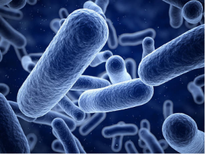
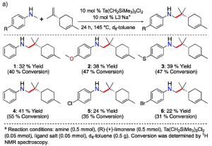
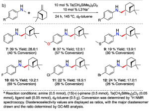
           Figure 2.The reaction of an enantiopure limonene with six different anilines (left). The reaction of an enantiopure pinene with six different anilines (right). Both reactions result in high regio- and diastereoselectivity.

Reference
Dipucchio RC, Rosca SC, Athavan G, Schafer LL. Exploiting Natural Complexity: Synthetic Terpenoid‐Alkaloids by Regioselective and Diastereoselective Hydroaminoalkylation Catalysis. ChemCatChem. 2019;11(16):3871–6.

-Young Cho

No Medicine to Cure

Can you imagine the world without medicine?

Nowadays, more and more bacteria begin to show resistance against antibiotics. Azomycin, a type of antibiotics used to treat multi-drug resistance is found to be used more and more frequently. What if bacteria start to grow resistance against Azomycin? However, it is actually nailed fact, the only question is when. In order to solve this problem, in 2019, Dr. Jason Hedges and Dr. Katherine Ryan of the University of British Columbia engaged in finding a new way to synthesize the nitroimidazole, the main component of azomycin.

source:https://www.reactgroup.org/toolbox/understand/how-did-we-end-up-here/few-antibiotics-under-development/

The study of antibiotics can be traced back to the 19th century. For these two centuries, antibiotics saved countless lives from all kinds of diseases. However, antibiotics can not kill all the bacteria. Every time when one bacteria survived from the massacre of the antibiotic, they grow the resistance against the antibiotic. Then it split, split and split. Finally, the survived bacteria become countless bacteria that can not be defeated by the antibiotic again. Thus, more and more bacteria begin to survive from the war with antibiotics, and more and more antibiotics become useless. Herein, the race between the evolving of antibiotics and evolving of bacteria begins. 

So, let’s take a look at what did Dr. Jason Hedges and Dr. Katherine Ryan do and what did they find.

By doing a lot of research, Dr. Jason Hedges and Dr. Katherine Ryan found that the development of nitroimidazole can be dated back to 1953 when azomycin was first found. And they noticed that when strain Streptomyces eurocidicus was produced, L-arginine is converted to azomycin. Therefore, they came up with a plan to synthesis nitroimidazoles by linking L-arginine to azomycin via in vitro reconstitution. 

In vitro reconstitution process of nitroimidazole. Source: Hedges and Ryan, 2019

Through the experiment, Dr. Jason Hedges and Dr. Katherine Ryan successfully synthesized nitroimidazole via in vitro reconstitution. But unfortunately, no azomycin was produced via Streptomyces cattleya. 

Although the experiment is failed to synthesis azomycin through Streptomyces cattleya, it still provides a lot of valuable information for further researchers. It points a direction on the biocatalytic pathway of azomycin synthesis and set the stage for the discovery of new antibiotics.

Reference:

Hedges, J. B.; Ryan, K. S. In Vitro Reconstitution of the Biosynthetic Pathway to the Nitroimidazole Antibiotic Azomycin. Angewandte Chemie International Edition 201958 (34), 11647–11651.


Yicheng Zhu


Biosynthetic Pathway Found to Synthesize Anaerobic Antibiotics!

Fig 1. Image of Azomycin. source

Scientists found the biosynthetic pathway to the Nitroimidazole Antibiotic Azomycin. A steppingstone towards the revolution of anaerobic bacterial infection treatments.

Scientists from the University of British Columbia found the biosynthetic pathway to the Nitroimidazole antibiotic Azomycin. The enzymatic mechanism from L-Arginine to Nitroimidazole has now been proved and present to the public. Their formal paper was published online on July 17th, 2019. Nitroimidazole is an essential component for the modern antibiotics, it is crucial to know its synthetic pathway for further pharmaceutical studies. The result of their study set the stage for further development of important anaerobic antibiotic Azomycin.

What is it? Why do we need to know about this?

Nitroimidazole is an essential antibiotic specifically to treat anaerobic bacterial infections. They are widely used to treat diseases such as Amoebiasis, Parasitic infections, skin infections, diarrhea and so on. The low redox potential of anaerobic bacteria cells allowed nitroimidazole to act as the electron sink and form the radical species. The resultant radical species would induce the bacteria cells’ death by damaging their DNA. Antibiotic is the most powerful “weapon” to fight against bacterial infections. However, according to the World Health Organization, there are more than 700000 people die every year due to antibiotic resistance. Despite the several decade’s usages of Nitroimidazole antibiotics, the drug resistance of it still remains low relatively. Thus, Nitroimidazole antibiotics are increasingly used to treat multi-drug resistant bacteria as well.


Previous research established that L-Arginine is converted to azomycin by 2-aminioimidazole. They determined that the intermediate of the reaction is 4-hydroxy-2-ketoarginine (2). Furthermore, they also observed the accumulation of pyruvate(3) side products and 2-aminoimidazole(5) from the intermediate(2). However, the actual enzymatic synthetic pathway has not been determined detailly yet. Jason and Katherine in the research group determined that PLP-dependent enzymes, RohP,RohR,RohQ and RohS plays esstential role in the catalytic pathway of the reaction. Researchers examined the in vitro activity of RohP, RohR, RohQ and RohS. They put in these enzymes separately and stepwise to different reactants. For example, in order to test whether RohR could catalyze a retro-aldol cleavage of 2 into 3 and guanidinoacetaldehyde (4), they added purified RohR instead of RohP. Then according to activity analysis and also the mass spectrum, the result shows that RohP yields a bigger portion of 2.

Fig 2. Reaction scheme from L-Arginine to Nitroimidazole. Source

Antibiotics are the most powerful “weapon” to kill bacteria in modern pharmaceutical studies. As early as in the 20th century, the observation of penicillin saved millions of people injured in the World War. Yet, the enormous benefits of antibiotics cause the consequences of drug-resistance. Azomycin, consider as a low-resistance antibiotic, it is crucial to understand its enzymatic reaction mechanism. Reaction mechanism allows scientist to have a more detail interpretation of the synthesis. It is crucial to find the catalytic cycle of the reaction, in order to allow scientists to develop and derive further study.

The study done by Jason and Katherine at the University of British Columbia provides the public with a steppingstone in future nitroimidazole anti-biotics study. Their study expanded people’s knowledge of the biosynthetic pathway to nitro-compounds. It also makes bacteria engineering to produce nitroaromatic compounds possible. This study will open the new door in enzymatic synthesis and biochemistry synthesis.

Cited article:

Hedges, J. B.; Ryan, K. S. In Vitro Reconstitution of the Biosynthetic Pathway to the Nitroimidazole Antibiotic Azomycin. Angewandte Chemie International Edition 201958 (34), 11647–11651.