Category Archives: Biological Chemistry

Origin of the Coronavirus

Recently, the outbreak of new coronavirus has seriously affected people’s lives in countries and even around the world. New coronaviruses can be transmitted through air and contact. Symptoms include fever, cough, and difficulty breathing. In order to completely solve this infectious disease, it is very important to find the primary case.

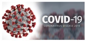
Source:https://www.heywood.org/education/covid19-coronavirus-updates

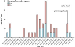
Initially, Chinese scientists believed that the source of the virus was the South China Seafood Market in Wuhan, China. When people eat wild animals that contain the virus, the non-pathogenic version of the virus jumps from animal hosts to humans and then evolves into the current pathogenic state in the population. For example, the RBD structure of some coronaviruses from pangolins, armadillo mammals found in Asia and Africa, is very similar to SARS-CoV-2. Coronavirus from pangolin may have been transmitted to humans either directly or through an intermediary host such as a civet or ferret.

Source:http://m.cyol.com/content/2020-03/05/content_18413469.htm

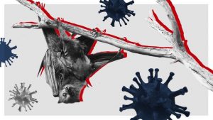
Most people in this epidemic believed that this new coronavirus should be derived from flying mammals such as bats. Many people also criticized them, and even many people targeted the bats directly, thinking that those who eat bats Caused the disease. However, this is actually questionable. Although it is possible that bats can directly infect humans, so far, most of the time, bats are not the direct source of infection but are adapted to spread to humans through the intermediate source of infection. This can be seen from the case report published in the top medical journal “The Lancet”.

Source:https://news.sky.com/story/how-did-coronavirus-start-scientists-tackle-the-conspiracy-theories-11959500

Researchers from institutions such as the Xishuangbanna Tropical Botanical Garden of the Chinese Academy of Sciences recently published a paper in the form of a preprint, saying that they analyzed the genomic data of 93 new coronavirus samples in 12 countries on four continents and found that they contained 58 haplotypes, which are related to the South China Seafood Market The associated patient sample haplotypes were H1 or its derivative types, while the more “older” haplotypes such as H3, H13, and H38 came from outside the South China seafood market, confirming that the new crown virus of the South China seafood market was transmitted from elsewhere In perspective.

Source:https://meaww.com/coronavirus-did-chinese-officials-downplay-extent-of-deadly-wuhan-outbreak-in-early-stages

Finding an “index case” is equivalent to finding a weapon that can cut the epidemic from the source. But in the long history of humans fighting the epidemic, few “index cases” have been found. On the one hand, the timeline of the index case and first case are not necessarily the same, which makes the process of tracing like a needle in a haystack. In addition, the chain of evidence tracking “index case” is difficult to finalize and will always be repeatedly overturned and readjusted.

To this day, the epidemic of AIDS, Ebola, SARS and so on has never clearly identified the “index case” in the strict sense. From the perspective of the development of the global epidemic, it is still of great significance to curb the development of the epidemic while researching and developing effective drugs and vaccines and controlling the development of the epidemic in a timely manner. However, the new Coronavirus may be the same as AIDS and SARS. There is no way to accurately find the first human it infected.     

Yicheng Zhu

No Medicine to Cure

Can you imagine the world without medicine?

Nowadays, more and more bacteria begin to show resistance against antibiotics. Azomycin, a type of antibiotics used to treat multi-drug resistance is found to be used more and more frequently. What if bacteria start to grow resistance against Azomycin? However, it is actually nailed fact, the only question is when. In order to solve this problem, in 2019, Dr. Jason Hedges and Dr. Katherine Ryan of the University of British Columbia engaged in finding a new way to synthesize the nitroimidazole, the main component of azomycin.

source:https://www.reactgroup.org/toolbox/understand/how-did-we-end-up-here/few-antibiotics-under-development/

The study of antibiotics can be traced back to the 19th century. For these two centuries, antibiotics saved countless lives from all kinds of diseases. However, antibiotics can not kill all the bacteria. Every time when one bacteria survived from the massacre of the antibiotic, they grow the resistance against the antibiotic. Then it split, split and split. Finally, the survived bacteria become countless bacteria that can not be defeated by the antibiotic again. Thus, more and more bacteria begin to survive from the war with antibiotics, and more and more antibiotics become useless. Herein, the race between the evolving of antibiotics and evolving of bacteria begins. 

So, let’s take a look at what did Dr. Jason Hedges and Dr. Katherine Ryan do and what did they find.

By doing a lot of research, Dr. Jason Hedges and Dr. Katherine Ryan found that the development of nitroimidazole can be dated back to 1953 when azomycin was first found. And they noticed that when strain Streptomyces eurocidicus was produced, L-arginine is converted to azomycin. Therefore, they came up with a plan to synthesis nitroimidazoles by linking L-arginine to azomycin via in vitro reconstitution. 

In vitro reconstitution process of nitroimidazole. Source: Hedges and Ryan, 2019

Through the experiment, Dr. Jason Hedges and Dr. Katherine Ryan successfully synthesized nitroimidazole via in vitro reconstitution. But unfortunately, no azomycin was produced via Streptomyces cattleya. 

Although the experiment is failed to synthesis azomycin through Streptomyces cattleya, it still provides a lot of valuable information for further researchers. It points a direction on the biocatalytic pathway of azomycin synthesis and set the stage for the discovery of new antibiotics.

Reference:

Hedges, J. B.; Ryan, K. S. In Vitro Reconstitution of the Biosynthetic Pathway to the Nitroimidazole Antibiotic Azomycin. Angewandte Chemie International Edition 201958 (34), 11647–11651.


Yicheng Zhu


Biosynthetic Pathway Found to Synthesize Anaerobic Antibiotics!

Fig 1. Image of Azomycin. source

Scientists found the biosynthetic pathway to the Nitroimidazole Antibiotic Azomycin. A steppingstone towards the revolution of anaerobic bacterial infection treatments.

Scientists from the University of British Columbia found the biosynthetic pathway to the Nitroimidazole antibiotic Azomycin. The enzymatic mechanism from L-Arginine to Nitroimidazole has now been proved and present to the public. Their formal paper was published online on July 17th, 2019. Nitroimidazole is an essential component for the modern antibiotics, it is crucial to know its synthetic pathway for further pharmaceutical studies. The result of their study set the stage for further development of important anaerobic antibiotic Azomycin.

What is it? Why do we need to know about this?

Nitroimidazole is an essential antibiotic specifically to treat anaerobic bacterial infections. They are widely used to treat diseases such as Amoebiasis, Parasitic infections, skin infections, diarrhea and so on. The low redox potential of anaerobic bacteria cells allowed nitroimidazole to act as the electron sink and form the radical species. The resultant radical species would induce the bacteria cells’ death by damaging their DNA. Antibiotic is the most powerful “weapon” to fight against bacterial infections. However, according to the World Health Organization, there are more than 700000 people die every year due to antibiotic resistance. Despite the several decade’s usages of Nitroimidazole antibiotics, the drug resistance of it still remains low relatively. Thus, Nitroimidazole antibiotics are increasingly used to treat multi-drug resistant bacteria as well.


Previous research established that L-Arginine is converted to azomycin by 2-aminioimidazole. They determined that the intermediate of the reaction is 4-hydroxy-2-ketoarginine (2). Furthermore, they also observed the accumulation of pyruvate(3) side products and 2-aminoimidazole(5) from the intermediate(2). However, the actual enzymatic synthetic pathway has not been determined detailly yet. Jason and Katherine in the research group determined that PLP-dependent enzymes, RohP,RohR,RohQ and RohS plays esstential role in the catalytic pathway of the reaction. Researchers examined the in vitro activity of RohP, RohR, RohQ and RohS. They put in these enzymes separately and stepwise to different reactants. For example, in order to test whether RohR could catalyze a retro-aldol cleavage of 2 into 3 and guanidinoacetaldehyde (4), they added purified RohR instead of RohP. Then according to activity analysis and also the mass spectrum, the result shows that RohP yields a bigger portion of 2.

Fig 2. Reaction scheme from L-Arginine to Nitroimidazole. Source

Antibiotics are the most powerful “weapon” to kill bacteria in modern pharmaceutical studies. As early as in the 20th century, the observation of penicillin saved millions of people injured in the World War. Yet, the enormous benefits of antibiotics cause the consequences of drug-resistance. Azomycin, consider as a low-resistance antibiotic, it is crucial to understand its enzymatic reaction mechanism. Reaction mechanism allows scientist to have a more detail interpretation of the synthesis. It is crucial to find the catalytic cycle of the reaction, in order to allow scientists to develop and derive further study.

The study done by Jason and Katherine at the University of British Columbia provides the public with a steppingstone in future nitroimidazole anti-biotics study. Their study expanded people’s knowledge of the biosynthetic pathway to nitro-compounds. It also makes bacteria engineering to produce nitroaromatic compounds possible. This study will open the new door in enzymatic synthesis and biochemistry synthesis.

Cited article:

Hedges, J. B.; Ryan, K. S. In Vitro Reconstitution of the Biosynthetic Pathway to the Nitroimidazole Antibiotic Azomycin. Angewandte Chemie International Edition 201958 (34), 11647–11651.

A new technique that can cut gene of RNA virus?

Clustered Regularly Interspaced Short Palindromic Repeats (CRISPR), referred  as gene magic scissors, is a kind of tool that scientists can find in the immune system of bacteria to edit genes in other organisms is undoubtedly a revolutionary technology, which has greatly improved the efficiency of gene editing work.

In recent years, CRISPR-based gene screening has successfully helped scientists identify genes that play a key role in sickle cell anemia, cancer immunotherapy, lung cancer metastasis, and many other diseases.

However, the scope of these gene screenings is limited, and they can only edit and target DNA. This brings many restrictions since in many regions of the human genome, DNA may not work as a target; for some other organisms, such as RNA viruses including coronaviruses and influenza viruses.

On March 16, an important paper was published in the journal Nature-Biotechnology, reporting a new CRISPR screening technology that can target RNA. In the paper, researchers describe an enzyme called Cas13 that can be used for CRISPR screening techniques that target RNA instead of DNA.

Cas13 traveling along RNA (Source)

Cas13 is a type VI CRISPR enzyme, which is a class of RNA targeting proteins with nuclease activity that have only been discovered in recent years. They can knock out target genes without altering the genome. This property makes Cas13 a promising therapeutic tool that can affect gene expression without permanently altering the genome sequence.

Using Cas13, the researchers obtained an optimized platform for large-scale parallel genetic screening of human cells at the RNA level. Using this gene screening platform, researchers can learn about RNA regulation from all aspects and the functions used to identify non-coding RNAs (RNA molecules that cannot encode proteins).By targeting thousands of different sites in human RNA transcripts, they have developed a machine learning-based predictive model that can quickly identify the most effective Cas13 guide RNA (gRNA).

For example, one of the findings is about which regions of gRNA are more important when identifying target RNAs. They used thousands of gRNAs containing one, two, and three single bases that didn’t match the bases of the target RNA, and identified a key “seed” region that guides and targets CRISPR Mismatches between them are extremely sensitive. This is a very useful discovery for gRNA design.

Since a typical human cell can express approximately 100,000 RNAs, accurate targeting of Cas13’s predetermined targets is critical for screening and therapeutic applications. The “seed” area not only deepens our understanding of Cas13 off-target, it can also be used to study next-generation biosensors to more accurately distinguish between close relative RNA species.

Recently, researchers also applied their gRNA prediction model to the raging coronavirus Sars-CoV-2. We know that COVID-19 is caused by this coronavirus that contains RNA instead of DNA genome. The researchers said that using this new model, they have identified the best gRNAs for future detection and treatment.

Overall, the new study increased the data points of mammalian cells in Cas13 by more than two orders of magnitude, which is of great significance for advancing genomics and precision medicine.


-Xinyue Yang

-Posted on Mar. 23, 2020

Let’s talk to our cells!

We are one step closer to achieve communication with those 37.2 trillion tiny components that make up our bodies. If you have ever wondered how the human body is capable of doing impressive amounts of chemical work without us even thinking about it, now you can understand it! Our bodies are efficient in converting energy, and communication among our cells is key to the understanding of all the basic processes that govern our life.

Cells often communicate via receptors made of sugars, that are exposed outside of their membranes. Such processes are often carried out by tiny sugar molecules that interact with those in their specific target. Recently, a team of researchers from The University of British Columbia published a synthetic method for modified sugars with incredible potential. In other words, it is now possible to obtain reliable materials to applications in cellular communication, metabolism and other biochemical processes.


Figure 1: Structural representations of the transformation of sugars carried out by the researchers. Adapted from ACS Catalysis


This method was developed by using clones of genetically modified bacteria to express enzymes that are capable of modifying the sugar in accordance to the interest of the researcher. To achieve this, the scientists screened a library of 175 genes of the species E.coli that encoded variations of enzymes that can be used to catalyze selective chemical reactions in sugars for creating glycosidic bonds.

Computational representation of a hydrolase, an enzyme that breaks sugars. Adapted from Wikipedia

Enzymes are proteins that provide a path of a biochemical reaction to occur more efficiently. They are relatively easy to obtain and work with; however, they are specific to their target substrates which limit the extent in which their capabilities can be exploited. The scientists solved this issue by modifying the internal composition of the enzymes to improve the diversity of products in a process called selective mutagenesis. With the aid of this technique, the investigators obtained all variants that were tested in this experiment.

Schematic representation of bacterial transformation and cloning. Adapted from Griffiths et a (2000)

As the use of biotechnology increases, the understanding of our microscopic world becomes a major tool for scientific development. In this case, E.coli cells are essential since bacteria are inoculated with synthetic versions of genes that encode these enzymes and are then used as living machineries for protein production.

It is worth to mention that transformations of sugars were already reported. Nonetheless, previous methods rely on the use of expensive reagents as catalysts, which represent  a major cost and are not widely accessible. This new approach opens a significant area in biochemical research. As technology improves to newer and more accessible  methods, the diversification of these enzymes could develop new approaches for interaction with cell receptors that could enable us to understand what our cells have to say.

-Aron Engelhard

Journal Source:

Armstrong, Z.; Liu, F.; Chen, H.-M.; Hallam, S. J.; Withers, S. G. Systematic Screening of Synthetic Gene-Encoded Enzymes for Synthesis of Modified Glycosides. ACS Catalysis 20199 (4), 3219–3227.


Life Saving Improvements to Blood Transfusions

Previously thought impossible, researchers have found a way to create O type blood from other blood types. Since O is a universal donor, these other blood types can now be used as universal donors, potentially saving countless lives.

A team of biochemists from the University of British Columbia, lead by doctor Stephen Withers, have turned A positive and A negative type blood into the universal O type blood. These findings were published in June of 2019 in the journal “Nature Microbiology.”[1]

Before these findings, the 8 major blood types were not all compatible with each other. This meant that if a blood transfusion were needed for a patient, a specific type of blood maybe be needed, with no other type working.

A chart of what blood types can donate or receive other blood types.[2]

This problem has existed since blood transfusions were first scientifically achieved, and scientists have been looking for a solution for just as long. It turns out the solution was hiding right under our noses; inside our stomachs, to be specific!

Inside the human gut are thousands of microscopic bacteria which we use to digest food and convert it into energy. As it turns out, these bacteria are very good at safely interacting with the human body in helpful ways. The researchers extracted these microorganisms through human feces and found they could be used in exactly the way they were hoping.

“Why would they be looking in our stomachs for this solution?” you might ask. The Withers group were on a hunt for a special kind of protein called an enzyme. Enzymes are produced by the body with a very specific task, and that task varies based on what the body wants in to do. In the below picture, notice how the enzyme matches perfectly the substrate. This is because enzymes are made for specific purposes, and only match specific molecules in the body. Since our gut has the ability to process blood and turn it into energy, Withers and his team decided to see if these enzymes could be harnessed for their research.  As it turns out, they were completely correct.

An enzyme interacting with a specific molecule (known as the substrate) in the body.[3]

Future Impacts

 In the United States and Canada alone, 4.5 million patients need blood transfusions every year.[4] This high demand means that often times, the supply cannot meet the demand. With this new discovery, more blood types are compatible, which means more people can be helped.

While the process has been completed in the lab, it has yet to be scaled up to convert massive amounts of blood at a time.  This may take some time to accomplish. However, it is impossible to quantify exactly how many people this new method will help, or even how many lives it will save. One thing is for certain,  is that the world of blood donations will forever feel the impact of these findings. 

 References:

  1. Rahfeld, P., Sim, L., Moon, H., Constantinescu, I., Morgan-Lang, C., Steven, J. H., Kizhakkedathu, J. N., Withers, S. G.; An enzymatic pathway in the human gut microbiome that converts A to universal O type blood. Nature Microbiology. 2019, 1475-1485.
  2. Canadian Blood Services. Do You Know Your Blood Type? https://blood.ca/en/blood/donating-blood/whats-my-blood-type (accessed March 21, 2020)
  3. Western Oregon University: Chemistry. CH450 and CH451: Biochemistry – Defining Life at the Molecular Level. Chapter 6: Enzyme Principles and Biotechnological Applications. https://wou.edu/chemistry/courses/online-chemistry-textbooks/ch450-and-ch451-biochemistry-defining-life-at-the-molecular-level/chapter-7-enzyme-kinetics/ (accessed Mar 21, 2020)
  4. Community Blood Bank of Northwest Pennsylvania and Western New York. 56 facts about blood. https://fourhearts.org/facts/ (accessed March 22, 2020)

Making Different Sugars with Enzyme (Pac-Man) In Our Body

Although eating too much sugar can lead to health complications, a normal intake of sugar has its benefits. This is because sugars take part not only in many cellular activities as an energy source but also in cell-cell communication as a communicator. In 2019, a research team led by Dr. Stephen Withers at the University of British Columbia made different sugars by using enzymes (Pac-Man-like molecules) in bacteria. This finding enables the building of many sugars that are commonly hard to find in nature.

What are sugars?

All sugars are made of hexagonal building blocks as shown in red in Figure 1. Two common building blocks are glucose and fructose. To make different sugars with only two common building blocks, we can vary the number and arrangement of the building blocks. For example, cellulose has three long threads that are arranged almost parallel to each other. In contrast, starch has one long thread that adopts a helical structure.

Now we know that different sugars can be made by varying the kinds of building blocks, the numbers of building blocks and the arrangements of building blocks. What we haven’t talked about is how the building blocks are linked. There are two issues with linking the building blocks. First, building blocks are much smaller than our cells. Getting two building blocks together is as hard as fishing a needle from the Pacific Ocean. Besides, getting two building blocks together takes time because they prefer to be alone. How can our mother nature solve these problems to keep us alive?

Figure 1. Sugars are built differently. Some sugars are longer and more complex than others. Source: riasparklebiochemistry

Here comes the rescue…Pac-Man in the cells!

The illustrations of enzymes are like Pac-Man. However, different from Pac-Man that eats any “food” in different shapes, enzymes recognize different substrates and those substrates only! Because of this substrate-enzyme specificity, linking two building blocks together is much easier. As shown in Figure 2, the enzyme will attract two specific building blocks, making them closer to each other and eventually join.

Figure 2. Different enzymes recognize different substrates. Source: Wikipedia

Besides, enzymes can speed up the “hugging” process between the substrates (building blocks), making the formation of a long sugar favorable.

Figure 3. Enzymes speed up reactions. Source: pinimg.com

Manipulation of enzymes to build different sugars for cell-cell communication

Knowing that enzymes have many advantages, Dr. Wither and his team looked for enzymes that speed up the linkage of sugar building blocks in E. Coli, bacteria that live inside our digestive tracts. They chose eight enzymes out of the 175 sugar-specific enzymes in E. Coli. These eight enzymes were the most specific to the sugar building blocks they were interested in. However, after further investigation, the researchers found that the enzymes helped speed up the breakage of the linkage(s) between sugar building blocks instead of the formation of the linkage(s).

Figure 4. Some enzymes speed up the breakage of linkages while others speed up the formation of linkages.
Source: Canstock.com

To solve this problem, the researchers reverse-engineered these enzymes from speeding up breakage to speeding up linkage by changing parts of the enzymes. Now different sugars can be made! As mentioned above, cell-cell communication relies on sugars. This is because two cells adhere via the interaction between extracellular sugars and specific cell-surface receptors. Once these cells adhered to each other, they can talk to each other about their internal cellular condition/environment and trigger a corresponding response. This has great implications in triggering an immune response to a viral attack. By engineering more sugars on the surface of the cells, cells in our immune system can more quickly talk to each other and fight off infection.


Journal Source:

Armstrong, Z.; Liu, F.; Chen, H.-M.; Hallam, S. J.; Withers, S. G. Systematic Screening of Synthetic Gene-Encoded Enzymes for Synthesis of Modified Glycosides. ACS Catalysis 20199 (4), 3219–3227.

-Pricia Ouyang

Novel method of converting any blood type to the universal O

A journal article published by Nature in June 2019, studied a novel method in converting type A blood to the universal type O blood using bacteria found in poop! The Withers’ research group at the University of British Columbia used the DNA of bacteria found in the faecal samples to develop this efficient method.

HOW ARE BLOOD TYPES CLASSIFIED?

There are four main blood types: A, B, AB, O.

Blood type is determined by the type of antigens and antibodies present in the blood. Antibodies are part of your body’s defense system, if they sense germs (or any other foreign substance) they alert your immune system which stimulates a biological response to destroy said germs.

Antigens are proteins that are directly attached to red blood cells that are used to identify blood as A, B, AB or O. Antigens are also attached to foreign substances, and antibodies use them as indicators that a substance does not belong in the body. Figure 1 shows how antibodies interact with antigens.

Figure 1: Image of antibody interacting with an antigen. Obtained from Wikipedia commons.

For example, if you have type A blood then your red blood cells will have A antigens. However, your blood will have type B antibodies. This means that the immune system will attack any substance with B antibodies – including type B blood. This explains why individuals who get blood transfusions can only accept their same blood type, unless it is the universal type O.

Universal type O blood doesn’t have any antigens so anyone can accept O blood as their antibodies will not attack the red blood cells. Figure 2 shows the different blood groups and how they are classified.

Figure 2: Image of different blood types depicting what antigens and antibodies they contain. Obtained from Wikipedia commons.

RESULTS OF THE STUDY

Figure 3 shows an in depth depiction of the difference in antigens between the blood groups.

Figure 3: In depth image depicting the different constituents of antigens on type A, O and B blood. Obtained from journal article “An enzymatic pathway in the human gut microbiome that converts A to universal O type blood”

The researchers of this study, synthesized  enzymes from the bacterial DNA found in poop. Enzymes are a type of protein that aid in the reactivity of a reaction, essentially meaning they help a reaction occur faster by breaking down substances. Figure 4 demonstrates how enzymes break down substances known as substrates.

Figure 4: Image depicting how enzymes break down substances into two products. Obtained from Wikipedia commons.

In this case, the researches synthesized an enzyme that break down the bond labelled ‘3-α’ in type A and B blood cells (Figure 3). Once this bond is broken down, you are left with type O blood. The enzyme that Dr. Stephen Withers group identified, breaks down this bond 30 times faster than the previous candidate.

WHAT ARE THE IMPLICATIONS?

In January 2020, the American Red Cross announced that it has a ‘critical’ shortage of type O blood.

With this new method, hospitals and relief organizations, such as the American Red Cross, can convert type A and B blood to the universal O. It is vital that hospitals and relief organizations have a constant supply of O blood in case they need to do blood transfusions without knowing the blood type of the patient. It could also completely eliminate the need for blood type compatibility, if the research leaves the lab.

This enzyme is 30 times faster and, thus, more efficient than the previous candidate. This means that less of this enzyme is required to convert the same amount of blood, drastically decreasing the cost of production.

Overall, not only is this new method interesting to the science community but is is also incredibly important to the healthcare system.


-Chantell Jansz

Bacteria: Friend or Foe?

Did you know that not all bacteria is bad? In some cases, they cause diarrhea, stomach ulcers, and even intestinal diseases. However, what if I told you scientists have found a way to manufacture antibiotics that are used to treat these bacterial infections from bacteria themselves?

Scanning Electron Microscope view of bacteria. Retrieved from: NYTIMES

Dr. Jason Hedges and Dr. Katherine Ryan of the University of British Columbia took a look into finding new enzymatic pathways for synthesizing nitroimidazole, which is a component in the antibiotic, azomycin.

So what is the point of this whole process and why do we even want to use bacteria to synthesize antibiotics?

By finding more ways to develop antibiotics from bacteria, this improves our knowledge on biosynthetic pathways. This is beneficial not only for the scientific community, but for the public as well. As bacteria develop resistances to antibiotics over time, the discovery of new antibiotics would be able to treat more patients suffering from bacterial infections.

How is this done?

Now how could something that sounds so complex be done? Let us take a look at their process step-by-step.

Like all scientists do, background research was performed to see how previous scientists went about finding ways to develop antibiotics from bacteria. To do so, a bioinformatics search was performed. Bioinformatics is essentially ‘googling’ information about a certain topic, but in this case, they would be using a scientific database such as the National Centre for Biotechnology Information (NCBI).

A cryptic gene cluster was found in the bacterial strain Streptomyces cattleya. This along with various enzymes were the main points of interest. Their goal was to use L-arginine; a fundamental building block of proteins, and find a way to convert this into nitroimizadole (a component of the antibiotic, azomycin

Theoretically, a blueprint on how L-arginine would be converted to nitroimidazole was developed. However, experiments must be conducted to see if the pathway would work in real life, and not just on paper.

Figure 1 – Biosynthetic pathway towards nitroimidazole. Retrieved from: Hedges and Ryan, 2019

Through experimentation, the pathway as shown in figure 1 was deemed to have synthesized nitroimidazole successfully. The next step was to determine whether or not azomycin could be synthesized from Streptomyces cattleya. Unfortunately, they were unsuccessful in detecting any levels of nitroimidazole in the bacteria samples. They concluded that potentially a different molecule had been synthesized, or that this specific gene cluster is silent (inactivate).

Although Hedges and Ryan were unable to find a definitive pathway to synthesizing azomycin utilizing bacteria, their work was able to disprove aa few reaction schemes in the scientific community, allowing for further research to be conducted.

Science is not always about success. In science, you must fail in order to succeed. Their work provides a stepping stone into further scientific research such a finding other biosynthetic pathways in the synthesis of other antibiotics.


Literature Cited:

Hedges, J. B.; Ryan, K. S. In Vitro Reconstitution of the Biosynthetic Pathway to the Nitroimidazole Antibiotic Azomycin. Angewandte Chemie International Edition 201958 (34), 11647–11651.

-Jackson Kuan

Assemble Sugars with the Assistance of a “Secret Weapon”—Enzyme

People eat sugar every day, but do you know scientists can make whatever types of sugar they want? A group of researchers led by Dr Stephen Withers from the University of Columbia found an efficient method for creating new sugars. They selected a powerful type of biological catalyst called enzymes, which can assemble certain types of sugar molecules faster and cleaner than other chemical catalysts. This method has potential applications in drug development for diseases such as diabetes and obesity.

Sugars are referred to as a type of molecules that consist of units of hydrocarbons assembled in a long chain. The picture below shows various sugar molecules. Each hexagon represents a sugar unit, and different sugars have different numbers of units. Sugars with only one unit are called monosaccharides. Glucose and fructose are common monosaccharides. Our table sugar has one glucose combined with one fructose and is called disaccharide (of course!). Polysaccharides consist of starch, cellulose, and glycogen (sugar stored in your body). Sugars also exist on the cell surface and act as the receptor for many drug molecules. Therefore, knowing the properties of different sugars and how to synthesize them is an essential topic in modern biology and chemistry.

Figure 1. Sugar in daily life vs. Sugar in chemistry

Despite sugars are important to human, making the desired type of sugar molecules is a tricky problem. The reason is that many sugar units have a unique geometry. To maintain the biological functions of sugars, we also need to keep its original shape. Most of the synthetic chemical methods can assemble the sugar unit in the desired order, but cannot retain the geometry. To solve this problem, Withers and his group decided to use a “secret weapon” in biology—enzymes.

Enzymes are a special type of proteins widely existing in all organisms. They can accelerate the chemical reactions in our body and sustain normal metabolic processes. More importantly, enzymes are highly specific to particular sugar geometry. In other words, they only react with sugars that fit their structures and yield product that also has one specific structure. The type of enzymes accelerates sugar assembly is called glycosynthase, and the type accelerates disassembly is called glycoside hydrolase. Now, using enzymes seem to be promising, but where to find the enzymes we want?

To find the desired enzyme more quickly, scientists used a technique called metagenomics which allows them to sample the genes of millions of microorganisms without the need for individual culture. Instead of directly searching for enzymes that can link the sugar together, the first step is to find enzymes (glycoside hydrolase) which break sugar bond (Surprise!). Researchers used bacteria as factories to produce the enzymes and collect them together. Of course, we want enzyme glycosynthase that LINK sugar bonds. The next step is to reconstruct those enzymes such that they can assemble the sugar correctly. Researchers change the reaction centre of the glycoside hydrolase by muting some of the critical structures. By doing so, some of the glycoside hydrolases betrayed their original duties and started to assemble the sugar unit. Figure 2 shows the overall procedures for the experiment.

Abstract Image

Figure 2. Experimental procedure. Sugars are shown as chair-like hexagons. The aim is to link the sugars to various substrate molecules (shown in different colours).

Eventually, Withers and his group found eight types of enzymes that are specific to the assembly of different sugar molecules, which is almost impossible using traditional methods. As discussed before, sugars construct the receptors for drug and other signal molecules in our body. Understanding how to synthesize sugar will help scientists build new medicines targeted to specific body cells. Diseases such as diabetes and obesity that are related to sugars will also be better understood in the future.

Reference:

Armstrong, Z.; Liu, F.; Chen, H.-M.; Hallam, S. J.; Withers, S. G. Systematic Screening of Synthetic Gene-Encoded Enzymes for Synthesis of Modified Glycosides. ACS Catalysis 2019, 9 (4), 3219–3227.

https://pubs.acs.org/doi/abs/10.1021/acscatal.8b05179