Category Archives: Biological Chemistry

Characterizing Asthma Attacks Using Gene Expression

Analyzing gene expression levels can be a way to distinguish patients with severe and non-severe asthma. Lower rRNA expression levels of histone deacetylase 2 (HDAC2), (erythroid-derived 2)-like 2 (Nrf2), and glucocorticoid-induced transcript 1 (GLCCI1) are observed in severe asthma patients compared to non-severe patients. These low levels indicate asthma exacerbation happens more frequently in severe asthma patients.

5-10% of people with asthma are affected with severe asthma. A study in 2019 examined HDAC2, Nrf2, and GLCCI1 genes to compare their mRNA expression levels with severe and non-severe asthma patients. In all 3 genes, lower expression levels is observed in severe asthma patients, as seen in Figure 1.

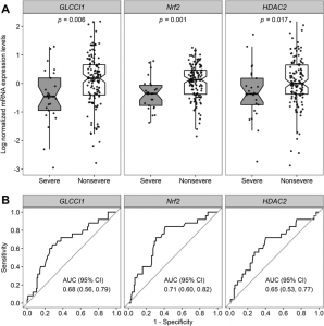
Fig 1. (A) mRNA expression levels of GLCCI1, Nrf2, and HDAC2 for severe and non-severe asthma (B) Receiver operating characteristic for the discrimination between severe and non-severe asthma (Retrieved from: Hirai)

Samples were retested if the variations were greater than 5%. With a 95% confidence interval and p values less than 0.05, indicating statistical significance, the coefficients were calculated for all 3 genes.

Patients were re-evaluated after 1 year to identify exacerbations that may occur. It was shown that exacerbations occurred in 44% of severe asthma patients and only 9.4% of non-severe asthma patients. This is directly related to the mRNA expression levels. Patients with a much lower expression level are more likely to have asthma exacerbations.

These results can be quantified to predict future exacerbation. This helps with the amount of corvicalsteroids needed for treatment. This is especially important for severe asthma patients, as corvicalsteroids are less effective compared to non-severe patients.

Observing gene expression is a method to distinguish between severe and non-severe asthma patients. It can help with reducing exacerbations using appropriate treatment.

-Wilson Wong

Eat with your… Environment?

We have all heard about eating with our eyes first, but no one ever talks about how our environment affects our meals. Mother Nature Network (MNN) indicate that your environment plays big factor in your perception of food. Whether it’s lighting, furniture, or noise, they all play a role.

Figure 1 – Chocolate Ice Cream Retrived from: HandletheHeat

This study published in October 2019 explored temporal changes in how chocolate ice cream was perceived when eaten at different locations. Each participant had their electrophysisological properties, emotions, and temporal changes in flavour monitored, with 5 minute breaks inbetween each measurement. The participants were randomly assigned different environments such as a university study area, a bus stop, a cafe, or a sensory testing laboratory.

Figure 2 – The 4 locations in which tests were conducted. A – Sensory testing laboratory B – University study area C – Bus stop D – Cafe Retrieved from: Figure 2 of Xu et al.

Electrophysiological Responses

3 electrophysiological responses were measured, including skin conductance (SC), blood volume pulse (BVP), and heart rate (HR). They found that SC and HR was significantly influenced by different environments. Using the Tukey-Kramer test, they found that eating chocolate ice cream in the study space compared to the laboratory significantly increases SC (F(3,156) = 3.149, p < 0.05). Furthermore, the HR was significantly lower after consumption in the study area compared to a bus stop (F(3,156) = 2.673, p < 0.05).

Figure 3 – Electrophysiological response measurements. n= 160 (50 males/110 females) Retrieved from: Figure 10 Xu et al.

Emotional Response

In a pilot study, the emotional responses were reported among 97 individuals. Positive emotions were noted such as happiness, cheerfulness, and joy. In addition, negative emotions were noted as well, such as tenseness, unhappiness, and anxiousness. Using a Cochran Q-test, they found that a significant number of negative emotions were associated with the bus stop compared to the other 3 environments. Furthermore, a significant number of positive emotions were expressed after consuming chocolate ice cream at a cafe or university compared to a bus stop.

Figure 4 – Both positive and negative emotions associated with eating chocolate ice cream in 4 different environments. Data adapted from: Xu et al.

Taste

The dominance of different attributes were measured and converted to a percentage of time it spent as a dominant factor. Sweetness the dominant attribute across all environments (46% lab, 33% university, 48% cafe, 38% bus stop). Interestingly, the dominance of sweetness subsided overtime, and other attributes became dominant. Other factors such as creaminess, roastedness, and bitterness was noted as well. At the bus stop, bitterness became the most dominant factor after sweetness, while the other 3 locations reported either creaminess, cocoa, or vanilla flavours were dominant.

How do I improve my next meal?

Next time you’re at the dinner table, try some of these tricks to improve the taste of your meal. By listening to higher pitched music, sour and sweet flavours are highlighted, while lower pitched music enhances bitter flavours. Even something as simple as the way food is arranged on the plate will impact its flavour.

-Jackson Kuan

Orvig Group at UBC Creates Novel Molecule for Diagnostic Nuclear Medicine

You may be aware of the role physicists and doctors play in diagnostic nuclear medicine, however you may not know that chemists also play a significant role in this area of science!

In 2019, Chris Orvig of the Medicinal Inorganic Chemistry Group at the University of British Columbia (UBC) created a new organic molecule for medical imaging. They also determined that their novel organic molecule has superior properties to similar molecules currently being used.

The molecule created by the inorganic chemistry group at UBC is simply known as H2hox, a hexadentate chelating ligand. What exactly does that mean? Let’s break it down piece-by-piece.

A ligand is a type of molecule that can bind onto a metal ion, like sodium (Na+) or calcium (Ca2+). In the case of H2hox, the metal ion it’s binding to is Gallium (Ga3+) because it is used in medical imaging.

The word chelating comes from the latin root word chela, which means claw. This is because chelating ligands have multiple points of attachment to a metal ion, similar to a crab’s claw, making them significantly stronger binders to metal ions.

The word hexadentate comes from the latin root words hexa, which means six and dent, which means tooth. So a hexadentate chelating ligand has six attachment points, or teeth, that can grab onto a desired metal ion.

Image sources (left to right): Research Gate, Orvig et al..


So why is H2hox used in medical imaging?

Molecules such as H2hox are used in a form of medical imaging known as Positron-emission tomography (PET). John Hopkins Medicine defines PET imaging as “using a scanning device (a machine with a large hole at its center) to detect photons (subatomic particles) emitted by a radionuclide in the organ or tissue being examined”.

PET imaging is primarily used to diagnose health issues related to biochemical processes occurring inside our cells, such as cancer. The radionuclide, or radioactive atom, of choice for H2hox is Gallium ions. Since ions alone cannot be used in imaging, due to their poor mobility through our cells and tissues, they are packaged together with small organic molecules, such as H2hox, before injection into human tissue.

So what makes H2hox better than the current available options?

H2hox is an advantageous ligand for Gallium PET medical imaging because…

  • It can be easily synthesized (made) through only 1 reaction step.
  • It has a strong affinity to Gallium, exhibiting significant radiolabeling (binding to Ga3+) in only 5 minutes with low amounts of ligand and under mild conditions (room temperature)
  • The combined ligand and ion have excellent stability in vitro (inside cells) and in vivo (inside a beaker).

These combined properties make H2hox an effective and convenient molecule for Gallium PET imaging. Furthermore, Orvig’s research will act as a launching-off point for the development of even better ligands to improve the quality and ease of PET imaging and diagnosis.

I hope this news article educated you about medicinal inorganic chemistry through describing its role in medical imaging.


Literature cited:

Wang, X.; De Guadalupe Jaraquemada-Peláez, M.; Cao, Y.; Pan, J.; Lin, K. S.;                       Patrick, B. O.; Orvig, C. H2hox: Dual-Channel Oxine-Derived Acyclic                       Chelating Ligand for 68Ga Radiopharmaceuticals. Inorg. Chem.                                 [Online] 2019, 58, 2275-2285.                                                                                          https://pubs.acs.org/doi/10.1021/acs.inorgchem.8b01208 (accessed                      March 22, 2020).


-Mark Rubinchik

The Power of Acceptance

When life throws us curveballs, we’re often told to hit them out of the park. However, in a recent study, researchers at Yale University found that when individuals were presented with negative stimuli, those who merely accepted their situations experienced less pain and unpleasant emotion than those who reacted naturally to the stimuli.

First, the researchers introduced participants to the concept of mindfulness, which practices the acceptance of a situation. Then, participants were placed in two different groups: one subject to high heat on the forearm, and the other subject to negative images.

Through brain scans, the team observed that participants who practiced mindfulness had reduced activity in regions of the brain concomitant to pain response and negative emotion upon stimuli compared to those who reacted. Furthermore, the participants self-reported that they experienced significantly less negative affects when they accepted their situations.

Figure 1. Participants (n=16) experienced more negative affects upon seeing negative vs. neutral images (a), and upon feeling hot vs. warm temperatures (b) when they chose to react instead of accepting. The * indicates p<0.05, *** indicates p<0.0001, and error bars indicate standard error. (Credits: Kober et al. (2020))

Mindfulness is already practiced by patients suffering from chronic pain and depression, but these findings show the power that acceptance has even on individuals who have never meditated, and the team believes that mindfulness is a good way to temporarily regulate the intensity of pain and negative emotions.

HOW DOES MINDFULNESS WORK?

Scientists are still unsure of how meditation elicits responses in the brain; perhaps mindfulness allows us to feel more in control of our circumstances when we’re having negative experiences, or it reminds us that we have enough strength to make it through whatever. Nonetheless with this technique added to our repertoire, the next time life throws us a curveball we will be more prepared to deal with it.

-Athena Wang

Vaccination and Herd Immunity

Herd immunity is often generated through vaccination or widespread infection. For the current Covid-19 pandemic, many scientists and experts advocate social distancing to avoid overwhelming hospitals while buying more time for the inventions of vaccines and treatments. Why is vaccination favored by scientists and medical experts than a widespread infection? How is herd immunity achieved through vaccination?

What is herd immunity?

Herd immunity refers to a means of protecting a whole community from disease by immunizing a critical portion of its populace. Vaccination protects the vaccinated person but also the people who are not immunized. However, to achieve herd immunity, we need a certain percentage of people in a community to be vaccinated.

Herd immunity, the result of a high immunization rate. Source: The National Institute of Allergy and Infectious Disease (NIAID)

To reach the herd immunity threshold, different vaccination coverages which depend on the basic reproduction number (Ro) are required. Vaccination coverage is the estimated percentage of people who have received specific vaccines. For example, measles, a highly contagious virus, has a Ro value between 12 and 18. This high Ro value calls for a high vaccine coverage which is 92-94%. In other words, to reach the herd immunity threshold, at least 92% of the population needs to be vaccinated.

The higher the vaccine coverage the better…

Does it mean that measles will die out as long as 92% of the population is vaccinated against measles? The answer is no. Dr. Plans-Rubio, an epidemiology expert in Europe, found a significant negative correlation (P<0.05) between the incidence of measles in 2017–2018 in different countries of the European Union and measles vaccination coverage with herd immunity levels in the target measles vaccination population during 2015–2017. According to Dr. Plans-Rubio, low percentages of measles vaccination coverage with two doses of vaccine and the resulting low herd immunity levels explained measles incidence and persistence of measles in the European Union in 2017-2018. To eliminate the measles virus in the European Union, W.H.O must improve routine measles vaccination coverage and conduct supplementary measles vaccination campaigns.

Linear correlation coefficient p
Coverage with two doses of measles vaccine − 0.533 0.003
Coverage with one dose of measles vaccine 0.523 0.004
Coverage with first dose of measles vaccine − 0.332 0.079
Coverage with second dose of measles vaccine − 0.559 0.002
Prevalence of individuals with vaccine-induced measles protection (Iv) − 0.580 0.001
Herd immunity gap (94.5 − Iv)a − 0.580 0.001

(Table source: European Journal of Clinical Microbiology & Infectious Diseases)

Relating to Covid-19 pandemic

Without measles vaccines, we would not have lowered the mortality rate of measles and reached herd immunity in most countries. The novel coronavirus, similar to measles, is also contagious. To lower the mortality rate of Covid-19 and reach herd immunity, the corresponding vaccine is required. Hence, every single one of us should practice social distancing to avoid overwhelming our healthcare system while scientists strive to invent the corresponding vaccine.


Reference:

Plans-Rubio Pedro. Low percentages of measles vaccination coverage with two doses of vaccine and low herd immunity levels explain measles incidence and persistence of measles in the European Union in 2017–2018. European Journal of Clinical Microbiology & Infectious Diseases, 2019; 38, 1719-1729. DOI: https://link-springer-com.ezproxy.library.ubc.ca/article/10.1007%2Fs10096-019-03604-0#Sec2

-Pricia

Sugar Chemistry: A Pathway to Antibiotics

We’ve all heard it endlessly as kids. Don’t eat too much sugar, it’s bad for you. However, what if I told you that sugars aren’t all that bad and in fact, careful changes to its chemistry can lead to life-saving drugs, such as antibiotics! Just last year, researchers at the University of British Columbia, led by Stephen Withers, found a unique way to tinker with sugars’ chemical structures by using molecules in bacteria. The same bacteria found in our poop!

A view of E. Coli, the bacteria that was used by Wither’s and his team. Credits: The Philadelphia Inquirer

Sweet…but what are they?

Before we go further, let’s start with a simple question: What exactly are sugars? Sugars are molecules shaped like hexagons which are often joined to other molecules known as “acceptors”. In a way we are kind of like sugars; we find someone we like, confess how we feel, and they accept our love! Right? Wrong. As we all know the last part rarely happens and this is the same in sugars, as chemists have yet to find easy ways to join the sugar and acceptors. Luckily for sugars (and unluckily for us), Mother Nature has come up with some solutions, using helper molecules known as enzymes.

Curious as to what type of sugars chemists work with? There’s no ambiguity here, chemists use the same molecules found in sugar cubes. Yes! The ones you put in your coffee. Credits: The Verge

a solution under our noses…

Instead of struggling to find ways of joining sugars and acceptors, Wither’s team thought: Why not just use these enzymes? In other words, hijack Mother Nature. To make their idea a reality, they extracted sugar-specific enzymes from E. Coli, a bacterium that lives inside the human digestive tract. Their efforts gave them 175 sugar-specific enzymes, and from this they chose 8 enzymes that were most specific to the type of sugars and acceptors they were interested in.

“With the 8 enzymes in hand, Withers and his team could now easily make these sugar-acceptor linkages” is what I would like to report; however, things are never so simple. It turns out that the sugar-specific enzymes they got from E. Coli did the exact opposite of what they wanted. Instead of forming sugar-acceptor linkages, they were specialized in breaking them.

Unsurprisingly the savvy researchers expected this and already had a reliable strategy to reverse-engineer these enzymes from linkage breakers to linkage makers. You may be wondering how they re-purposed something to work completely opposite of what it was intended for. To reconcile this, think of this example: hammers. If you’re feeling angry one day you would likely use the hammer to smash things. However, if you’re feeling innovative one day, the hammer would help you build things by hammering in nails. These enzymes are similar; an enzyme that breaks sugar bonds differs very little from one that builds sugar bonds.

more than just a bond…

Sugars go way beyond than just satisfying your sugar fix. They are molecules essential to the maintenance and regulation of not only your body, but in most living things! Because they are found everywhere, including infectious bacteria, sugar-based molecules serve as effective antibiotics, however making these drugs are difficult. Why? Well as mentioned before, chemists have trouble making these sugar-acceptor bonds; however, the research done by Wither’s team show that this will not remain the case. On a lighter note, they also created a sugar-based molecule that had nothing to do with health; detergent. This just further shows that these bonds are far-reaching and relevant in many contexts.

Story source

Armstrong, Z.; Liu, F.; Chen, H.-M.; Hallam, S. J.; Withers, S. G. Systematic Screening of Synthetic Gene-Encoded Enzymes for Synthesis of Modified Glycosides. ACS Catalysis 20199 (4), 3219–3227.

-Kenny Lin

Gold: Precious in a Different Way

Let’s face it, to most people gold is just an over-glorified rock with no real value; however, that’s not the case at all! Just this month, researchers from University College London have created a novel light-activated coating that kills infectious bacteria. The key ingredient? Gold.

upgrading with gold…

The invention of a bacteria-killing coating sounds ingenious; however, Hwang’s team was actually not the first to come up with this idea. Previous studies have already shown that coatings incorporating the chemical crystal violet can adequately kill bacteria. The problem was that the coating had to be light-activated by UV rays, which harm the skin by promoting skin cancer.

This was exactly the problem Hwang’s team looked to solve; to make a coating that did not require harmful wavelengths of light. They overcame this challenge by incorporating small clusters of gold into a polymer containing crystal violet. The result? Now this new coating could effectively eliminate bacteria upon activation with low intensity white light – the level of light found in offices.

Concentration of bacteria (CFU/mL) across three conditions after 6 hours exposure to low-intensity white light. Star indicates bacterial concentration is undetected. Sample size = 6 per treatment, error bars are standard deviation. Adapted from Hwang et al.’s data

The figure above perfectly illustrates their result. Statistical analyses show that bacterial concentration does not significantly differ between the violet crystal and control (no coating) condition. This indicates that low-intensity white light cannot activate the bacterial-killing function in the violet crystal coating. What’s interesting is that addition of gold with the violet crystal, reduces the bacterial concentration significantly to near zero values, indicating successful activation.

More than a novelty…

The results of Hwang’s study are truly impactful. It is well known that hospitals are a hotbed for infectious bacteria. In fact, 27% of surfaces in hospital rooms are contaminated with bacteria even after regular and thorough cleaning. As such, applying the coating on these surfaces will definitely reduce the chances of contracting a hospital-related disease. Who would have thought? Not only is gold more than just a hunk of rock, it can also save lives.

-Kenny Lin

Inexpensive and rapid test to detect Lyme disease

A Great Challenge

Lyme disease is the most common vector-borne infectious disease in North America and Europe. Caused by the spirochete bacterium Borrelia burgdorferi, it is characterized by a rash in infected skin and leads to major symptoms if left untreated. Though many tests have been developed to diagnose this disease, the currently available tests are expensive and lack sensitivity (true positive rate) when it comes to the early stages of the infection.


Fortunately, a team of scientists from The University of California, Los Angeles, has recently developed a new inexpensive and trustful way of detecting this infection. They claim that this new procedure does not need previous training to be implemented, and that its sensitivity can be greater than 90%.

Figure 1: Lyme disease testing procedure. Adapted from ACSNANO

How does it work?

Figure 2: Illustration of the complexed reactions that lead to identification of the lyme disease. Adapted from ACSNANO

This novel test consists of a paper based multicomplex vertical flow assay, where small paper layers are covered in various disease-specific target proteins that interact with different antigens present in human samples. The protein-antigen interaction results in an observable change in colour. Upon the completion of the test, they generate a colour pattern that can be analyzed by a computer or even a smartphone. This allows possible diagnosis of the disease within minutes and increases specificity (true negative rate) and sensitivity in its early stages. The test has also been optimized with positive and negative controls to avoid false diagnoses, and it is enclosed in a 3D printed case for easy handling.


Major Improvements to Technology

Figure 3: Reported data on test sensitivity, specificity and Area Under the Curve. Adapted from ACSNANO

Previously used examinations could cost up to 400 USD per test, and their average time for diagnosis is currently over 24 hours. They also have very low sensitivity to the early stages of the disease, with values of less than 50% being reported. As mentioned by the authors, this assessment has a material cost of 0.42 USD per test which greatly reduces costs for diagnoses. They also report values of sensitivity of over 90% in early stages of Lyme disease and a specificity value of 87%. Nonetheless, this team of researchers have demonstrated the correct diagnosis of the disease in a matter of minutes making this process efficient, easy and available to the public at a reduced cost.


-Aron Engelhard

New method found to build artificial blood vessels!

Figure1, yellow: Number of People died on the kidney transplant waitlist, blue: Number of people removed from the waitlist classified as “too sick”. Source


As the research shows, there are more than 114000 people in the United State on the waiting list of life-saving organ transplants. On average, there will be one new name added to the list every ten minutes, and 20 people die because of the lack of available organs every day. Research team from the University of Minnesota Medical School has published their new founding in Nature Biotechnology on March 11st, 2020. Their research proved the possibility to build artificial blood vessels in a pig, which has the potential to provide ultimate blood vessels for human organ transplants.

How did they come up with the idea?

“There is so many chronic and terminal diseases, and many people are not able to participate in organ transplantation,” said Daniel, a cardiologist who accepted heart transplantation before, “About 98 percent of people are not going to be eligible for a heart transplant, so there’s been a huge effort in trying to come up with strategies to increase the donor pool. Our approach looked at a pig.” Due to the physiological similarity between human and pigs, scientists have done similar studies of using pig insulin to treat human diabetes. These historical researches give scientists confidence and also an existing platform to study.

What is the benefit of choosing pigs?

Figure 2:Acute rejection Anti-CD3 Donor DC. Source

Transplant rejection is a process of transplanted tissue rejected by the recipient’s immune system. According to the research done previously, there are 50-80% of patients would have at least one rejection episode. This symptom is due to recipient’s body cannot adapt to a “foreign” tissue and the immune system would fight against the transplanted tissue. Transplant rejection always causes severe consequences, and a lot of patients would have to remove the tissue immediately.

Daniel Garry, the leader of the research team came up with an idea to avoid transplant rejection. They took mature cells scraped off from a patient’s skin, reprogram these cells and eventually inject them into a pig embryo. This process would develop patient’s own genetic information in pig’s body. Thus, patients will get their own “blood vessels” which can greatly avoid transplant rejection. Mary, coworker of Garry said: “These blood vessels would be engineered and could be utilized in these patients to prevent those kinds of life-long handicaps, if you will.”

The first phase of their study has been approved by the University of Minnesota’s Stem Cell Research Oversight committee. “While it is a first phase, there’s pretty solid proof of concept,” Mary said. “We believe that we’ve proven that there’s no off-target effects of these cells, so we’re ready to move forward to later gestational stages.” This study will eventually benefit millions of people who are suffering from transplantation surgery. Also, this study shines lights and give hopes to modern organ transplantation studies.

-Vicky Gu

Surgery can activate cancer cells, but aspirin stops metastasis

When patients are diagnosed with breast cancer, the cancer cells have already metastasized to another part of the body. However, the number of cancer cells involved in this process is negligible, and current equipment cannot detect them. Cancer cells after metastasis remain inactive, which seems unlikely to threaten patients’ health. Nevertheless, those dormant cancer cells are time bombs. One way to set them off, surprisingly, is through cancer surgery.

Recent research led by Dr. Robert Weinberg of the Whitehead Institute found the mechanisms that may explain why surgeries activate the hidden cancer cells. They designed a set of comparison experiments based on mice that injected with breast cancer cells and observed how breast cancer developed in different conditions.

To simulate the postoperative recovery process, scientists implanted sterile sponges in the mice injected with breast cancer cells. This “unnatural” design may be controversial, but it maintains all animals experiencing the same experimental conditions.

“Weinberg gets some pushback because he works on artificial systems, but this is often the only way to expose fundamental principles of biology.” said biologist Sui Huang, professor of the Institute for Systems Biology, who was convinced by this experiment.[1]

Figure 1. (A) Schematic illustrating the experimental design. Mice had been previously wounded by sponge implantation at one or two distant sites. (B) Tumor diameter during the one-month experiment. (C) Tumor incidence as a function of time (n = 9 to 10 per group) for the experimental and control group. Data are plotted as means ± SEM. P values were calculated using the Mann-Whitney test (P < 0.05). Source: Translational Medicine Science

One month after the surgery, researchers tested the number of cancer cells that remained in mice’s bodies. Figure 1 summarizes the results: for those that accepted the surgery, 60% of mice developed tumors in other parts of the body. While in the comparison group, the value is only 15%. Based on the results from 270 mice, Weinberg concluded that surgery could accelerate the cancer cell metastasis and even facilitate tumor formation.

The reason for the effect, as explained in the paper, has to do with the immune system. During the surgical wound recovery process, the inflammatory response restricts the immune system. Therefore, the “guard cells” cannot effectively monitor the cancer cells, resulting in metastasis and tumor formation.

Figure 2. Tumor diameter after the injection of cancer cells into previously unwounded (left) or wounded (right) mice treated with saline or meloxicam (n = 15 mice per group). Data are plotted as means ± SEM. P values were calculated the Mann-Whitney test P < 0.0005. Source: Translational Medicine Science

The good news is common pain killers, such as aspirin, can efficiently inhibit this process. Scientists found that many nonsteroidal anti-inflammatory drugs can effectively suppress tumor formation resulting from surgical wounds. Figure 2 shows that the wounded mice had a constant tumor size at around 2mm after given meloxicam, while the comparison developed tumors at average 5mm. Note the experiment only tested meloxicam; aspirin was also proved to be effective in the follow-up research.

Although the results are quite delightful, whether we can apply the same experiment to humans remains unclear. Weinberg pointed out that the aim of the investigation is not telling people not to trust lumpectomy or other tumor surgeries but develop a more effective treatment for postoperative recovery. He hoped that this research would promote further experiment on human and test whether drugs like aspirin has the same effect in the human body.