Category Archives: Chemistry News

Novel method of converting any blood type to the universal O

A journal article published by Nature in June 2019, studied a novel method in converting type A blood to the universal type O blood using bacteria found in poop! The Withers’ research group at the University of British Columbia used the DNA of bacteria found in the faecal samples to develop this efficient method.

HOW ARE BLOOD TYPES CLASSIFIED?

There are four main blood types: A, B, AB, O.

Blood type is determined by the type of antigens and antibodies present in the blood. Antibodies are part of your body’s defense system, if they sense germs (or any other foreign substance) they alert your immune system which stimulates a biological response to destroy said germs.

Antigens are proteins that are directly attached to red blood cells that are used to identify blood as A, B, AB or O. Antigens are also attached to foreign substances, and antibodies use them as indicators that a substance does not belong in the body. Figure 1 shows how antibodies interact with antigens.

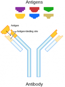
Figure 1: Image of antibody interacting with an antigen. Obtained from Wikipedia commons.

For example, if you have type A blood then your red blood cells will have A antigens. However, your blood will have type B antibodies. This means that the immune system will attack any substance with B antibodies – including type B blood. This explains why individuals who get blood transfusions can only accept their same blood type, unless it is the universal type O.

Universal type O blood doesn’t have any antigens so anyone can accept O blood as their antibodies will not attack the red blood cells. Figure 2 shows the different blood groups and how they are classified.

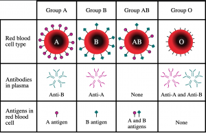
Figure 2: Image of different blood types depicting what antigens and antibodies they contain. Obtained from Wikipedia commons.

RESULTS OF THE STUDY

Figure 3 shows an in depth depiction of the difference in antigens between the blood groups.

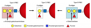
Figure 3: In depth image depicting the different constituents of antigens on type A, O and B blood. Obtained from journal article “An enzymatic pathway in the human gut microbiome that converts A to universal O type blood”

The researchers of this study, synthesized  enzymes from the bacterial DNA found in poop. Enzymes are a type of protein that aid in the reactivity of a reaction, essentially meaning they help a reaction occur faster by breaking down substances. Figure 4 demonstrates how enzymes break down substances known as substrates.

Figure 4: Image depicting how enzymes break down substances into two products. Obtained from Wikipedia commons.

In this case, the researches synthesized an enzyme that break down the bond labelled ‘3-α’ in type A and B blood cells (Figure 3). Once this bond is broken down, you are left with type O blood. The enzyme that Dr. Stephen Withers group identified, breaks down this bond 30 times faster than the previous candidate.

WHAT ARE THE IMPLICATIONS?

In January 2020, the American Red Cross announced that it has a ‘critical’ shortage of type O blood.

With this new method, hospitals and relief organizations, such as the American Red Cross, can convert type A and B blood to the universal O. It is vital that hospitals and relief organizations have a constant supply of O blood in case they need to do blood transfusions without knowing the blood type of the patient. It could also completely eliminate the need for blood type compatibility, if the research leaves the lab.

This enzyme is 30 times faster and, thus, more efficient than the previous candidate. This means that less of this enzyme is required to convert the same amount of blood, drastically decreasing the cost of production.

Overall, not only is this new method interesting to the science community but is is also incredibly important to the healthcare system.


-Chantell Jansz

Bacteria: Friend or Foe?

Did you know that not all bacteria is bad? In some cases, they cause diarrhea, stomach ulcers, and even intestinal diseases. However, what if I told you scientists have found a way to manufacture antibiotics that are used to treat these bacterial infections from bacteria themselves?

Scanning Electron Microscope view of bacteria. Retrieved from: NYTIMES

Dr. Jason Hedges and Dr. Katherine Ryan of the University of British Columbia took a look into finding new enzymatic pathways for synthesizing nitroimidazole, which is a component in the antibiotic, azomycin.

So what is the point of this whole process and why do we even want to use bacteria to synthesize antibiotics?

By finding more ways to develop antibiotics from bacteria, this improves our knowledge on biosynthetic pathways. This is beneficial not only for the scientific community, but for the public as well. As bacteria develop resistances to antibiotics over time, the discovery of new antibiotics would be able to treat more patients suffering from bacterial infections.

How is this done?

Now how could something that sounds so complex be done? Let us take a look at their process step-by-step.

Like all scientists do, background research was performed to see how previous scientists went about finding ways to develop antibiotics from bacteria. To do so, a bioinformatics search was performed. Bioinformatics is essentially ‘googling’ information about a certain topic, but in this case, they would be using a scientific database such as the National Centre for Biotechnology Information (NCBI).

A cryptic gene cluster was found in the bacterial strain Streptomyces cattleya. This along with various enzymes were the main points of interest. Their goal was to use L-arginine; a fundamental building block of proteins, and find a way to convert this into nitroimizadole (a component of the antibiotic, azomycin

Theoretically, a blueprint on how L-arginine would be converted to nitroimidazole was developed. However, experiments must be conducted to see if the pathway would work in real life, and not just on paper.

Figure 1 – Biosynthetic pathway towards nitroimidazole. Retrieved from: Hedges and Ryan, 2019

Through experimentation, the pathway as shown in figure 1 was deemed to have synthesized nitroimidazole successfully. The next step was to determine whether or not azomycin could be synthesized from Streptomyces cattleya. Unfortunately, they were unsuccessful in detecting any levels of nitroimidazole in the bacteria samples. They concluded that potentially a different molecule had been synthesized, or that this specific gene cluster is silent (inactivate).

Although Hedges and Ryan were unable to find a definitive pathway to synthesizing azomycin utilizing bacteria, their work was able to disprove aa few reaction schemes in the scientific community, allowing for further research to be conducted.

Science is not always about success. In science, you must fail in order to succeed. Their work provides a stepping stone into further scientific research such a finding other biosynthetic pathways in the synthesis of other antibiotics.


Literature Cited:

Hedges, J. B.; Ryan, K. S. In Vitro Reconstitution of the Biosynthetic Pathway to the Nitroimidazole Antibiotic Azomycin. Angewandte Chemie International Edition 201958 (34), 11647–11651.

-Jackson Kuan

Assemble Sugars with the Assistance of a “Secret Weapon”—Enzyme

People eat sugar every day, but do you know scientists can make whatever types of sugar they want? A group of researchers led by Dr Stephen Withers from the University of Columbia found an efficient method for creating new sugars. They selected a powerful type of biological catalyst called enzymes, which can assemble certain types of sugar molecules faster and cleaner than other chemical catalysts. This method has potential applications in drug development for diseases such as diabetes and obesity.

Sugars are referred to as a type of molecules that consist of units of hydrocarbons assembled in a long chain. The picture below shows various sugar molecules. Each hexagon represents a sugar unit, and different sugars have different numbers of units. Sugars with only one unit are called monosaccharides. Glucose and fructose are common monosaccharides. Our table sugar has one glucose combined with one fructose and is called disaccharide (of course!). Polysaccharides consist of starch, cellulose, and glycogen (sugar stored in your body). Sugars also exist on the cell surface and act as the receptor for many drug molecules. Therefore, knowing the properties of different sugars and how to synthesize them is an essential topic in modern biology and chemistry.

Figure 1. Sugar in daily life vs. Sugar in chemistry

Despite sugars are important to human, making the desired type of sugar molecules is a tricky problem. The reason is that many sugar units have a unique geometry. To maintain the biological functions of sugars, we also need to keep its original shape. Most of the synthetic chemical methods can assemble the sugar unit in the desired order, but cannot retain the geometry. To solve this problem, Withers and his group decided to use a “secret weapon” in biology—enzymes.

Enzymes are a special type of proteins widely existing in all organisms. They can accelerate the chemical reactions in our body and sustain normal metabolic processes. More importantly, enzymes are highly specific to particular sugar geometry. In other words, they only react with sugars that fit their structures and yield product that also has one specific structure. The type of enzymes accelerates sugar assembly is called glycosynthase, and the type accelerates disassembly is called glycoside hydrolase. Now, using enzymes seem to be promising, but where to find the enzymes we want?

To find the desired enzyme more quickly, scientists used a technique called metagenomics which allows them to sample the genes of millions of microorganisms without the need for individual culture. Instead of directly searching for enzymes that can link the sugar together, the first step is to find enzymes (glycoside hydrolase) which break sugar bond (Surprise!). Researchers used bacteria as factories to produce the enzymes and collect them together. Of course, we want enzyme glycosynthase that LINK sugar bonds. The next step is to reconstruct those enzymes such that they can assemble the sugar correctly. Researchers change the reaction centre of the glycoside hydrolase by muting some of the critical structures. By doing so, some of the glycoside hydrolases betrayed their original duties and started to assemble the sugar unit. Figure 2 shows the overall procedures for the experiment.

Abstract Image

Figure 2. Experimental procedure. Sugars are shown as chair-like hexagons. The aim is to link the sugars to various substrate molecules (shown in different colours).

Eventually, Withers and his group found eight types of enzymes that are specific to the assembly of different sugar molecules, which is almost impossible using traditional methods. As discussed before, sugars construct the receptors for drug and other signal molecules in our body. Understanding how to synthesize sugar will help scientists build new medicines targeted to specific body cells. Diseases such as diabetes and obesity that are related to sugars will also be better understood in the future.

Reference:

Armstrong, Z.; Liu, F.; Chen, H.-M.; Hallam, S. J.; Withers, S. G. Systematic Screening of Synthetic Gene-Encoded Enzymes for Synthesis of Modified Glycosides. ACS Catalysis 2019, 9 (4), 3219–3227.

https://pubs.acs.org/doi/abs/10.1021/acscatal.8b05179

Sugar Chemistry: A Pathway to Antibiotics

We’ve all heard it endlessly as kids. Don’t eat too much sugar, it’s bad for you. However, what if I told you that sugars aren’t all that bad and in fact, careful changes to its chemistry can lead to life-saving drugs, such as antibiotics! Just last year, researchers at the University of British Columbia, led by Stephen Withers, found a unique way to tinker with sugars’ chemical structures by using molecules in bacteria. The same bacteria found in our poop!

A view of E. Coli, the bacteria that was used by Wither’s and his team. Credits: The Philadelphia Inquirer

Sweet…but what are they?

Before we go further, let’s start with a simple question: What exactly are sugars? Sugars are molecules shaped like hexagons which are often joined to other molecules known as “acceptors”. In a way we are kind of like sugars; we find someone we like, confess how we feel, and they accept our love! Right? Wrong. As we all know the last part rarely happens and this is the same in sugars, as chemists have yet to find easy ways to join the sugar and acceptors. Luckily for sugars (and unluckily for us), Mother Nature has come up with some solutions, using helper molecules known as enzymes.

Curious as to what type of sugars chemists work with? There’s no ambiguity here, chemists use the same molecules found in sugar cubes. Yes! The ones you put in your coffee. Credits: The Verge

a solution under our noses…

Instead of struggling to find ways of joining sugars and acceptors, Wither’s team thought: Why not just use these enzymes? In other words, hijack Mother Nature. To make their idea a reality, they extracted sugar-specific enzymes from E. Coli, a bacterium that lives inside the human digestive tract. Their efforts gave them 175 sugar-specific enzymes, and from this they chose 8 enzymes that were most specific to the type of sugars and acceptors they were interested in.

“With the 8 enzymes in hand, Withers and his team could now easily make these sugar-acceptor linkages” is what I would like to report; however, things are never so simple. It turns out that the sugar-specific enzymes they got from E. Coli did the exact opposite of what they wanted. Instead of forming sugar-acceptor linkages, they were specialized in breaking them.

Unsurprisingly the savvy researchers expected this and already had a reliable strategy to reverse-engineer these enzymes from linkage breakers to linkage makers. You may be wondering how they re-purposed something to work completely opposite of what it was intended for. To reconcile this, think of this example: hammers. If you’re feeling angry one day you would likely use the hammer to smash things. However, if you’re feeling innovative one day, the hammer would help you build things by hammering in nails. These enzymes are similar; an enzyme that breaks sugar bonds differs very little from one that builds sugar bonds.

more than just a bond…

Sugars go way beyond than just satisfying your sugar fix. They are molecules essential to the maintenance and regulation of not only your body, but in most living things! Because they are found everywhere, including infectious bacteria, sugar-based molecules serve as effective antibiotics, however making these drugs are difficult. Why? Well as mentioned before, chemists have trouble making these sugar-acceptor bonds; however, the research done by Wither’s team show that this will not remain the case. On a lighter note, they also created a sugar-based molecule that had nothing to do with health; detergent. This just further shows that these bonds are far-reaching and relevant in many contexts.

Story source

Armstrong, Z.; Liu, F.; Chen, H.-M.; Hallam, S. J.; Withers, S. G. Systematic Screening of Synthetic Gene-Encoded Enzymes for Synthesis of Modified Glycosides. ACS Catalysis 20199 (4), 3219–3227.

-Kenny Lin

Inexpensive and rapid test to detect Lyme disease

A Great Challenge

Lyme disease is the most common vector-borne infectious disease in North America and Europe. Caused by the spirochete bacterium Borrelia burgdorferi, it is characterized by a rash in infected skin and leads to major symptoms if left untreated. Though many tests have been developed to diagnose this disease, the currently available tests are expensive and lack sensitivity (true positive rate) when it comes to the early stages of the infection.


Fortunately, a team of scientists from The University of California, Los Angeles, has recently developed a new inexpensive and trustful way of detecting this infection. They claim that this new procedure does not need previous training to be implemented, and that its sensitivity can be greater than 90%.

Figure 1: Lyme disease testing procedure. Adapted from ACSNANO

How does it work?

Figure 2: Illustration of the complexed reactions that lead to identification of the lyme disease. Adapted from ACSNANO

This novel test consists of a paper based multicomplex vertical flow assay, where small paper layers are covered in various disease-specific target proteins that interact with different antigens present in human samples. The protein-antigen interaction results in an observable change in colour. Upon the completion of the test, they generate a colour pattern that can be analyzed by a computer or even a smartphone. This allows possible diagnosis of the disease within minutes and increases specificity (true negative rate) and sensitivity in its early stages. The test has also been optimized with positive and negative controls to avoid false diagnoses, and it is enclosed in a 3D printed case for easy handling.


Major Improvements to Technology

Figure 3: Reported data on test sensitivity, specificity and Area Under the Curve. Adapted from ACSNANO

Previously used examinations could cost up to 400 USD per test, and their average time for diagnosis is currently over 24 hours. They also have very low sensitivity to the early stages of the disease, with values of less than 50% being reported. As mentioned by the authors, this assessment has a material cost of 0.42 USD per test which greatly reduces costs for diagnoses. They also report values of sensitivity of over 90% in early stages of Lyme disease and a specificity value of 87%. Nonetheless, this team of researchers have demonstrated the correct diagnosis of the disease in a matter of minutes making this process efficient, easy and available to the public at a reduced cost.


-Aron Engelhard

Enhancing Safety Gloves

Safety gloves do not protect you from every chemical or dangerous substance. The glove deteriorates and makes it easier for chemicals to penetrate through and onto the skin. One way to tackle this problem is by implementing a self-healing material, which can be used for rubber gloves.

Researchers at the Central Institute for Labour Protection in Poland tested  polyamide, cotton–polyamide, and cotton fabrics, onto methyl vinyl silicone rubber containing inorganic silsesquioxane, which are used for rubber gloves. Its resistance to chemical substances, abrasions, and punctures were analyzed. Using SEM, the surface is observed for any damages or self-healing behaviour for each rubber material containing each type of fabric.

A cross-section of a rubber material with textile reinforcements (Source:

A test to determine the resistance of chemical substance with methyl vinyl silicone rubber with silsesquioxanes, and one coated with cotton was carried by using 2-propanol. This studied the breakthrough time of the 2-propanol through the 2 different materials at various conditioning times. The idea is to simulate the effectiveness of the self-healing mechanism.

Permeation times of 2-propanol into 2 different materials (Adapted: )

The data shows an increase in permeation time when coated with cotton, and when conditioned at 70 degrees Celsius for 24, 48, 72 hours. This means coating the material with textile reinforcements increases the resistance of chemical substances from penetrating. A similar trend was obtained when testing different textile compositions through puncture and abrasion tests.

The result concludes the effectiveness of the textile reinforcement in the self-healing process, qualitatively and quantitatively. This study brought improvements to lessen the stress chemists have on the quality of their own safety gloves. Safety gloves should never be a safety concern.

-Wilson Wong

Where Fluke meets Fortune: How Chance Lead to Discovering Novel Green Chemistry Reactions

Dr. Petri Turhanen from the University of Eastern Finland discovered that Dowex, a cation exchange resin, opens up an untapped area of green chemistry – the scientific initiative to find chemical reactions that produce the least waste. The best part? It wasn’t on purpose.

While working on an organic synthesis project in 2015, Turhanen noticed that the cation exchange resin he was using, Dowex, produced an unintended byproduct in the presence of sodium iodide (NaI), an iodide (I) source. Further analysis unveiled that the byproduct was the result of an iodide addition reaction. This is a reaction where a double bond between two carbon atoms is converted into a single bond with a new atom on each carbon, one hydrogen and one iodine.

The novel and green iodide addition reaction discovered by Dr. Petri Turhanen

The source of this unique reactivity comes from the polymer known as Dowex. Dowex is a solid resin made of polystyrene sulfonate. Its main use is as a cation exchange resin, a type of solid that is able to exchange cations, such as H+, for other cations, such as Na+ or K+.

Why is this reaction significant? Iodinated molecules serve multiple purposes. They are often intermediate molecules in organic synthesis, acting as a precursor to building up larger, more complex organic molecules such as pharmaceuticals. Furthermore, radioactive iodine isotopes attached to organic molecules are used as tags in medical imaging.

The industrial processes used to iodinate compounds require toxic starting materials, harmful solvents and high temperatures. These include hazardous, or even carcinogenic, compounds such as iodine, hydrogen peroxide, trimethylsulfonium iodate and iodine monochloride and heavy metals catalysts. To contract, Dowex has low toxicity and can be reused after the reaction is complete.

Comparison of iodide addition reactions

Since the first experiment in 2015, Turhanen has expanded the library of reactions possible in the presence of Dowex, such as esterifications and the conversion ethylene to a di-iodide species. Continued organic synthesis initiatives such as Turhanen’s will pave the way for a greener future of science.


-Mark Rubinchik

Antibiotics found to kill bacteria in a new way!


Fig1.Antibiotics source

Antibiotic is the most powerful “weapon” to fight against bacterial infections. However, according to the World Health Organization, there are more than 700000 people die every year due to antibiotic resistance. On Feb 13th, 2020, Research team from the David Braley center for Antibiotic Discovery, University of McMaster posted an article on nature. Newly found corbomycin and complestatin would kill bacteria in a brand-new way. The discovery of these new groups of antibiotics would be the clinical candidate in the fight against antimicrobial resistance.

Fig2,Antibiotic resistance strategies in bacteria. source:Courtesy of E. Gullberg.


Antibiotics are the revolution of the pharmaceutical study in the 20th century. They are the most important type of antibacterial agent which either kills or inhibit the growth of bacterial cell walls. Alexander Fleming discovered modern antibiotic medicine – penicillin in 1928, which saved thousands of people’s life.

What does old antibiotics also bring you?

The enormous benefits of antibiotics also lead to new problems such as over-usage and resistance. Bacteria soon formed resistance toward these antibiotics and caused the ineffectiveness of the medicine. The resistance of antibiotics had become a new-rise problem. The World Health Organization announced: “serious threat is no longer a prediction for the future, it is happening right now in every region of the world and has the potential to affect anyone.”

Fig2.image of corbomycin

Fig2.image of corbomycin. source

The newly found corbomycin and complestatin have brand new way to attack bacteria. It is discovered from a glycopeptide family, and the new approach appears no significant resistance toward bacteria. “Antibiotics like penicillin kill bacteria by preventing the building of the wall, but the antibiotics that we found actually work by doing the opposite – they prevent the wall form being broken down. This is critical for cell to divide.” Said Beth Culp, a PhD candidate in biochemistry at McMaster.

Why do we know about these?

“We hypothesized that if the genes that made these antibiotics were different, maybe the way they killed the bacteria was also different”, Culp explained. The “unique approach” to kill bacteria is a new mechanism that is worthy of studying. Scientists might be able to find the new family of antibiotics which have a completely different way to attack bacteria. These new antibiotics will be the revolution of modern biochemistry which will be powerful to fight against antibiotic-resistant.


Scientists believed that the observation of corbomycin and complestatin would open the “new door” in the field of antibiotics. People will be able to investigate more antiobiotics to fight against resistance in the glycopeptide family. This study will eventually benefit thousands of people suffering in antibiotic-resistant and give them hope to survive!

–Vicky Gu


Revised: Should you be worried?—An Outbreak of Novel Coronavirus!

Since December 2019, an unexplained pneumonia epidemic has occurred in Wuhan City, Hubei Province, China. An investigation found that these were related to Wuhan’s “South China Seafood Market”. Wuhan organized a multi-disciplinary expert consultation survey and used laboratory testing to identify pneumonia in Wuhan as viral pneumonia. On January 8, 2020, a new coronavirus was initially identified as the pathogen of the epidemic. With the outbreak of this novel coronavirus, it is crucial to know how this virus spread and evolved, more importantly, how we can take precautions against it.

The spread of the novel Coronavirus

Sources: National Health Commission of the People’s Republic of China; local governments. Note: Data as of 9 p.m E.T., Jan. 27

The outbreak of this infectious disease was first occured in Wuhan in December, 2019 and then spreaded globally since the huge flow og people in Wuhan during lunar new year. According to the New York Timesthere are more than 4,500 people in Asia infected the coronavirus as well as many other are suspected. At least 106 people have died as of Jan. 27, 20.

Evolutionary sources of coronavirus  and molecular pathways for infecting humans

To analyze the evolutionary source and possible natural host of the novel coronavirus, the researchers in this paper analyzed genetic evolution by comparing the novel coronavirus with collected large amount of coronavirus data. It was found that the novel coronavirus of Wuhan belongs to Betacoronavirus which is a RNA virus that parasitizes and infects higher animals (including humans). It is adjacent to the SARS virus and the SARS-like virus group in the position of the evolutionary tree. Therefore, Wuhan coronavirus and SARS or SARS-like coronavirus may share common ancestor. As the evolutionary neighbors and outgroups of Wuhan coronavirus have been found in various types of bats, it is speculated that the natural host of Wuhan coronavirus may also be bats and Wuhan coronavirus is likely to have unknown intermediate host vectors during the transmission from bat to human.

Phylogenetic tree (Source)

The authors used molecular computational simulation methods to perform structural docking studies on Wuhan coronavirus S-protein and human ACE2 protein, and found that although 4 of the 5 key amino acids that bind to ACE2 protein in Wuhan coronavirus S-protein have changed, the amino acids after the change have perfectly maintained the interaction between SARS virus S-protein and ACE2 protein. This result indicates that Wuhan coronavirus infects human respiratory epithelial cells through the molecular mechanism of S-protein interaction with human ACE2 protein, predicitng that Wuhan coronavirus has strong ability to infect humans.

Cα RMSD of 1.45 Å on the RBD domain compared to the SARS-CoV S-protein structure (Source)

Tips for prevention of coronavirus (source):

  • Wash your hands with soap for at least 20 seconds and avoid touching you mouth, nose and eyes with unwashed hands.
  • Keep a safe distance with people who are sick
  • Cover your cough or sneeze with tissue and throw the tissue in the trash
  • Clean frequently touched surfaces

-Xinyue Yang

Posted on Jan. 27th. 2020

Revised: Embarassed of Asian Glow? Don’t Worry, The Future is Promising

Ever find yourself beet red after one small drink? You’re not alone! Over one-third of East Asians and eight percent of the world population experience this awkward phenomenon; however, a solution is in the works. Just last month, researchers from Weill Cornell Medical College have solved this problem by experimenting with targeted gene therapy on mice.

What does asian glow look like? A before and after comparison. (Credits: Wikimedia Commons)

The Dangers of Asian Glow

Apart from causing embarrassment, asian glow comes with more serious consequences than just flushing red. The red glow is caused by a deficiency in the ALDH2 enzyme, a key component in detoxifying alcohol. When you drink alcohol, your body converts the alcohol into acetaldehyde. Normally, acetaldehyde is further converted to a safer compound by ALDH2; however in individuals with asian glow, ALDH2 does not function, causing acetaldehyde to build up. Since acetaldehyde is a cancer-causing agent, its accumulation drastically increases the risk of developing esophageal cancer by six to ten folds.

Conversion of alcohol to acetate is stopped in people with asian glow. This leads to toxic buildup of acetaldehyde. (Credits: Me – created with Notability)

A Glowing Solution…

Matsumura’s team reasoned if a lack of ALDH2 enzyme was the problem, maybe they could simply add it back in.

“We hypothesized that a one-time administration of a […] virus […] expressing the human ALDH2 coding sequence […] would correct the deficiency”

They tested their idea on three strains of mice: mice with functional ALDH2, mice lacking ALDH2, and mice with a non-functional version of ALDH2. The latter two simulated the asian flush syndrome seen in humans. After introducing all the mice with the ALDH2 gene and feeding them alcohol, the researchers carefully monitored acetaldehyde levels in the blood.

Their hard-work paid off! In the two strains deficient for ALDH2 function, acetaldehyde levels and abnormal behavior associated with alcohol consumption were back to near-normal levels. Furthermore, they found that one dose was enough to confer persistent and long-term protection.

From Mice to Humans: A Complicated Decision

Matsumura’s team emphasize that a long-lasting treatment for ALDH2 deficiency currently does not exist. Although making the jump from mice to humans will be challenging, they assure that virus-mediated gene therapy shows the most promise in becoming an effective therapy. The million-dollar question is whether the risks of the glow outweigh the benefits of reduced alcohol consumption seen in affected individuals. To this Matsumura’s team say:

“the overall burden […] on human health, particularly […] cancer, supports […] gene therapy.”

What do you think?

-Kenny Lin