Let’s talk to our cells!

We are one step closer to achieve communication with those 37.2 trillion tiny components that make up our bodies. If you have ever wondered how the human body is capable of doing impressive amounts of chemical work without us even thinking about it, now you can understand it! Our bodies are efficient in converting energy, and communication among our cells is key to the understanding of all the basic processes that govern our life.

Cells often communicate via receptors made of sugars, that are exposed outside of their membranes. Such processes are often carried out by tiny sugar molecules that interact with those in their specific target. Recently, a team of researchers from The University of British Columbia published a synthetic method for modified sugars with incredible potential. In other words, it is now possible to obtain reliable materials to applications in cellular communication, metabolism and other biochemical processes.


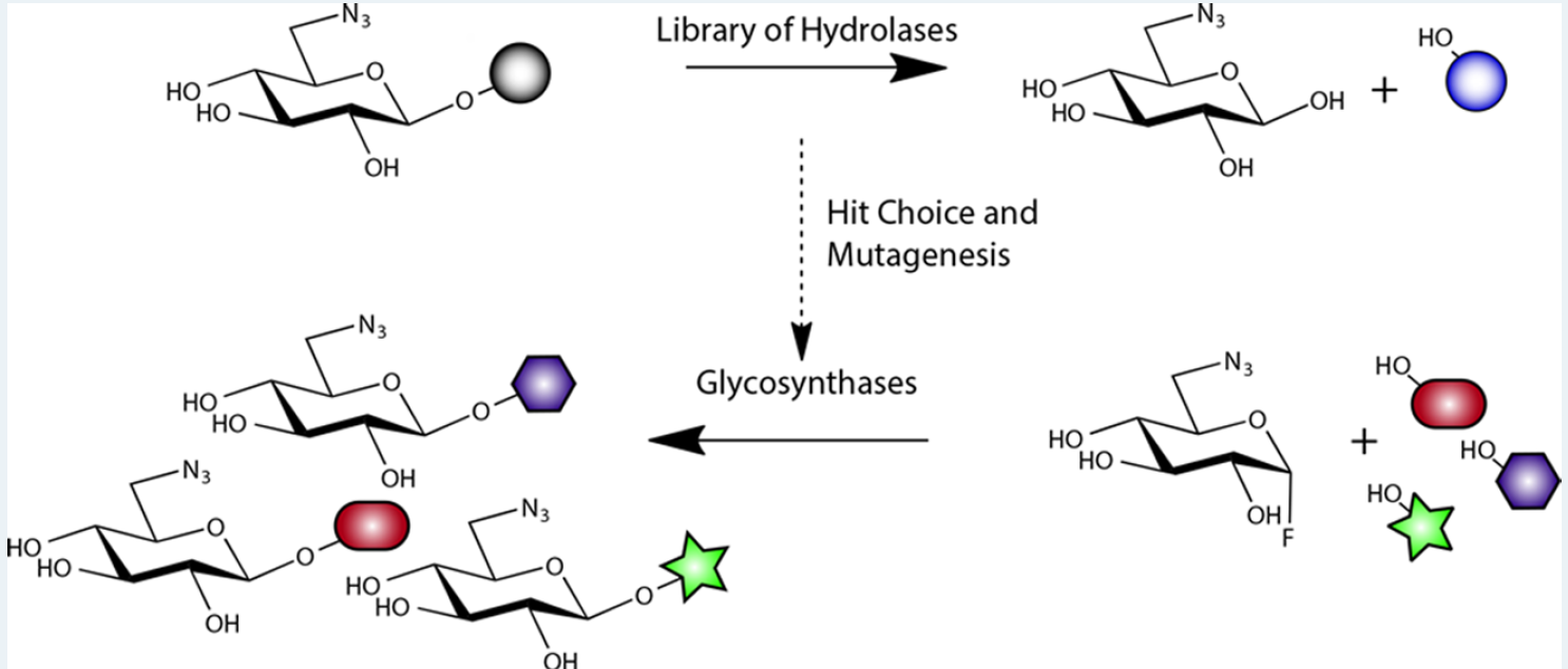
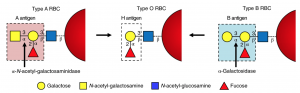
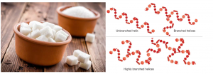
Figure 1: Structural representations of the transformation of sugars carried out by the researchers. Adapted from ACS Catalysis


This method was developed by using clones of genetically modified bacteria to express enzymes that are capable of modifying the sugar in accordance to the interest of the researcher. To achieve this, the scientists screened a library of 175 genes of the species E.coli that encoded variations of enzymes that can be used to catalyze selective chemical reactions in sugars for creating glycosidic bonds.

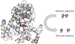
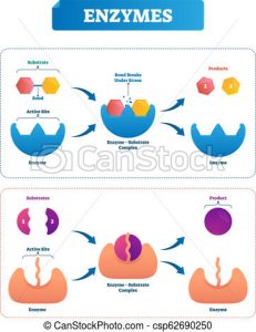
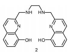
Computational representation of a hydrolase, an enzyme that breaks sugars. Adapted from Wikipedia

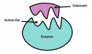
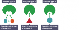
Enzymes are proteins that provide a path of a biochemical reaction to occur more efficiently. They are relatively easy to obtain and work with; however, they are specific to their target substrates which limit the extent in which their capabilities can be exploited. The scientists solved this issue by modifying the internal composition of the enzymes to improve the diversity of products in a process called selective mutagenesis. With the aid of this technique, the investigators obtained all variants that were tested in this experiment.

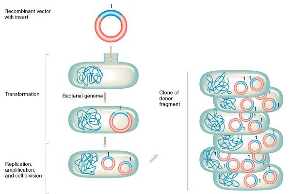
Schematic representation of bacterial transformation and cloning. Adapted from Griffiths et a (2000)

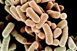
As the use of biotechnology increases, the understanding of our microscopic world becomes a major tool for scientific development. In this case, E.coli cells are essential since bacteria are inoculated with synthetic versions of genes that encode these enzymes and are then used as living machineries for protein production.

It is worth to mention that transformations of sugars were already reported. Nonetheless, previous methods rely on the use of expensive reagents as catalysts, which represent  a major cost and are not widely accessible. This new approach opens a significant area in biochemical research. As technology improves to newer and more accessible  methods, the diversification of these enzymes could develop new approaches for interaction with cell receptors that could enable us to understand what our cells have to say.

-Aron Engelhard

Journal Source:

Armstrong, Z.; Liu, F.; Chen, H.-M.; Hallam, S. J.; Withers, S. G. Systematic Screening of Synthetic Gene-Encoded Enzymes for Synthesis of Modified Glycosides. ACS Catalysis 20199 (4), 3219–3227.


Life Saving Improvements to Blood Transfusions

Previously thought impossible, researchers have found a way to create O type blood from other blood types. Since O is a universal donor, these other blood types can now be used as universal donors, potentially saving countless lives.

A team of biochemists from the University of British Columbia, lead by doctor Stephen Withers, have turned A positive and A negative type blood into the universal O type blood. These findings were published in June of 2019 in the journal “Nature Microbiology.”[1]

Before these findings, the 8 major blood types were not all compatible with each other. This meant that if a blood transfusion were needed for a patient, a specific type of blood maybe be needed, with no other type working.

A chart of what blood types can donate or receive other blood types.[2]

This problem has existed since blood transfusions were first scientifically achieved, and scientists have been looking for a solution for just as long. It turns out the solution was hiding right under our noses; inside our stomachs, to be specific!

Inside the human gut are thousands of microscopic bacteria which we use to digest food and convert it into energy. As it turns out, these bacteria are very good at safely interacting with the human body in helpful ways. The researchers extracted these microorganisms through human feces and found they could be used in exactly the way they were hoping.

“Why would they be looking in our stomachs for this solution?” you might ask. The Withers group were on a hunt for a special kind of protein called an enzyme. Enzymes are produced by the body with a very specific task, and that task varies based on what the body wants in to do. In the below picture, notice how the enzyme matches perfectly the substrate. This is because enzymes are made for specific purposes, and only match specific molecules in the body. Since our gut has the ability to process blood and turn it into energy, Withers and his team decided to see if these enzymes could be harnessed for their research.  As it turns out, they were completely correct.

An enzyme interacting with a specific molecule (known as the substrate) in the body.[3]

Future Impacts

 In the United States and Canada alone, 4.5 million patients need blood transfusions every year.[4] This high demand means that often times, the supply cannot meet the demand. With this new discovery, more blood types are compatible, which means more people can be helped.

While the process has been completed in the lab, it has yet to be scaled up to convert massive amounts of blood at a time.  This may take some time to accomplish. However, it is impossible to quantify exactly how many people this new method will help, or even how many lives it will save. One thing is for certain,  is that the world of blood donations will forever feel the impact of these findings. 

 References:

  1. Rahfeld, P., Sim, L., Moon, H., Constantinescu, I., Morgan-Lang, C., Steven, J. H., Kizhakkedathu, J. N., Withers, S. G.; An enzymatic pathway in the human gut microbiome that converts A to universal O type blood. Nature Microbiology. 2019, 1475-1485.
  2. Canadian Blood Services. Do You Know Your Blood Type? https://blood.ca/en/blood/donating-blood/whats-my-blood-type (accessed March 21, 2020)
  3. Western Oregon University: Chemistry. CH450 and CH451: Biochemistry – Defining Life at the Molecular Level. Chapter 6: Enzyme Principles and Biotechnological Applications. https://wou.edu/chemistry/courses/online-chemistry-textbooks/ch450-and-ch451-biochemistry-defining-life-at-the-molecular-level/chapter-7-enzyme-kinetics/ (accessed Mar 21, 2020)
  4. Community Blood Bank of Northwest Pennsylvania and Western New York. 56 facts about blood. https://fourhearts.org/facts/ (accessed March 22, 2020)

Making Different Sugars with Enzyme (Pac-Man) In Our Body

Although eating too much sugar can lead to health complications, a normal intake of sugar has its benefits. This is because sugars take part not only in many cellular activities as an energy source but also in cell-cell communication as a communicator. In 2019, a research team led by Dr. Stephen Withers at the University of British Columbia made different sugars by using enzymes (Pac-Man-like molecules) in bacteria. This finding enables the building of many sugars that are commonly hard to find in nature.

What are sugars?

All sugars are made of hexagonal building blocks as shown in red in Figure 1. Two common building blocks are glucose and fructose. To make different sugars with only two common building blocks, we can vary the number and arrangement of the building blocks. For example, cellulose has three long threads that are arranged almost parallel to each other. In contrast, starch has one long thread that adopts a helical structure.

Now we know that different sugars can be made by varying the kinds of building blocks, the numbers of building blocks and the arrangements of building blocks. What we haven’t talked about is how the building blocks are linked. There are two issues with linking the building blocks. First, building blocks are much smaller than our cells. Getting two building blocks together is as hard as fishing a needle from the Pacific Ocean. Besides, getting two building blocks together takes time because they prefer to be alone. How can our mother nature solve these problems to keep us alive?

Figure 1. Sugars are built differently. Some sugars are longer and more complex than others. Source: riasparklebiochemistry

Here comes the rescue…Pac-Man in the cells!

The illustrations of enzymes are like Pac-Man. However, different from Pac-Man that eats any “food” in different shapes, enzymes recognize different substrates and those substrates only! Because of this substrate-enzyme specificity, linking two building blocks together is much easier. As shown in Figure 2, the enzyme will attract two specific building blocks, making them closer to each other and eventually join.

Figure 2. Different enzymes recognize different substrates. Source: Wikipedia

Besides, enzymes can speed up the “hugging” process between the substrates (building blocks), making the formation of a long sugar favorable.

Figure 3. Enzymes speed up reactions. Source: pinimg.com

Manipulation of enzymes to build different sugars for cell-cell communication

Knowing that enzymes have many advantages, Dr. Wither and his team looked for enzymes that speed up the linkage of sugar building blocks in E. Coli, bacteria that live inside our digestive tracts. They chose eight enzymes out of the 175 sugar-specific enzymes in E. Coli. These eight enzymes were the most specific to the sugar building blocks they were interested in. However, after further investigation, the researchers found that the enzymes helped speed up the breakage of the linkage(s) between sugar building blocks instead of the formation of the linkage(s).

Figure 4. Some enzymes speed up the breakage of linkages while others speed up the formation of linkages.
Source: Canstock.com

To solve this problem, the researchers reverse-engineered these enzymes from speeding up breakage to speeding up linkage by changing parts of the enzymes. Now different sugars can be made! As mentioned above, cell-cell communication relies on sugars. This is because two cells adhere via the interaction between extracellular sugars and specific cell-surface receptors. Once these cells adhered to each other, they can talk to each other about their internal cellular condition/environment and trigger a corresponding response. This has great implications in triggering an immune response to a viral attack. By engineering more sugars on the surface of the cells, cells in our immune system can more quickly talk to each other and fight off infection.


Journal Source:

Armstrong, Z.; Liu, F.; Chen, H.-M.; Hallam, S. J.; Withers, S. G. Systematic Screening of Synthetic Gene-Encoded Enzymes for Synthesis of Modified Glycosides. ACS Catalysis 20199 (4), 3219–3227.

-Pricia Ouyang

Novel method of converting any blood type to the universal O

A journal article published by Nature in June 2019, studied a novel method in converting type A blood to the universal type O blood using bacteria found in poop! The Withers’ research group at the University of British Columbia used the DNA of bacteria found in the faecal samples to develop this efficient method.

HOW ARE BLOOD TYPES CLASSIFIED?

There are four main blood types: A, B, AB, O.

Blood type is determined by the type of antigens and antibodies present in the blood. Antibodies are part of your body’s defense system, if they sense germs (or any other foreign substance) they alert your immune system which stimulates a biological response to destroy said germs.

Antigens are proteins that are directly attached to red blood cells that are used to identify blood as A, B, AB or O. Antigens are also attached to foreign substances, and antibodies use them as indicators that a substance does not belong in the body. Figure 1 shows how antibodies interact with antigens.

Figure 1: Image of antibody interacting with an antigen. Obtained from Wikipedia commons.

For example, if you have type A blood then your red blood cells will have A antigens. However, your blood will have type B antibodies. This means that the immune system will attack any substance with B antibodies – including type B blood. This explains why individuals who get blood transfusions can only accept their same blood type, unless it is the universal type O.

Universal type O blood doesn’t have any antigens so anyone can accept O blood as their antibodies will not attack the red blood cells. Figure 2 shows the different blood groups and how they are classified.

Figure 2: Image of different blood types depicting what antigens and antibodies they contain. Obtained from Wikipedia commons.

RESULTS OF THE STUDY

Figure 3 shows an in depth depiction of the difference in antigens between the blood groups.

Figure 3: In depth image depicting the different constituents of antigens on type A, O and B blood. Obtained from journal article “An enzymatic pathway in the human gut microbiome that converts A to universal O type blood”

The researchers of this study, synthesized  enzymes from the bacterial DNA found in poop. Enzymes are a type of protein that aid in the reactivity of a reaction, essentially meaning they help a reaction occur faster by breaking down substances. Figure 4 demonstrates how enzymes break down substances known as substrates.

Figure 4: Image depicting how enzymes break down substances into two products. Obtained from Wikipedia commons.

In this case, the researches synthesized an enzyme that break down the bond labelled ‘3-α’ in type A and B blood cells (Figure 3). Once this bond is broken down, you are left with type O blood. The enzyme that Dr. Stephen Withers group identified, breaks down this bond 30 times faster than the previous candidate.

WHAT ARE THE IMPLICATIONS?

In January 2020, the American Red Cross announced that it has a ‘critical’ shortage of type O blood.

With this new method, hospitals and relief organizations, such as the American Red Cross, can convert type A and B blood to the universal O. It is vital that hospitals and relief organizations have a constant supply of O blood in case they need to do blood transfusions without knowing the blood type of the patient. It could also completely eliminate the need for blood type compatibility, if the research leaves the lab.

This enzyme is 30 times faster and, thus, more efficient than the previous candidate. This means that less of this enzyme is required to convert the same amount of blood, drastically decreasing the cost of production.

Overall, not only is this new method interesting to the science community but is is also incredibly important to the healthcare system.


-Chantell Jansz

A Novel Molecule for Nuclear Medicine

Cancer is complicated, there’s no doubt about that. From its onset to its invasive prevalence, preventing the progression of each stage seems like a never-ending battle. Be that as it may, with the advent of new chemical entities, this fight has become a lot easier.

In 2019, the Orvig group in the University of British Columbia were able to design and create a new molecule with a variety of applications. One of which is in cancer imaging, which is what makes their compound particularly astonishing. Further, the authors have reported that their chemical species is stable in, and is removed quite easily from the body. Though to understand the importance of their findings, it is vital to start at the very beginning.

Dr. Orvig (Top row, left) and his medicinal inorganic chemistry group

Within the realm of diagnostic medicine, imaging is a vital facet. The goal of imaging is to ultimately produce a viable image of the body. Positron emission tomography (PET) is an imaging method used to generate images of the chemical changes that happen in tissue. These scans often rely on the properties of the radiation emitted by a radioactive isotope. In this instance, the radioisotope used is Gallium-68 (68Ga), which is a readily available and versatile tracer species. Though, to harness the tracer capabilities of 68Ga, it would have to exist in the form of a compound. This is where the Orvig group plays an important role.

They were able to synthesize a ligand with the ability to interact with the 68Ga. A ligand is essentially a molecule that can bind to a metal center, forming a metal complex. This leads to the generation of a stable species that can exist in solution and thus can move around the body. What the group was able to show was that this novel ligand had a high affinity for 68Ga even at low concentrations. Their ligand is an acyclic hexadentate ligand, named H2hox. Though the name is inherently complicated, it does make sense once it is explained in some depth.

The Orvig group’s hexadentate chelating ligand

A hexadentate ligand is a ligand that coordinates to the metal center at 6 different positions. What this means is that there are 6 different points on the one molecule that bond with the metal center. Furthermore, the ligand is said to chelate the metal center. This term is generally given to a ligand that binds to a central metal atom at two or more points. Fortunately, this ligand is advantageous in a number of different ways.

The authors have stated that this compound is easy to synthesize, removing a number of potentially challenging synthetic strategies typically associated with 68Ga chelating species. Their initial starting materials were also inexpensive and are actually available online. To put this into perspective relative to some of the other ligand synthesis methods, it’s like baking a box cake versus baking a cake from scratch. The former is simple and quite easy to do, while the latter is a lot harder and is a lot more intensive. Ease of synthesis is an important feature, as it can affect the commercial applicability of the chelating ligand. Fortunately, their synthesis strategy is also mild.

What was also found is that their ligand is more stable than other 68Ga chelating ligand species. One advantage reported is with respect to acids and bases, where it was found that the complex is actually stable in both. It was found that there exists a single species, [68Ga(H2hox)]+, within a pH range of 1-11. Moreover, the group assessed the conditional stability of the complex. Again, these findings reinforce the advantages of their chelating ligand, H2hox.

Stability studies conducted in the sternum of mice and dynamic imaging PET studies also suggest that their compound is stable in the body and in a vial. This impressive aspect correlates with the enhanced stability of the H2hox metal complex. It was also removed from the mouse relatively quickly.

As a result of their success, their chelating species surpasses any ligand currently used as a 68Ga chelator. Not only have they managed to add to the current library of chelators, but they have also developed a convenient toolkit radiopharmaceutical compound.

Reference:

Wang, X.; De Guadalupe Jaraquemada-Peláez, M.; Cao, Y.; Pan, J.; Lin, K. S.; Patrick, B. O.; Orvig, C. H2hox: Dual-Channel Oxine-Derived Acyclic Chelating Ligand for 68Ga Radiopharmaceuticals. Inorg. Chem. [Online] 2019, 58, 2275-2285. (Accessed: March 23, 2020).

https://pubs.acs.org/doi/abs/10.1021/acs.inorgchem.8b01208

– Akash Panjabi

Bacteria: Friend or Foe?

Did you know that not all bacteria is bad? In some cases, they cause diarrhea, stomach ulcers, and even intestinal diseases. However, what if I told you scientists have found a way to manufacture antibiotics that are used to treat these bacterial infections from bacteria themselves?

Scanning Electron Microscope view of bacteria. Retrieved from: NYTIMES

Dr. Jason Hedges and Dr. Katherine Ryan of the University of British Columbia took a look into finding new enzymatic pathways for synthesizing nitroimidazole, which is a component in the antibiotic, azomycin.

So what is the point of this whole process and why do we even want to use bacteria to synthesize antibiotics?

By finding more ways to develop antibiotics from bacteria, this improves our knowledge on biosynthetic pathways. This is beneficial not only for the scientific community, but for the public as well. As bacteria develop resistances to antibiotics over time, the discovery of new antibiotics would be able to treat more patients suffering from bacterial infections.

How is this done?

Now how could something that sounds so complex be done? Let us take a look at their process step-by-step.

Like all scientists do, background research was performed to see how previous scientists went about finding ways to develop antibiotics from bacteria. To do so, a bioinformatics search was performed. Bioinformatics is essentially ‘googling’ information about a certain topic, but in this case, they would be using a scientific database such as the National Centre for Biotechnology Information (NCBI).

A cryptic gene cluster was found in the bacterial strain Streptomyces cattleya. This along with various enzymes were the main points of interest. Their goal was to use L-arginine; a fundamental building block of proteins, and find a way to convert this into nitroimizadole (a component of the antibiotic, azomycin

Theoretically, a blueprint on how L-arginine would be converted to nitroimidazole was developed. However, experiments must be conducted to see if the pathway would work in real life, and not just on paper.

Figure 1 – Biosynthetic pathway towards nitroimidazole. Retrieved from: Hedges and Ryan, 2019

Through experimentation, the pathway as shown in figure 1 was deemed to have synthesized nitroimidazole successfully. The next step was to determine whether or not azomycin could be synthesized from Streptomyces cattleya. Unfortunately, they were unsuccessful in detecting any levels of nitroimidazole in the bacteria samples. They concluded that potentially a different molecule had been synthesized, or that this specific gene cluster is silent (inactivate).

Although Hedges and Ryan were unable to find a definitive pathway to synthesizing azomycin utilizing bacteria, their work was able to disprove aa few reaction schemes in the scientific community, allowing for further research to be conducted.

Science is not always about success. In science, you must fail in order to succeed. Their work provides a stepping stone into further scientific research such a finding other biosynthetic pathways in the synthesis of other antibiotics.


Literature Cited:

Hedges, J. B.; Ryan, K. S. In Vitro Reconstitution of the Biosynthetic Pathway to the Nitroimidazole Antibiotic Azomycin. Angewandte Chemie International Edition 201958 (34), 11647–11651.

-Jackson Kuan

Assemble Sugars with the Assistance of a “Secret Weapon”—Enzyme

People eat sugar every day, but do you know scientists can make whatever types of sugar they want? A group of researchers led by Dr Stephen Withers from the University of Columbia found an efficient method for creating new sugars. They selected a powerful type of biological catalyst called enzymes, which can assemble certain types of sugar molecules faster and cleaner than other chemical catalysts. This method has potential applications in drug development for diseases such as diabetes and obesity.

Sugars are referred to as a type of molecules that consist of units of hydrocarbons assembled in a long chain. The picture below shows various sugar molecules. Each hexagon represents a sugar unit, and different sugars have different numbers of units. Sugars with only one unit are called monosaccharides. Glucose and fructose are common monosaccharides. Our table sugar has one glucose combined with one fructose and is called disaccharide (of course!). Polysaccharides consist of starch, cellulose, and glycogen (sugar stored in your body). Sugars also exist on the cell surface and act as the receptor for many drug molecules. Therefore, knowing the properties of different sugars and how to synthesize them is an essential topic in modern biology and chemistry.

Figure 1. Sugar in daily life vs. Sugar in chemistry

Despite sugars are important to human, making the desired type of sugar molecules is a tricky problem. The reason is that many sugar units have a unique geometry. To maintain the biological functions of sugars, we also need to keep its original shape. Most of the synthetic chemical methods can assemble the sugar unit in the desired order, but cannot retain the geometry. To solve this problem, Withers and his group decided to use a “secret weapon” in biology—enzymes.

Enzymes are a special type of proteins widely existing in all organisms. They can accelerate the chemical reactions in our body and sustain normal metabolic processes. More importantly, enzymes are highly specific to particular sugar geometry. In other words, they only react with sugars that fit their structures and yield product that also has one specific structure. The type of enzymes accelerates sugar assembly is called glycosynthase, and the type accelerates disassembly is called glycoside hydrolase. Now, using enzymes seem to be promising, but where to find the enzymes we want?

To find the desired enzyme more quickly, scientists used a technique called metagenomics which allows them to sample the genes of millions of microorganisms without the need for individual culture. Instead of directly searching for enzymes that can link the sugar together, the first step is to find enzymes (glycoside hydrolase) which break sugar bond (Surprise!). Researchers used bacteria as factories to produce the enzymes and collect them together. Of course, we want enzyme glycosynthase that LINK sugar bonds. The next step is to reconstruct those enzymes such that they can assemble the sugar correctly. Researchers change the reaction centre of the glycoside hydrolase by muting some of the critical structures. By doing so, some of the glycoside hydrolases betrayed their original duties and started to assemble the sugar unit. Figure 2 shows the overall procedures for the experiment.

Abstract Image

Figure 2. Experimental procedure. Sugars are shown as chair-like hexagons. The aim is to link the sugars to various substrate molecules (shown in different colours).

Eventually, Withers and his group found eight types of enzymes that are specific to the assembly of different sugar molecules, which is almost impossible using traditional methods. As discussed before, sugars construct the receptors for drug and other signal molecules in our body. Understanding how to synthesize sugar will help scientists build new medicines targeted to specific body cells. Diseases such as diabetes and obesity that are related to sugars will also be better understood in the future.

Reference:

Armstrong, Z.; Liu, F.; Chen, H.-M.; Hallam, S. J.; Withers, S. G. Systematic Screening of Synthetic Gene-Encoded Enzymes for Synthesis of Modified Glycosides. ACS Catalysis 2019, 9 (4), 3219–3227.

https://pubs.acs.org/doi/abs/10.1021/acscatal.8b05179

Characterizing Asthma Attacks Using Gene Expression

Analyzing gene expression levels can be a way to distinguish patients with severe and non-severe asthma. Lower rRNA expression levels of histone deacetylase 2 (HDAC2), (erythroid-derived 2)-like 2 (Nrf2), and glucocorticoid-induced transcript 1 (GLCCI1) are observed in severe asthma patients compared to non-severe patients. These low levels indicate asthma exacerbation happens more frequently in severe asthma patients.

5-10% of people with asthma are affected with severe asthma. A study in 2019 examined HDAC2, Nrf2, and GLCCI1 genes to compare their mRNA expression levels with severe and non-severe asthma patients. In all 3 genes, lower expression levels is observed in severe asthma patients, as seen in Figure 1.

Fig 1. (A) mRNA expression levels of GLCCI1, Nrf2, and HDAC2 for severe and non-severe asthma (B) Receiver operating characteristic for the discrimination between severe and non-severe asthma (Retrieved from: Hirai)

Samples were retested if the variations were greater than 5%. With a 95% confidence interval and p values less than 0.05, indicating statistical significance, the coefficients were calculated for all 3 genes.

Patients were re-evaluated after 1 year to identify exacerbations that may occur. It was shown that exacerbations occurred in 44% of severe asthma patients and only 9.4% of non-severe asthma patients. This is directly related to the mRNA expression levels. Patients with a much lower expression level are more likely to have asthma exacerbations.

These results can be quantified to predict future exacerbation. This helps with the amount of corvicalsteroids needed for treatment. This is especially important for severe asthma patients, as corvicalsteroids are less effective compared to non-severe patients.

Observing gene expression is a method to distinguish between severe and non-severe asthma patients. It can help with reducing exacerbations using appropriate treatment.

-Wilson Wong

Eat with your… Environment?

We have all heard about eating with our eyes first, but no one ever talks about how our environment affects our meals. Mother Nature Network (MNN) indicate that your environment plays big factor in your perception of food. Whether it’s lighting, furniture, or noise, they all play a role.

Figure 1 – Chocolate Ice Cream Retrived from: HandletheHeat

This study published in October 2019 explored temporal changes in how chocolate ice cream was perceived when eaten at different locations. Each participant had their electrophysisological properties, emotions, and temporal changes in flavour monitored, with 5 minute breaks inbetween each measurement. The participants were randomly assigned different environments such as a university study area, a bus stop, a cafe, or a sensory testing laboratory.

Figure 2 – The 4 locations in which tests were conducted. A – Sensory testing laboratory B – University study area C – Bus stop D – Cafe Retrieved from: Figure 2 of Xu et al.

Electrophysiological Responses

3 electrophysiological responses were measured, including skin conductance (SC), blood volume pulse (BVP), and heart rate (HR). They found that SC and HR was significantly influenced by different environments. Using the Tukey-Kramer test, they found that eating chocolate ice cream in the study space compared to the laboratory significantly increases SC (F(3,156) = 3.149, p < 0.05). Furthermore, the HR was significantly lower after consumption in the study area compared to a bus stop (F(3,156) = 2.673, p < 0.05).

Figure 3 – Electrophysiological response measurements. n= 160 (50 males/110 females) Retrieved from: Figure 10 Xu et al.

Emotional Response

In a pilot study, the emotional responses were reported among 97 individuals. Positive emotions were noted such as happiness, cheerfulness, and joy. In addition, negative emotions were noted as well, such as tenseness, unhappiness, and anxiousness. Using a Cochran Q-test, they found that a significant number of negative emotions were associated with the bus stop compared to the other 3 environments. Furthermore, a significant number of positive emotions were expressed after consuming chocolate ice cream at a cafe or university compared to a bus stop.

Figure 4 – Both positive and negative emotions associated with eating chocolate ice cream in 4 different environments. Data adapted from: Xu et al.

Taste

The dominance of different attributes were measured and converted to a percentage of time it spent as a dominant factor. Sweetness the dominant attribute across all environments (46% lab, 33% university, 48% cafe, 38% bus stop). Interestingly, the dominance of sweetness subsided overtime, and other attributes became dominant. Other factors such as creaminess, roastedness, and bitterness was noted as well. At the bus stop, bitterness became the most dominant factor after sweetness, while the other 3 locations reported either creaminess, cocoa, or vanilla flavours were dominant.

How do I improve my next meal?

Next time you’re at the dinner table, try some of these tricks to improve the taste of your meal. By listening to higher pitched music, sour and sweet flavours are highlighted, while lower pitched music enhances bitter flavours. Even something as simple as the way food is arranged on the plate will impact its flavour.

-Jackson Kuan

Orvig Group at UBC Creates Novel Molecule for Diagnostic Nuclear Medicine

You may be aware of the role physicists and doctors play in diagnostic nuclear medicine, however you may not know that chemists also play a significant role in this area of science!

In 2019, Chris Orvig of the Medicinal Inorganic Chemistry Group at the University of British Columbia (UBC) created a new organic molecule for medical imaging. They also determined that their novel organic molecule has superior properties to similar molecules currently being used.

The molecule created by the inorganic chemistry group at UBC is simply known as H2hox, a hexadentate chelating ligand. What exactly does that mean? Let’s break it down piece-by-piece.

A ligand is a type of molecule that can bind onto a metal ion, like sodium (Na+) or calcium (Ca2+). In the case of H2hox, the metal ion it’s binding to is Gallium (Ga3+) because it is used in medical imaging.

The word chelating comes from the latin root word chela, which means claw. This is because chelating ligands have multiple points of attachment to a metal ion, similar to a crab’s claw, making them significantly stronger binders to metal ions.

The word hexadentate comes from the latin root words hexa, which means six and dent, which means tooth. So a hexadentate chelating ligand has six attachment points, or teeth, that can grab onto a desired metal ion.

Image sources (left to right): Research Gate, Orvig et al..


So why is H2hox used in medical imaging?

Molecules such as H2hox are used in a form of medical imaging known as Positron-emission tomography (PET). John Hopkins Medicine defines PET imaging as “using a scanning device (a machine with a large hole at its center) to detect photons (subatomic particles) emitted by a radionuclide in the organ or tissue being examined”.

PET imaging is primarily used to diagnose health issues related to biochemical processes occurring inside our cells, such as cancer. The radionuclide, or radioactive atom, of choice for H2hox is Gallium ions. Since ions alone cannot be used in imaging, due to their poor mobility through our cells and tissues, they are packaged together with small organic molecules, such as H2hox, before injection into human tissue.

So what makes H2hox better than the current available options?

H2hox is an advantageous ligand for Gallium PET medical imaging because…

  • It can be easily synthesized (made) through only 1 reaction step.
  • It has a strong affinity to Gallium, exhibiting significant radiolabeling (binding to Ga3+) in only 5 minutes with low amounts of ligand and under mild conditions (room temperature)
  • The combined ligand and ion have excellent stability in vitro (inside cells) and in vivo (inside a beaker).

These combined properties make H2hox an effective and convenient molecule for Gallium PET imaging. Furthermore, Orvig’s research will act as a launching-off point for the development of even better ligands to improve the quality and ease of PET imaging and diagnosis.

I hope this news article educated you about medicinal inorganic chemistry through describing its role in medical imaging.


Literature cited:

Wang, X.; De Guadalupe Jaraquemada-Peláez, M.; Cao, Y.; Pan, J.; Lin, K. S.;                       Patrick, B. O.; Orvig, C. H2hox: Dual-Channel Oxine-Derived Acyclic                       Chelating Ligand for 68Ga Radiopharmaceuticals. Inorg. Chem.                                 [Online] 2019, 58, 2275-2285.                                                                                          https://pubs.acs.org/doi/10.1021/acs.inorgchem.8b01208 (accessed                      March 22, 2020).


-Mark Rubinchik