Tag Archives: Biology

Let’s talk to our cells!

We are one step closer to achieve communication with those 37.2 trillion tiny components that make up our bodies. If you have ever wondered how the human body is capable of doing impressive amounts of chemical work without us even thinking about it, now you can understand it! Our bodies are efficient in converting energy, and communication among our cells is key to the understanding of all the basic processes that govern our life.

Cells often communicate via receptors made of sugars, that are exposed outside of their membranes. Such processes are often carried out by tiny sugar molecules that interact with those in their specific target. Recently, a team of researchers from The University of British Columbia published a synthetic method for modified sugars with incredible potential. In other words, it is now possible to obtain reliable materials to applications in cellular communication, metabolism and other biochemical processes.
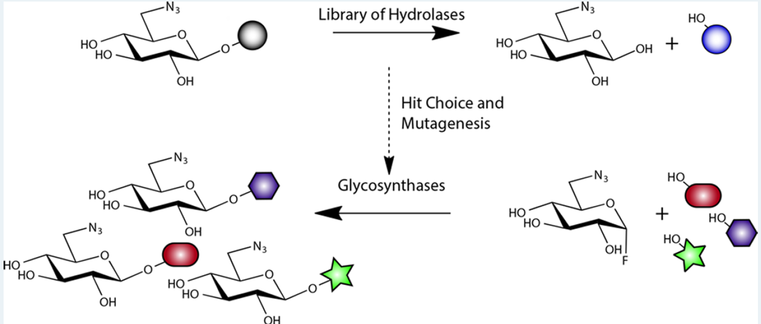
 

Figure 1: Structural representations of the transformation of sugars carried out by the researchers. Adapted from ACS Catalysis


This method was developed by using clones of genetically modified bacteria to express enzymes that are capable of modifying the sugar in accordance to the interest of the researcher. To achieve this, the scientists screened a library of 175 genes of the species E.coli that encoded variations of enzymes that can be used to catalyze selective chemical reactions in sugars for creating glycosidic bonds.
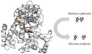
Computational representation of a hydrolase, an enzyme that breaks sugars. Adapted from Wikipedia

Enzymes are proteins that provide a path of a biochemical reaction to occur more efficiently. They are relatively easy to obtain and work with; however, they are specific to their target substrates which limit the extent in which their capabilities can be exploited. The scientists solved this issue by modifying the internal composition of the enzymes to improve the diversity of products in a process called selective mutagenesis. With the aid of this technique, the investigators obtained all variants that were tested in this experiment.
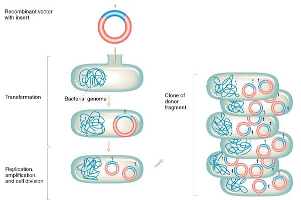
Schematic representation of bacterial transformation and cloning. Adapted from Griffiths et a (2000)

As the use of biotechnology increases, the understanding of our microscopic world becomes a major tool for scientific development. In this case, E.coli cells are essential since bacteria are inoculated with synthetic versions of genes that encode these enzymes and are then used as living machineries for protein production.

It is worth to mention that transformations of sugars were already reported. Nonetheless, previous methods rely on the use of expensive reagents as catalysts, which represent  a major cost and are not widely accessible. This new approach opens a significant area in biochemical research. As technology improves to newer and more accessible  methods, the diversification of these enzymes could develop new approaches for interaction with cell receptors that could enable us to understand what our cells have to say.

-Aron Engelhard

Journal Source:

Armstrong, Z.; Liu, F.; Chen, H.-M.; Hallam, S. J.; Withers, S. G. Systematic Screening of Synthetic Gene-Encoded Enzymes for Synthesis of Modified Glycosides. ACS Catalysis 20199 (4), 3219–3227.


New method found to build artificial blood vessels!

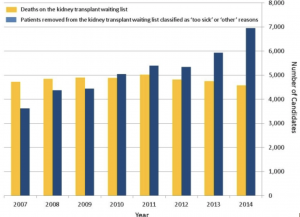
Figure1, yellow: Number of People died on the kidney transplant waitlist, blue: Number of people removed from the waitlist classified as “too sick”. Source


As the research shows, there are more than 114000 people in the United State on the waiting list of life-saving organ transplants. On average, there will be one new name added to the list every ten minutes, and 20 people die because of the lack of available organs every day. Research team from the University of Minnesota Medical School has published their new founding in Nature Biotechnology on March 11st, 2020. Their research proved the possibility to build artificial blood vessels in a pig, which has the potential to provide ultimate blood vessels for human organ transplants.

How did they come up with the idea?

“There is so many chronic and terminal diseases, and many people are not able to participate in organ transplantation,” said Daniel, a cardiologist who accepted heart transplantation before, “About 98 percent of people are not going to be eligible for a heart transplant, so there’s been a huge effort in trying to come up with strategies to increase the donor pool. Our approach looked at a pig.” Due to the physiological similarity between human and pigs, scientists have done similar studies of using pig insulin to treat human diabetes. These historical researches give scientists confidence and also an existing platform to study.

What is the benefit of choosing pigs?

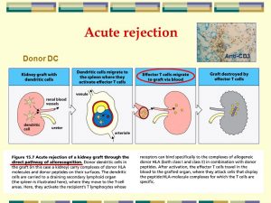
Figure 2:Acute rejection Anti-CD3 Donor DC. Source

Transplant rejection is a process of transplanted tissue rejected by the recipient’s immune system. According to the research done previously, there are 50-80% of patients would have at least one rejection episode. This symptom is due to recipient’s body cannot adapt to a “foreign” tissue and the immune system would fight against the transplanted tissue. Transplant rejection always causes severe consequences, and a lot of patients would have to remove the tissue immediately.

Daniel Garry, the leader of the research team came up with an idea to avoid transplant rejection. They took mature cells scraped off from a patient’s skin, reprogram these cells and eventually inject them into a pig embryo. This process would develop patient’s own genetic information in pig’s body. Thus, patients will get their own “blood vessels” which can greatly avoid transplant rejection. Mary, coworker of Garry said: “These blood vessels would be engineered and could be utilized in these patients to prevent those kinds of life-long handicaps, if you will.”

The first phase of their study has been approved by the University of Minnesota’s Stem Cell Research Oversight committee. “While it is a first phase, there’s pretty solid proof of concept,” Mary said. “We believe that we’ve proven that there’s no off-target effects of these cells, so we’re ready to move forward to later gestational stages.” This study will eventually benefit millions of people who are suffering from transplantation surgery. Also, this study shines lights and give hopes to modern organ transplantation studies.

-Vicky Gu

Revolution of Orthopedic Surgery – Bioactive Glass Composite Pore-forming Strategy

The use of adhesives to replace traditional invasive internal fixation tools (such as steel plates and nails) will revolutionize orthopedic surgery. The ideal bone cement should be able to immediately fix the fracture site, while providing a space and microenvironment suitable for bone cell growth and promoting fracture healing.

Cyanoacrylate (commonly known as universal glue) is currently the only medical glue with excellent instant adhesive strength and biocompatibility, but its polymerization product is non-degradable and cannot support the growth of new bone tissue through the adhesive layer, which hinders Because of bone healing, it cannot yet be used as a bone cement.

Recently, with the support of the Ministry of Science and Technology, the National Natural Science Foundation of China, and the Chinese Academy of Sciences, Qiu Dong’s group at the has proposed a bioactive glass composite pore-forming strategy to help cyanoacrylate bonding Agents are used for internal fixation of fractures to promote fracture healing. Bioactive glass has excellent osteoconductivity, osteoinduction, and can promote the regeneration of bone tissue.

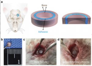
To prove that the bioactive pore‐forming adhesive is not only strong and instant bonded but also facilitates cell ingrowth and displays excellent bioactivity, they conducted bone‐fracture healing experiments. In the experiment, the bioactive pore‐forming adhesive (PSC1/PEG4/OCA5) was compared with commercial adhesive (OCA) and pore‐forming adhesive (PEG5/OCA5) by applying all to a circular bone piece which was fixed within freshly formed cranial fractures in a group of mice.

Figure 1. The scheme of a mouse cranial fracture with the craniotomy location (Source)

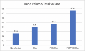
The stability of the circular bone piece and bone healing effect were evaluated at 12 weeks post‐operation. The ratio of bone volume (BV) within the circular gap over the total volume (TV) within the circular gap was quantified as shown in the figure 2 below. After 12 weeks, BV/TV of the PSC1/PEG4/OCA5 group was 0.76 ± 0.06, which was significantly higher than that of the PEG5/OCA5 group (0.47 ± 0.10), the OCA group (0.40 ± 0.06), and the group with no adhesive treatment (0.26 ± 0.05). These findings supported that the adhesive with pores for bone ingrowth was crucial for new bone formation.

Figure 2. quantitative analysis of new bone formation (Source)

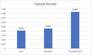
ARS (Alizarin Red S) can detect calcium which is a characteristic evidence to bone‐like structures. As shown in Figure 3, the Ca content in the bioactive pore‐forming adhesive (OD = 0.084 ± 0.008) was around two-fold higher than those in the merely pore‐forming adhesive (OD = 0.046 ± 0.004) and CA adhesive (OD = 0.041 ± 0.001). As a result, these findings were consistent with the statement that the PSC BG can promote osteogenic differentiation that is of importance for bone regeneration.

Figure 3. The optical density of eluents from ARS‐stained adhesives (Source)

The bioactive bone cement has good clinical transformation prospects and can provide new ideas and methods for clinical fracture treatment. At the same time, the above-mentioned composite pore-forming strategy can also be used for substances other than bioactive glass to increase matrix materials and functions. Material compatibility.


-Xinyue Yang

Posted on Mar.2nd, 2020