
Group 4: Jeremy Cheng, Klous Cui, Joanne Lin, and Layla Vera
Love Seafood? Then you’ll want to read this!

Seafood Platter
Global climate change is something that affects all of us, including our neighbouring marine life. Researchers investigate and prove that over fished species are more temperature sensitive and as the ocean temperature increases due to global warming, these fish species tend to move northward more drastically than other fish. Could we be losing our fish populations as a result of the combined effects of overfishing and global warming?

Global Climate Change Graph
It has been observed that over the past 30 years the global surface temperature has increased 0.2°C per decade (Hansen et al. 2006). This dramatic increase in temperature has been attributed to the increasing temperatures of the world’s oceans, which could be affecting our fish populations. In the recent article, “Environmental sensitivity of spatial shifts in marine fishes depends on latitudes and fishing effects” (Kuo et al. 2012), Ting-Chun Kuo, a current PhD candidate at the University of British Columbia, seeks to bring awareness to the changing global distributions of fish populations as a result of confounded effects of overfishing and rising ocean temperature. The following video clip is Ting-Chun Kuo describing her research and the message behind her results.

The Science
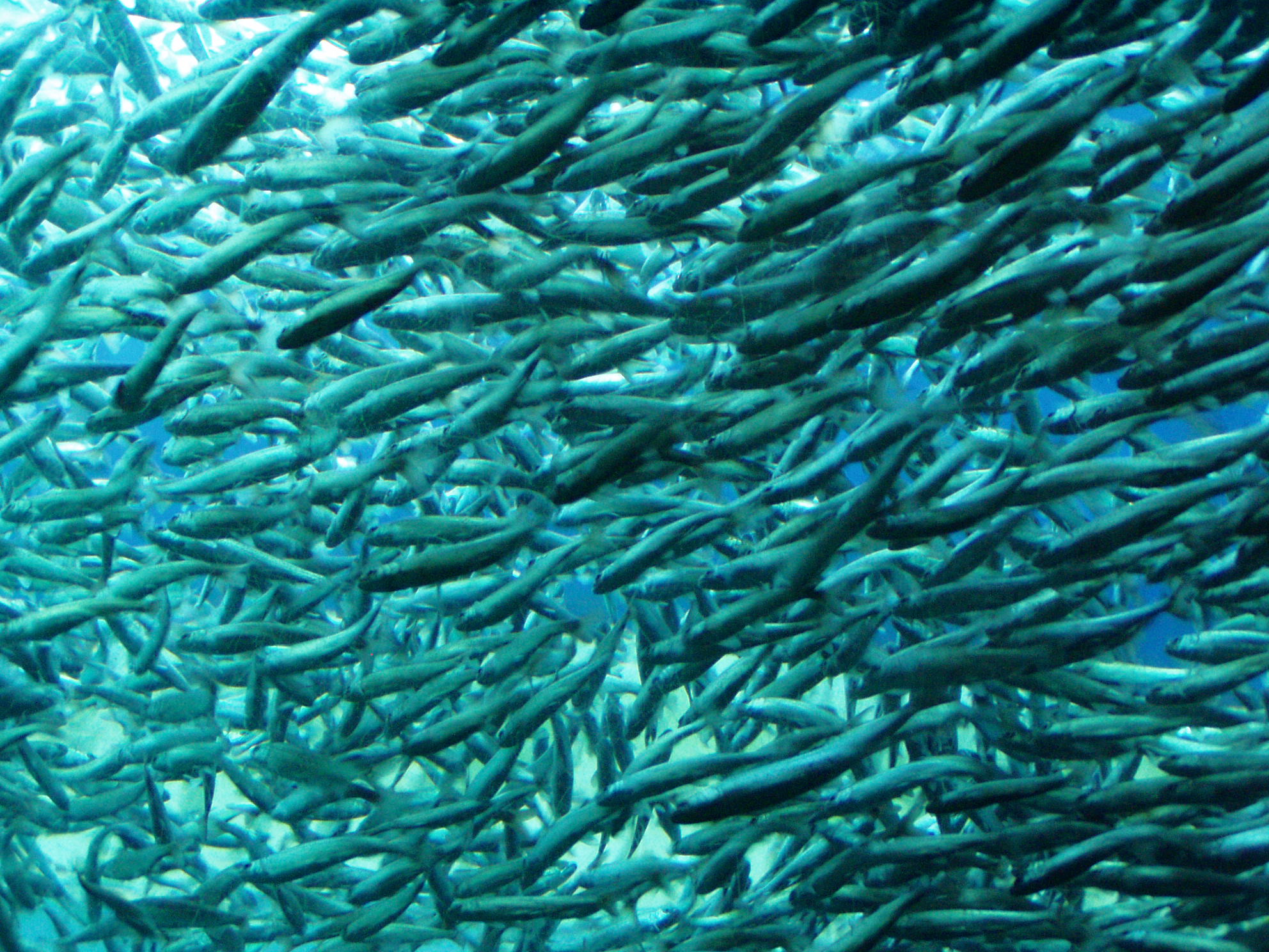
Swimming Fish
Ting-Chun Kuo based her research on data of fish species’ spatial distribution from four different databases at four different locations: the Bering Sea, the east and west coast of the USA (California and Massachusetts), and the North Sea. Each database used, observed over 13 species over a 30 year period. She performed a statistical analysis to compare and contrast the data from these databases and looked for a common trend. In particular, she wanted to find out how three factors, the fish species’ latitude of origin, life history traits, and

UBC PhD Zoology: Ting-Chun Kuo
extent of exploitation by commercial fishing, affect the species’ northward movement in response to increasing temperature. Her results suggest that over-exploited species (populations that have been disrupted by commercial fishing), are more greatly affected by the increasing marine temperatures caused by global warming. However, the results for the other two factors were inconclusive, yet she continues to investigate these two aspects today.
So now that you know, what should you do?
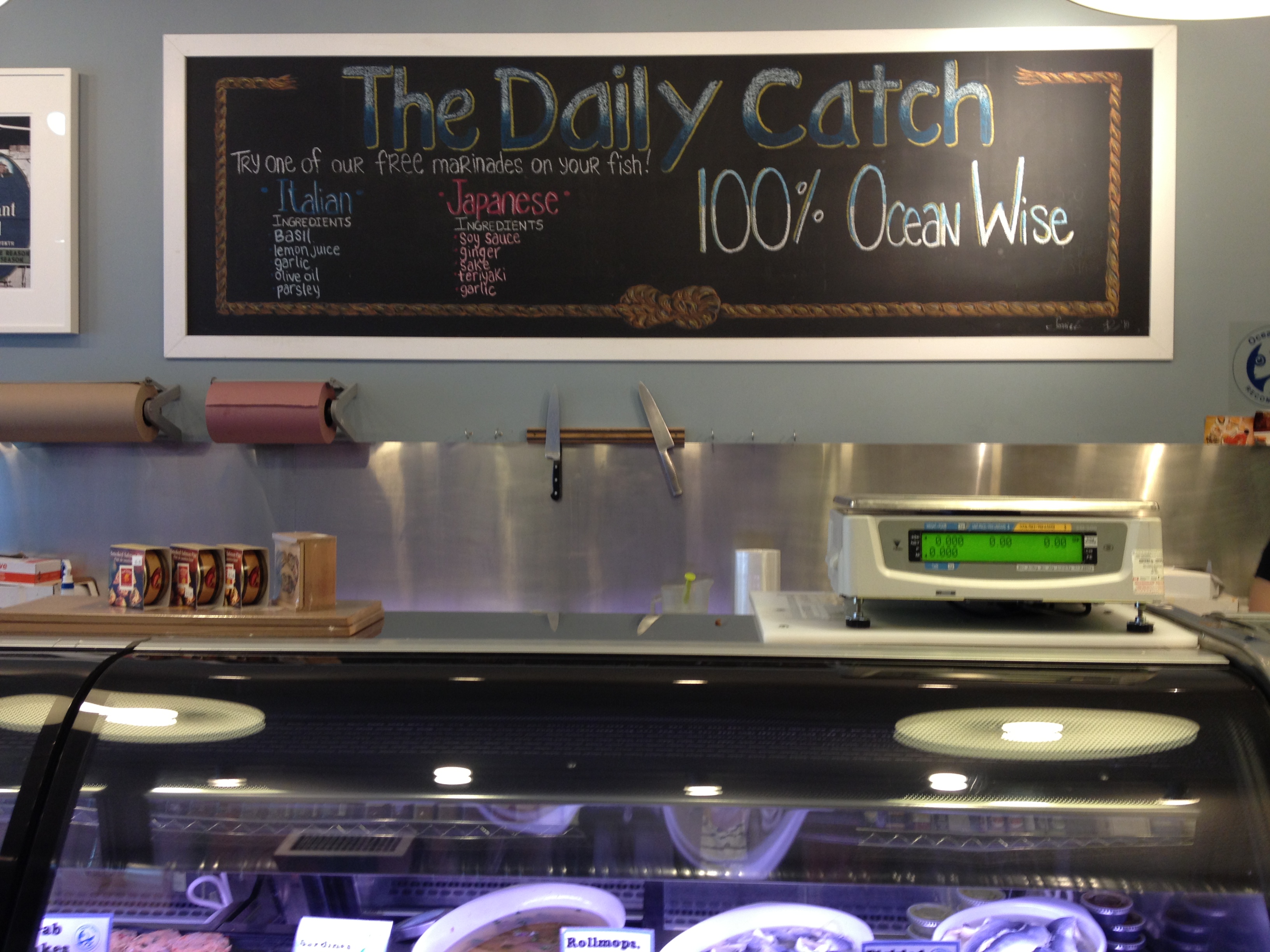
Daily Catch Seafood Company
Industry and traffic are just a few examples of how we, the main contributors to global climate change, are affecting the well-being of the numerous species with whom we share this Earth. As Vancouverites, we have the luxury of enjoying a wide range of seafood right from our very own coast on a daily basis. It is no one’s responsibility but our own to protect our fish resources to ensure they are available for future generations to enjoy. We know we cannot change the global climate overnight, but we are able to purchase and advocate for sustainable seafood to lessen the impact the increasing ocean temperatures has on our fish species.
Ocean Wise Logo
Local programs like Ocean Wise, are helping local businesses provide their customers with sustainable seafood products. An example of a local business jumping on board with the Ocean Wise Program is The Daily Catch

The Daily Catch Seafood Company
Seafood Company (1418 Commercial Drive, Vancouver, BC). The Daily Catch is owned by two BC boys, Dylan McCulloch and Ryan Johnson who both grew up on the Sunshine Coast. They are doing their part to contribute to the solution by only providing 100% Ocean Wise seafood products. These two ocean advocates are making it extremely easy for Vancouverites like you to contribute to the solution by making sustainable seafood available at a reasonable price. There really is no excuse not to be a part of the solution!
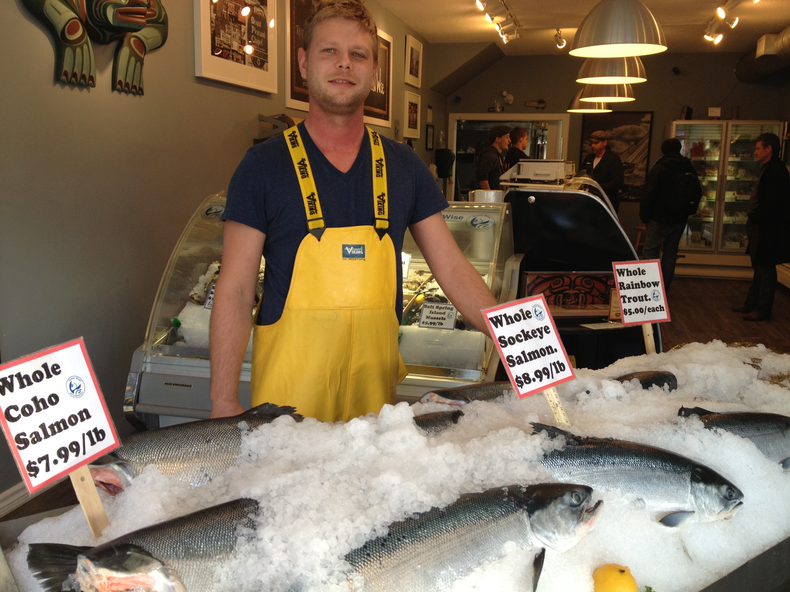
Ryan Johnson-Owner of the Daily Catch
Audio clip: Adobe Flash Player (version 9 or above) is required to play this audio clip. Download the latest version here. You also need to have JavaScript enabled in your browser.


