Category Archives: Biological Sciences

Concussions: Impacts on the Brain

The brain is a complex organ, and while researchers have made great strides in understanding its function and mechanisms, we still know relatively little about the consequences of damage to the delicate structure.

The brain is suspended in the skull cavity, and sharp accelerations can sometimes cause collisions between the unyielding bone of the skull and soft tissue, bruising the surface and damaging important neural connections within. This is known as a concussion, an injury common in contact sports where blows to the head are frequent.

A concussion can be harmful to anyone, but could the impact be greater on a developing brain, like that of a teenager’s?

Dr. Naznin Virji-Babul

Dr. Naznin Virji-Babul, a physical therapist and neuroscientist at the University of British Columbia, set out to discover the true extent of brain damage on concussed adolescents.

“The common perception of people is that your brain stops developing when you’re 3-5 years old. That’s not exactly true… the frontal areas of your brain are still developing when you’re a teenager,” she says, adding that this frontal area is what collides with the skull when a concussion occurs. The frontal and temporal lobes are most vulnerable to injury, and damage to these areas is associated with impairments of regular function. The study that her team conducted at UBC used Magnetic Resonance Imaging to gauge the extent of damage to the brain.

The study was conducted using a group of teen athletes, some of whom had experienced a sports-related concussion within the past two months and others who had not  Each athlete underwent an MRI scan which measured the rates of diffusion of the fluids within the brain.

The results were surprising to the team, who had expected to find clear evidence of damage, and lower rates of diffusion in the concussed group compared to the uninjured group.

“But it was completely opposite to what we had expected, we thought we would find a decrease in [one of the tests] but we found an increase… It was like solving a puzzle trying to find out what was so different.”

Diffusion Tensor Imaging scan for a healthy brain(left) and concussed brain(right)

Dr. Virji formulated a theory as to why the changes were different than expected: the neural damage in the brain was subtle, not outright breaking but causing smaller tears in the neuron which collect fluids and cause edema. This could throw off a diffusion tensor image but still indicates damage present in the brain long after the actual injury occurred.

“I wasn’t expecting to find changes in the kids who have had a concussion two months prior, but we still did… Kids and adolescents take longer to recover.”

These findings call to question the established guidelines concerning returning concussed athletes to play and school, as all of the concussed athletes scanned in the study had returned to their sport. Luckily, thanks to the findings of Dr. Virji and her team, new light is being shed on the nature of concussions on a teenager’s developing brain. This will hopefully lead to safer practices regarding the athletes care both during the game, and after.

Celebrating Hockey without injury

This podcast covers further the nature of this resistance by the public towards concussions in adolescents, and how the established safety measures are not adequate enough to prevent brain injury.

Audio clip: Adobe Flash Player (version 9 or above) is required to play this audio clip. Download the latest version here. You also need to have JavaScript enabled in your browser.

For more information, feel free to watch this video on the impact of Dr. Virji’s research.

YouTube Preview Image

By: Ammar Vahanvaty, Derrick Lee and Ashley Dolman


What if Extinction Wasn’t Permanent?

The gastric-brooding frog was first discovered in 1972 and was native to Queensland, Australia. This frog belongs to the genus Rheobatrachus which contains only two species. Both are now extinct. These two species were so fascinating because of their unique way of reproduction. The female would swallow the eggs after fertilization, carry them in her stomach and then orally give birth to the offspring. The average number of juveniles that was observed in the stomach of a female was 21-26.

Below is a picture of a female frog giving birth orally to one of her offspring:

www.rainforestinfo.org.au

While the offspring are in the mother’s stomach, she does not eat, and as her stomach grows larger in size, the lungs deflate to make room. In order to breathe, she must rely on gas exchange that occurs across the surface of her skin.

The gastric-brooding frog went extinct in 1983 and it has been suggested that this may have occurred due to a variety of reasons: habitat loss, pollution and/or pathogens. The other species in the genus Rheobatrachu, the Platypus frog, also went extinct in the early 80s.

According to this article, scientists in Australia are now trying to bring the gastric-brooding frog back from extinction through a cloning technique called somatic-cell nuclear transfer.

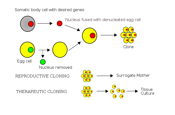
Basically, the researchers extracted cell nuclei from tissues of the gastric-brooding frog (which was collected in the 1970s and frozen) and implanted these nuclei into donor eggs. The donor eggs used in this case were from the Great Barred Frog, Mixophyes fasciolatu, which is distantly related to the gastric-brooding frog. The nuclei in the donor eggs were inactivated and replaced with the nuclei from the extinct species.  After this occurred, some of the eggs started to spontaneously divide and formed into early embryos. None of the embryos survived for more than a couple of days. Genetic tests were done to confirm that the genes in the embryos were those of the gastric-brooding frog.

Below is a simplistic diagram of how somatic-cell nuclear transfer works:

Wikipedia.org

Even though the experiment did not successful create an adult specimen of the extinct frog, the researchers are confident that it won’t be long until such a procedure will be able to be performed successfully.

The article further states that researchers from all over the world recently gathered in Washington for a conference in order to discuss the progress being made in the attempts to bring other extinct species back to life.

Possible candidates include the woolly mammoth and the dodo.

I find this so exciting!

Can you imagine having a zoo full of “extinct” animals, or even dinosaurs!?

Dragana Savic

Reference:

http://en.wikipedia.org/wiki/Gastric-brooding_frog

Liver kept alive for 24 hours outside of body

Hundreds of thousands of lives are leaning on the edge waiting for the right organ and a new life. However, donated liver still remains a scarce resource these days and many lives are lost while waiting for the right liver. What makes the situation worse, is that over 2000 livers were wasted every year in the US and Europe because they were damaged during transport by the ice pack and solution used as the only method of preservation. However, a new device has recently been unveiled in London, England by the University of Oxford that keeps a donated liver at body temperature before they reached the patient waiting for transplant.

Because of the fact that an available liver must be matched with an appropriate receiver, it’s rare that an available liver is in close proximity to the receiver’s hospital. Therefore a long transportation time is sometimes incurred during which the liver is easily damaged because it is kept out of it optimal living conditions. The new device, however, allows the donated liver to be kept for at least 24 hours, what’s even more amazing, is that the liver keeps on functioning while in the device.

A donor liver connected with the device

The device simply acts as a body and supply the liver with oxygenated blood and nutrients, but the trick is, how much and at what pressure. “The key is that the system ‘listens’ to the organ to find out how much blood to supply, and at what pressure,” says Stuart Kay, of Team Consulting in Cambridge, UK. There are multiple sensor for the flow of fluid, oxygen and carbon dioxide levels and sugar concentration in blood to create the optimum environment for the liver. Currently this device only works for donated livers, but the researchers at Cambridge are certain that it can be modified to fit other organs as well.

Check out this video on the actually hooking up of the liver and the device!

http://www.newscientist.com/video/2228569979001-device-keeps-liver-alive-outside-the-body.html

by Jonathan Lui

Panda Feces and Biofuel Production

These days that everyone is talking about climate change and increase in greenhouse gases, bio-fuels can be a good alternative to fossil fuels. Bio-fuels are liquid, gas or solid fuel that are produced from bio-masses such as corn, sugarcane, wheat, oil seed and so on. Production of these edible crops requires  high quality agricultural lands; using them for bio-fuel production can cause food scarcity, environmental concerns (replacing forest with crop lands) and economical costs. To minimize the negative effects of bio-fuel production on human beings, scientists are hoping to use  inedible super-tough plant materials known as lignocellulose  instead of using humans’ food source in production of bio-fuels. However breaking down lignocellulose in switch grass, corn stalks and wood chips requires high heat, pressure and harsh acids.

Who could imagine that solution to those limitations can be found in panda’s feces. There is an enzyme produced by bacteria found in panda’s gut that is strong enough to breakdown the tough plant materials. Researcher Ashli Brown and her colleagues have collected and analyzed panda’s feces and found several digestive bacteria that are similar to those found in termites that digest wood. Pandas eat 20 to 40 pounds of bamboo, leaves and stems everyday and have the ability to break down 95% of plant cellulose into nutrition. By isolating the bacteria in the giant panda’s gut, Brown is hoping to find the most powerful digestive enzyme that can be used in production of bio-fuels in order to genetically engineer this enzyme gene into yeast for massive production.

Using the enzyme contained within the giant panda’s gut in production of bio-fuels can be clearly faster, cleaner and less costly than the traditional method used today. This also can help in reduction of hunger by avoiding burning edible food source for bio-fuel production. Another point is that “The discovery also teaches a lesson about the importance of biodiversity and preserving endangered animals,” Brown said. I believe that every living thing such as plants or animals around us could be a beneficial source of products that may save our lives, so  we have to take action in saving those endangered plants and animals in order to save the beneficial resources that they provide us.

https://www.youtube.com/watch?v=flJCcyAUn24

Simin Yahyavi

Bee Stings Could Help Save People’s Lives!

Most people that have been stung by a bee would agree that it is a very painful and uncomfortable experience that they would not want to experience again. The pain takes days to go away, and the scar that a bee sting leaves may last weeks. Now though, it looks like a bee sting may not be as bad as it seems. Researchers at the Washington University School of medicine have found that a chemical found in the venom of a bee sting can destroy the human immunodeficiency virus (HIV). What is extremely important is the fact that even though the chemical can kill the HIV virus, regular cells are immune to this chemical and are not affected by it in any way.

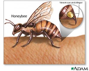
Image showing bee injecting venom. Taken from google images creative commons

Researchers believe that their findings will help them develop a vaginal gel that may help prevent the spreading of the HIV virus and be used for therapy. Although they don’t think that the gel may cure the disease completely, it may be used as a preventative measure in areas where HIV is particularly common. The active chemical that researchers believe is causing the observed effects is called melittin. This chemical works by poking holes in the nuclear envelope of the virus, causing large amounts of damage that cannot be easily repaired.

To obtain their findings, researchers loaded melittin into nanoparticles and administered them to patients. These kinds of particles have been found to very effectively treat tumors, so the scientists decided to test them to see if they would be affective in treating other types of diseases. The researchers saw that when the nanoparticles came into contact with HIV infected cells they were destroyed, while the nanoparticles simply “bounced off” the normal cells, which are much larger in size.

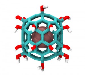
Nanoparticle incapsulating a chemical in the middle. This is the kind of mechanism the researchers used. Image from flicker user IBM Research

This is a major breakthrough in the world of science and medicine because HIV has been very hard to treat and prevent in the past. Also, HIV is a relatively common disease and many people suffer from it. It is approximated that about 36.1 million people around the world currently suffer from HIV. Furthermore, 21% of those that are infected in North America don’t even know that they are carriers of the deadly virus. Also, since the melittin loaded nanoparticles are so versatile, the researchers believe that this discovery could lead to the development of treatments for patients suffering from Hepatitis B and C.

Below is a short news report on this groundbreaking discovery:

YouTube Preview Image

So the next time you get stung by a bee and are panicking, calm down and just remember that the venom that the bee just injected you with is being used right now to potentially save millions of lives around the world.

Gagandeep Gill

Could Peptides Be The Cure For Depression?

For the first time ever, scientists at the UCLA have measured the release of the peptides hypocretin and melanin concentrating hormone (MCH) within humans. Hypocretin’s release is increased when the person is happy, but minimal when they are upset. MCH levels are maximized when the subject is asleep, but those levels diminished awake.

“The current findings explain the sleepiness of narcolepsy, as well as the depression that frequently accompanies this disorder” explains Jerome Siegel, professor of psychiatry and director of the Center for Sleep Research at UCLA. The measurable changes of these peptides’ release will eventually allow researchers to develop medications that target the changes in brain chemistry.

In 2000, Siegel was the first to show a biological cause of narcolepsy when he found that people who suffer from the illness had 95 percent less hypocretin nerve cells for in their brain than people who don’t have narcolepsy. Due to the strong affiliation between depression and narcolepsy, Siegel expanded his study by researching how hypocretin could be connected to depression.

Image Source: micah.sparacio.org

Siegel retrieved information on hypocretin and MCH levels from the patients who had intracranial depth electrodes implanted in their brains. The patient’s actions were recorded while they ate, fell asleep, woke up, were social with other patients, and watched television. Every 15 minutes, the patients’ activity was recorded along with the release levels of the peptides. Each waking hour, the patient filled out a questionnaire where they rated with mood and attitudes.

Siegel found that positive emotions, social interactions, and the act of waking up had higher hypocretin levels, while the levels of MCH were at their highest when the patient was falling asleep and were at their lowest when the patient wasn’t being social. “These results suggest a previously unappreciated emotional specificity in the activation of arousal and sleep in humans” Siegel explained. “The findings suggest that abnormalities in the pattern of activation of these systems may contribute to a number of psychiatric disorders”

Siegel mentioned that several drug companies are in the process of developing hypocretin antagonists to be used as sleeping pills, but this study means that not only will these drugs induce sleep, but they will also influence the person’s mood.

Recent studies have suggested that depression-fighting drugs, like selective serotonin reuptake inhibitors (SSRIs) may have the same effect as a placebo as they are not based on the changes in neurotransmitter levels. Because as many as 10% of the US population suffers from depression, I think this study is a breakthrough in our knowledge on depression and I hope that this will lead to the production of a drug that can balance the release of these neurotransmitters.

Kathleen Leask

The Chameleons of the Sea

By: Ammar Vahanvaty

Not many people can relate to the experience Marine Biologist Roger Hanlon had when he came upon a seemingly innocuous piece of kelp in the shallows of the Caribbean. At first glance, an ordinary piece of underwater flora. But as he approached something incredible happened; a piece of the kelp suddenly turned stark white and the next few moments found Roger Hanlon blasted with a face-full of ink. He had encountered an octopus, so well camouflaged as to be mistaken for kelp. The offending octopus shot off into the distance only to settle next to another patch of ocean floor and change colours again to match its new surroundings. This event both awed and intrigued Hanlon, who, to this day, is exploring the magnificent abilities octopi and its cephalopod cousins share; the ability to camouflage.

What is camouflage?

Camouflage is the ability of an individual to blend into its surroundings. The most notable example of this technique in nature is the chameleon, who, in addition to its bulbous eyes and whip-like tongue, is famous for its ability to change its skin color to match the background. But even though the name chameleon is almost synonymous with camouflage in the public eye, they are not the masters of this ability. That honor falls to the cephalopod family, whose members include the squid, octopus and cuttlefish. These animals trump the camouflage ability of chameleons by not only shifting colour much quicker and with more accuracy, but by also adopting the texture of the object they are mimicking. This is what intrigued Hanlon the most as he began his research into these animals.

How does octopus camouflage work?

The mechanics are simple enough, at the microscopic level, animals harbour pigment cells called chromatophores, which lead to the variety of colour hues displayed by reptiles and fish, and the skin colour of humans etc. For the most part, these chromataphores are autonomic, and cannot be altered once set. But it appears cephalopods have control of each pigment cell in their bodies, and can alter the composition of the pigments at any moment to effect widespread and large colour changes throughout the animal. That part is fairly well understood, but less understood is the octopus and cuttlefishes’ ability to alter the pattern of their skin.

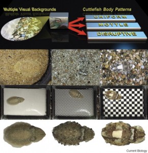
Hanlon and his team have identified three main forms of pattern generation for a cephalopod: uniform, mottled and disruptive. The bumps that allow an octopus to mimic the texture of the object they are up against appears to be formed by raising the papillae of the skin, much like controllable goose bumps. How exactly this process is achieved remains to be found.

The most vexing question though, is how does an octopus know what it is mimicking. Studies have shown that cephalopods are effectively colourblind, but they do not recognize the object’s colour through touch. So how do they do it? This is the question Hanlon is devoted to finding out, since  better understanding of this phenomenon could pave the way for advances in other fields. Who knows, an ‘invisibility cloak’ might not be so far off in our future.

For further reading into cephalopod coloration, Hanlon has created a nice primer to get started. 

YouTube Preview Image


An Unlikely Hero is the Key to Killing the Human Immunodeficiency Virus (HIV)

Each day, almost 7,000 people contract HIV around the world.  Many scientists have dedicated their entire lives to finding a cure for this virus and it seems that we have come one step closer to finding that cure. With a historical discovery coming out of Washington University, that number could soon be reduced to zero.

Researchers at Washington University School of Medicine in St. Louis have found that bee venom can effectively poke holes in the envelope surrounding the HIV virus and kill it. The potent toxin contained within bee venom is called melittin.

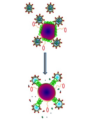
Nanoparticles, when infused with the toxin, can filter HIV through it’s surface and destroy it, while leaving normal cells unharmed due to their much larger size. When nanoparticles come into contact with normal cells, they simply bounce off. Joshua L. Hood, MD, PhD, explained, “The melittin forms little pore-like attack complexes and ruptures the envelope, stripping it off the virus.” The virus has to have a protective coat and there is no way for the virus to adapt when its essential physical property is under attack.

Image displays nanoparticles (purple) infused with melittin (green), with molecular bumpers (small red ovals) allowing them to bounce off of normal body cells. Fuse with HIV (small spiked circles) and destroy their envelope.
Photo credit: Joshua L. Hood, MD, PhD.

The following video by the Huffington Post further explains this topic.

Video source: Huffington Post

There is promising evidence that HIV infection in children will be eliminated. A baby born with HIV in Mississippi has apparently been cured. Its mother was diagnosed with HIV during labor, and the baby received intense care just after 30 hours of birth. This child shows no sign of the virus today.

Most anti-HIV drugs inhibit the virus’s ability to replicate, while doing nothing to stop initial infection. The development of a vaginal gel is the new revelation to prevent the spread of HIV, and in addition, an intravenous treatment to help those already infected and clear HIV from the blood stream.

Humans Will Breath Under Water

Have you ever tried to hold your breath for more than a minute under water? Well, if you had succeeded you were not reading this post! Holding breath under water has always been part of man’s dream. But it seems it will not be a dream for the future generations. According to a recent research  salamanders have developed the trait of producing oxygen themselves rather than taking it from atmosphere.

Baby under Water!


According to this study which was  conducted in Dalihousie University faculty of Zoology eggs of salamander , Ambystoma maculatum, were placed among chlamydomonad algae. The result of this experiment is that the DNA of the algae and the DNA of the salamander eggs were somehow combined. Algae is a nitrogen-dependent organism and produces oxygen gas as a form its waste. Salamanders on the other hand require oxygen to function and survive, and combining algae and the salamanders DNA would produce an organism which produces oxygen on its own and would not require oxygen  from atmosphere.

Salamander. Amphibian

Researchers have also found traces of this algae in mature salamander’s reproductive system which suggests that these algae are inherited to other generations.

What good is it to humans?

New technologies such as DNA Recombinant has enabled us to combine DNA strands of different species. In this technology, the wanted DNA is reproduced many times by molecular cloning in laboratory. These DNAs are then directed to the main DNA sequence by means of another organism and sequencing. Viruses are usually used in this step to deliver the multiplied DNA to the main DNA.

Formation of recombinant DNA requires a cloning vector, a DNA molecule that will replicate within a living cell. Vectors are generally derived from plasmids or viruses, and have the necessary sequence of DNA for replication. The inserted DNA may or may not express its codes depending on its place on the main DNA sequence. Thus, markers are used to place the DNA among functional genes.

Human DNA is packed with hundreds of viruses DNA s which have entered our DNA sequence along the course of evolution. This finding suggests that viral vectors can be used to alter human DNA sequence by means of insertion.  Recently, this technology has been used in the treatment course of of insulin-dependent diabetes. So, it’s not far beyond imagination if scientists mix algae DNA with that of human.

Soon the future of human beings will be altered with this technology and super humans will be produced! but until that day do not hold your breath for too long under water unless you are this guy:

References:

Canadian Journal of Zoology, 2008, 86(11): 1289-1298, 10.1139/Z08-115