Tag Archives: biomedical

Bee Stings Could Help Save People’s Lives!

Most people that have been stung by a bee would agree that it is a very painful and uncomfortable experience that they would not want to experience again. The pain takes days to go away, and the scar that a bee sting leaves may last weeks. Now though, it looks like a bee sting may not be as bad as it seems. Researchers at the Washington University School of medicine have found that a chemical found in the venom of a bee sting can destroy the human immunodeficiency virus (HIV). What is extremely important is the fact that even though the chemical can kill the HIV virus, regular cells are immune to this chemical and are not affected by it in any way.

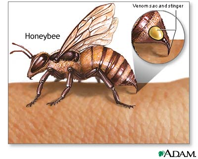
Image showing bee injecting venom. Taken from google images creative commons

Researchers believe that their findings will help them develop a vaginal gel that may help prevent the spreading of the HIV virus and be used for therapy. Although they don’t think that the gel may cure the disease completely, it may be used as a preventative measure in areas where HIV is particularly common. The active chemical that researchers believe is causing the observed effects is called melittin. This chemical works by poking holes in the nuclear envelope of the virus, causing large amounts of damage that cannot be easily repaired.

To obtain their findings, researchers loaded melittin into nanoparticles and administered them to patients. These kinds of particles have been found to very effectively treat tumors, so the scientists decided to test them to see if they would be affective in treating other types of diseases. The researchers saw that when the nanoparticles came into contact with HIV infected cells they were destroyed, while the nanoparticles simply “bounced off” the normal cells, which are much larger in size.

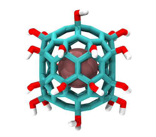
Nanoparticle incapsulating a chemical in the middle. This is the kind of mechanism the researchers used. Image from flicker user IBM Research

This is a major breakthrough in the world of science and medicine because HIV has been very hard to treat and prevent in the past. Also, HIV is a relatively common disease and many people suffer from it. It is approximated that about 36.1 million people around the world currently suffer from HIV. Furthermore, 21% of those that are infected in North America don’t even know that they are carriers of the deadly virus. Also, since the melittin loaded nanoparticles are so versatile, the researchers believe that this discovery could lead to the development of treatments for patients suffering from Hepatitis B and C.

Below is a short news report on this groundbreaking discovery:

YouTube Preview Image

So the next time you get stung by a bee and are panicking, calm down and just remember that the venom that the bee just injected you with is being used right now to potentially save millions of lives around the world.

Gagandeep Gill

The Promised Future of Prosthetics: Robotic Limbs

It must feel great to be able to feel a friend’s hand after seven years. Researchers at the University of Pittsburgh School of Medicine and UPMC have enabled a 30-year-old paralysed man to be able to move his robotic arm by means of electrodes placed in his brain. The tested patient has been paralysed for seven years after a motorcycle accident. “It feels like I’m robocop” says Tim Hemmes, the spinal patient. Researchers used the newly developed brain-computer interference (BCI) technology to make Tim he has hand again. The data released from Tim’s thoughts are then interpreted by an IBM designed processor. The analysed data are then put into command language for the robotic limb. “When Tim reached out to high-five me with the robotic arm, we knew this technology had the potential to help people who cannot move their own arms achieve greater independence,” said Dr. Wang, when watching a memorable scene in 2011.

Today, different types of bionics are being made. There exists bionic lenses, bionic arms and bionic legs. However, the accuracy of these devices are not perfect yet but the clinical cases are showing a promising future in this field.

Tim Hemmes’s case

How does it work?!

In order for patience to feel comfortable using the prosthetic limb, the designed limb’s weight should match the actual limb’s weight. This prevents researchers from producing gigantic robots. The next step in making a robotic limb is building an appropriate BCI which matches the right part of the brain. In order to do so, researchers use functional magnetic resonance imaging (fMRI) to find the right place for the conductors. Conductors take data orders from your brain and analyse those data using bio-computational algorithms to transform data into machine language. The robots then do the job for the patient.


The robots used should have the same functionality as the actual limb since it is believed the brain of the patient can only command in a certain manner. That certain manner matches with what the patient did with his/her actual limb and our brains are not trained for anything beyond what our limbs can do.

Below is a Ted talk showing the clinical accomplishments of robotic limbs:

Although it is very early to comment on this technology but it is pretty evident that soon this technology will become a solution for amputated limbs. Many different researched are also being conducted on robotic lenses but not a lot of successful cases have been reported yet.

References:

1. Di Pino G, Porcaro C, Tombini M, et al. A neurally-interfaced hand prosthesis tuned inter-hemispheric communication. Restorative Neurol Neurosci. 2012;30(5):407-418.

2. Di Pino G, Porcaro C, Tombini M, et al. A neurally-interfaced hand prosthesis tuned inter-hemispheric communication. Restorative Neurol Neurosci. 2012;30(5):407-418.

3. Guymer R. The challenge and the promise of the bionic eye. the bionic vision australia project. Clin Exp Ophthalmol. 2012;40:123-124.

4. Li Hu, Yang Jian-yu, Su Peng-cheng, Wang Wan-shan. Computer aided modeling and pore distribution of bionic porous bone structure. J Cent South Univ. 2012;19(12):3492-3499.

5. Mironov V, Boland T, Trusk T, Forgacs G, Markwald RR. Organ printing: Computer-aided jet-based 3D tissue engineering. Trends Biotechnol. 2003;21(4):157-161.

The promising future of transplantation

Are you wearing a donor wrist band? Well! Take it off. Soon no one will need your organs any-more . ‘Printing‘ the organs is a new technology which can revolutionize all transplantation procedures performed in today’s medical world. Tissue engineering technology or ‘printing’ is a new way of producing human organs by means of computers and organ printers. 

Organ Printing or cell printing are very recent ideas which were introduced to the world of biotechnology in 1987. The technology has been rapidly developing ever since. Synthetic blood vessels are the first body-parts which were made by this technology.

Organ Printer

Luke Massee is the first patient who has experienced this new technology successfully. Luke was born with dysfunctional kidneys a condition know as CKD.  He was chosen over tens of candidates after 10 years of investigations. His case proved that not only this procedure is possible but also safe and cheap to use.

What is organ printing?

organ printing is a biomedical version of rapid prototyping technology which is based on tissue fluidity. Computer-assisted printers put natural component of an organ together in the right shape and form.

How does it work?

“It’s like making a cake” said Anthony Atala of Wake Forest Institute for Regenerative Medicine.  A 3D scan of the wanted organ is captured first. Then, a sample from the recipient tissue is taken in order to make the organ with the right material. ‘Printer’ starts producing the organ layer by layer in the final step.  Thus, the procedure of organ printing can be divided into three main steps: preprocessing, processing and postprocessing. In preprocessing computer-aided design ( CAD) or blue print of the organ is done. in processing step, materials are put together by means of tissue scaffold. Printers play the main role in this step. postprinting is the final step and organ is double checked for functionality.

The progress of the stem cell technology has also greatly contributed to progress of the organ printing technology. Stem cells can be used to produce any organ in the body. They can be used for the tissue culturing and the produced culture can later on be used in producing the organ.

Anthony Atala: Printing a human kidney

Even though there has been one successful case of organ transplantation, there is still a lot not understood about human body and I believe it will take a long time for this technology to become accessible for everyone.  And until the day that science can solve every problem about our mysterious bodies it is much wiser to keep your donor wrist band on!

Refrences:

1. Mironov V, Boland T, Trusk T, Forgacs G, Markwald RR. Organ printing: Computer-aided jet-based 3D tissue engineering. Trends Biotechnol. 2003;21(4):157-161.

http://www.sciencedirect.com/science/article/pii/S0167779903000337#