Tag Archives: Medical Sciences

Help! I can’t stop eating!

A child eating pizza. Image from Wikimedia Commons

One slice of pizza turns into five boxes of pizza and twenty hours later… you’re still eating! Imagine an insatiable hunger and a love for food turned deadly. When eating becomes your worst enemy, Prader-Willi Syndrome may be the culprit.

What is Prader-Willi Syndrome?

Prader-Willi Syndrome (PWS) is a rare genetic disorder in which an individual feels hungry all the time. So much to the point where they are found constantly eating, and can continue eating even after they’re full. These individuals can literally eat to the point of death.

PWS was first described in 1956 by Swiss doctors Andrea Prader, Alexis Labhart, and Heinrich Willi. Anyone can develop PWS, and it was found that this disorder affects nearly 1 in every 15,000 births. As a result, PWS is one of the leading causes of childhood obesity.

Symptoms of a Deadly Appetite

The most common symptom of PWS is chronic hunger. Other symptoms can include: poor muscle tone during infancy, early-onset obesity, limited growth, delayed motor and verbal skills, behaviour and mental disorders, and curvature of the spine.

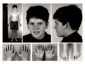
A 15-year-old child with typical PWS facial features. Note the presence of mild truncal obesity. Image from Wikimedia Commons

How does this happen?

PWS is a genetic disorder, which means that children inherit this disorder from their parents. Specifically, from an abnormality in a chromosome that comes from the father. This abnormality arises when a part of the gene is missing or malfunctioning. When this occurs, the hypothalamus (the part of the brain that controls hunger and thirst and releases hormones that promote growth) stops working which results in an inability to regulate food intake.

Is there a cure?

Unfortunately, there is no known cure for PWS. In fact, most of the research to date has only been targeted towards developing treatments for the disorder. For example, doctors may prescribe a growth hormone to treat PWS that not only increases height, but also decreases body fat, increases muscle mass, improves weight distribution, increases stamina, and increases bone mineral density.

Ultimately, the inability to regulate food intake remains one of the biggest obstacles that prevent individuals with PWS from living independently. There is still no effective medication that aids in regulating appetite. Nevertheless, those with PWS can still live a long and fulfilling life with the right guidance and support, as seen with Katie in the video below. Her documentary on living life with PWS gives us a better insight into the disorder, and presents a new meaning to the saying “you are what you eat”:

Documentary of Katie, a child living with PWS. Video from Youtube.

-Christina Rayos

Recording the Cell? New technologies further uncover mysteries surrounding the cell.

Does anyone really know what life is like inside of a cell? Sure, we can all say that the mitochondria is the powerhouse of the cell, and we’ve learned mitosis more time than we can count, but do we really know about the intricacies of day to day cellular processes? Historically, answer has been an overwhelming no, but that is something the researchers behind CAMERA are hoping to change.

CAMERA, or CRISPR-mediated analog multievent recording aperture is a tool developed by David Liu and Weixin Tang of Harvard university to record the molecular interactions within a cell, all of which are stored on the cell’s DNA. This new discovery allows scientists to observe and therefore clarify the processes that contribute to such things as the emergence of cancer, aging, environmental damage, and even embryonic development. CAMERA is only one of the many developments based off of the gene cutting technology known as CRISPR-Cas9.

Thyroid Cancer Cell Line. Courtesy of NASA’s Marshall Space Flight Centre and Flickr Commons. 

What is CRISPR-Cas9 you ask? Well, it’s basically a really small pair of scissors, so small that it can even cut DNA. CRISPR-Cas9, or CRISPR for short, is a technology based off of the natural defence mechanisms found in bacteria that have been reengineered for editing genomes. It has the ability to cut the double helix strand of DNA allowing for researchers to easily alter DNA sequences and modify gene expression. Some of the major implications of this include the possible correction of genetic defects, and the treatment and prevention of cancer and other diseases.

Video recreating a CRISPR-mediated genome editing. Courtesy of McGovern Institute for Brain Research at MIT .

So how did scientists develop a cellular recording device from this cutting tool? When CRISPR cuts a DNA strand to alter the sequence, the strand will naturally repair itself but in doing so can occasionally add in errors that make the targeted gene inactive. These random errors can sometimes be used as markers, mapping out the cell’s pattern of differentiation. Liu and Tang took this information and set out to regulate it thereby creating a more detailed, continuous record of a cell’s life, documenting not only its responses to external factors but the severity of the response and how long it lasts.

Flowchart of CRISPR mediated gene alterations. Image courtesy of Flickr Commons

At this point in time, CAMERA, is able to document cellular responses to light exposure, antibiotics, viral infections, and internal molecular interactions in as few as 10 cells. As well, it can record multiple events at once making it an impressive candidate for future medical technologies involved in screening embryos for a wide variety of mutations during development. Despite these impressive feats, Liu and Tang are still working towards pinpointing the recording down to one cell, allowing scientists to one day observe the processes of each cell individually and efficiently isolating any mutations. Another big step is proving it works to the same detailed extent when placed in the body of a living mammal as it does in a small cell group in a petri dish. There is still a lot to be done before we can confidently say we know how cells operate but CAMERA is a step in the right direction.

-Tenanye Haglund

Recording the Cell? New technologies further uncover the mystery of the cell

Does anyone really know what life is like inside of a cell? Sure, we can all say that the mitochondria is the powerhouse of the cell, and we’ve learned mitosis more time than we can count, but do we really know about the intricacies of day to day cellular processes? Historically, answer has been an overwhelming no, but that is something the researchers behind CAMERA are hoping to change.

CAMERA, or CRISPR-mediated analog multievent recording aperture is a tool developed by David Liu and Weixin Tang of Harvard university to record the molecular interactions within a cell, all of which are stored on the cell’s DNA. This new discovery allows scientists to observe and therefore clarify the processes that contribute to such things as the emergence of cancer, aging, environmental damage, and even embryonic development. CAMERA is only one of the many developments based off of the gene cutting technology known as CRISPR-Cas9.

Thyroid Cancer Cell Line. Courtesy of NASA’s Marshall Space Flight Centre and Flickr Commons.

What is CRISPR-Cas9 you ask? CRISPR-Cas9, or CRISPR for short, is a technology based off of the natural defense mechanisms found in bacteria that have been reengineered for editing genomes. It has the ability to cut the double helix strand of DNA allowing for researchers to easily alter DNA sequences and modify gene expression. Some of the major implications of this include the possible correction of genetic defects, and the treatment and prevention of cancer and other diseases.

So how did scientists develop a cellular recording device from this cutting tool? When CRISPR cuts a DNA strand to alter the sequence, the strand will naturally repair itself but in doing so can occasionally add in errors that make the targeted gene inactive. These random errors can sometimes be used as markers, mapping out the cell’s pattern of differentiation. Liu and Tang took this information and set out to regulate it thereby creating a more detailed, continuous record of a cell’s life, documenting not only its responses to external factors but the severity of the response and how long it lasts.

CRISPR mediated DNA splitting. Courtesy of Flickr Commons.

At this point in time, CAMERA, is able to document cellular responses to light exposure, antibiotics, viral infections, and internal molecular interactions in as few as 10 cells. As well, it can record multiple events at once making it an impressive candidate for future medical technologies involved in screening embryos for a wide variety of mutations during development. Despite these impressive feats, Liu and Tang are still working towards pinpointing the recording down to one cell, allowing scientists to one day observe the processes of each cell individually and efficiently isolating any mutations. Another big step is proving it works to the same detailed extent when placed in the body of a living mammal as it does in a small cell group in a petri dish. There is still a lot to be done before we can confidently say we know how cells operate but CAMERA is a step in the right direction.

-Tenanye Haglund