Monthly Archives: November 2016

Gene Regulation Occurs At Many Levels

One concept about gene regulation that I have learned through this course.

One concept about gene regulation that I have learned through this course as well as BIOL 441 (Cell Biology of Intracellular Trafficking) this year, is that gene regulation can be very difficult to study because it occurs at so many different levels. These different levels of control can exist within intracellular processes, but can also differ between cells depending upon signals they receive from the environment. The first, and arguably the most significant level of control is that achieved at the transcriptional level within the nucleus. Even at the transcriptional level alone, there are many ways that a gene can be regulated. From altering the number of copies of RNA that are transcribed, to the variation in timing of when the gene is actually transcribed. The next level of regulation is at translation or when the gene transcript is converted into a protein. Here, the ribosome complex “reads” the information encoded by the mRNA transcript, and it builds an entire protein out of the amino acids in the cytoplasm. Regulation through the number of ribosomes working on each transcript can be achieved at this level.

In BIOL 463, we focussed a lot on epigenetic regulation of genes. Epigenetic regulation is any functionally relevant changes to the genome that occur, but do not involve a change in the nucleotide sequence. Examples of epigenetic mechanisms are DNA methylation and histone modification, which alter how genes are expressed without altering the DNA sequence itself. Gene expression can be controlled through the action of repressor proteins that attach to silencer regions of the DNA. In BIOL 441, I learned about another mechanism that can indirectly regulate gene expression by affecting the amount of transcript or protein present in a certain cell compartment. This mechanism is intracellular trafficking. For example, if a transcript or protein acts in the extracellular environment, up regulating its secretion and release from vesicles could increase its effect without directly changing gene transcription or translation levels. Another way gene expression can be regulated is by having any protein or regulatory molecule involved be turned on or off by the activity of an enzyme.

This is why its important to test for levels of your molecule of interest at each level. Ex: You may notice that the phenotype of two individuals is the same, but there is a possibility that transcription differs greatly between the two and that the trafficking of the molecule is simply being held up at one point, in one individual.

Development of Egg Cells Through iPS Cell Experiments

If I could develop anything through an iPS cell experiment, what would it be? Why?

If I could develop anything through in iPS cell experiment, I would love to find a way to generate egg cells for females. Specifically, infertile females or those with low fertility. Personally, having problems with fertility and being able to have a child of my own has always been one of my greatest fears. Being able to have a child who shares much of my genetics would be incredible. In addition, one of my best friends is unable to produce fertile eggs because of the treatment she received for cancer when she was 11, and she has experienced a lot of hardship in accepting the idea that she will never be able to carry a baby and have a child who shares her own genetics. These are a couple of reasons I, and I think many people would be supportive of the generation of egg cells from a woman iPS cells. Right now, one of the common options for women who cannot produce viable eggs is to purchase eggs that have been donated by another woman. In this case, the egg can be fertilized by your partner’s sperm in vitro (in a laboratory), and then later implanted into your own, or a carrier’s uterus. Although this method works, it still does not solve the problem of women who cannot produce viable eggs being able to share their genetic material with their child. For their case, only the DNA of their partner can be transferred to the child, but DNA in the egg cell will come from the donor female. I brought this idea up in class when Dr. Kalas asked students what we would do if we could conduct any experiment with iPS cells. One of the problems with my idea that was mentioned was that it is very difficult to generate reproductive cells or gametes from iPS cells. Hopefully one day this technique can be possible so that women can always have the opportunity to have children that share their DNA.

PS: This was achieved successfully in mice for the first time this year. Study published just over a month ago, check it out!:

http://www.sciencemag.org/news/2016/10/mouse-egg-cells-made-entirely-lab-give-rise-healthy-offspring

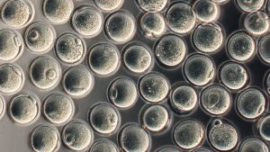
Egg cells derived in the lab from embryonic stem cells.

O. Hikabe et. al., Nature 538, 7625 (20 October 2016) © MacMillian Publisher Ltd.

Final Project Challenges

You have been working on developing a novel, testable question on a gene regulation-related topic. What is the most challenging aspect of it and why?

For my final project, I have been working on developing a novel testable question about a gene called pho88. When choosing a topic, I wanted to select a gene that very little was known about so that I could have a very wide array of experiments and options to choose from. Pho88 is gene related to the phosphate transport pathway in yeast, but as no research was done specifically on this gene, and therefore it is definitely poorly understood. Unexpectedly, this aspect of it has made it quite difficult for me to narrow my focus down to one testable question about it because so much could still be discovered about it. Specifically, I have found that the pho88 mutant has many phenotypic similarities to pho86, another phosphate transport gene. Both of these proteins have proven to localize in the ER membrane of yeast, suggesting possible interaction or interaction in the same pathway. More importantly however, an up regulation in certain phosphate transport genes was seen in the double mutant of pho88 and pho86 under high phosphate conditions. This affect on gene regulation is the opposite of what occurs in wild-type yeast, and therefore it suggests that pho88 and/or pho86 could influence gene regulation. This is the knowledge gap I will be investigating in my project. However, since it is known that pho86 interacts with other phosphate transport proteins in yeast (such as pho84), I also want to test interactions between pho88 and these proteins, as well as pho88 with pho86. All of these observations have sparked my interest in pho88 and what its role is in the phosphate transport pathway, but they have also made it difficult for me to choose only one or two experiments to “conduct” in my project.

Pasque and Plath (2015) Review Paper

In class assignment: Pasque and Plath (2015) review paper

In collaboration with one or two classmates, discuss the following and record your answers to five of the following questions (point form is fine).

What are the main differences between mouse ES cells and human iPS cells in terms of XCI?

Mice ES cells first maintain two X chromosomes and then undergo one round of random X chromosome inactivation, however they can respond to external factors that induce X chromosome reactivation (XCR). By contrast, human IPS cells can never reactivate X chromosomes once they have been inactivated.

What does what we know about XC reactivation (XCR) suggest about the roles of pluripotency factors in maintaining pluripotency and preventing/hindering cell determination?

X chromosome reactivation can occur in an inactivated chromosome, however, it is necessary that multiple factors and signals need to be present to bring the chromosome back in the reactivated state. Similar to this mechanism, it is highly likely that many pluripotency factors must be present and working together in order to maintain the pluripotent state in a cell. The default pathway would be for the cell to undergo detemination, but the pluripotency factors prevent this.

Given what is known about XCR during ES cells reprogramming, what do you think happens to the rest of the chromosomes during this process? Try to give some concrete examples.

Pasque et al. found that a dramatic reorganization of the epigenome occurs during the reprogramming of somatic cells to iPSCs. Changes in Xi-specific, as well as global  chromatin states, noncoding RNA expression, and pluripotency-associated factor expression occur. This could suggest that increases in gene expression on the other non-Xi chromosomes could also occur during XCR.

The authors describe a lot of observations they (and others) made about XCR (see for example the second column on page 77). Some include causal relationships, but many are purely descriptive. Select one “step” of XCR described there and propose an experiment to investigate cause-effect relationships between two factors.

Descriptive Relationship: The enrichment of the PCR2 protein EZH2 on the Xi, not seen in the starting mouse fibroblasts, appears after the mesenchymal to epithelial transition, before pluripotency gene activation, then disappears in fully reprogrammed iPSCs. Pasque et al. speculate that recruitment of EZH2 to the Xi during reprogramming is not required for XCR, but instead represents an intermediate reprogramming stage in which cells are in a de-differentiated state that precedes pluripotency

Investigation of Causality: Complete a knockdown experiment of EZH2 and then observe the Xi. If it undergoes XCR, this shows that EZH2 is not necessary for reprogramming. If the Xi does not undergoe XCR and remains active, EZH2 is necessary for XCR.

What do you think is the key factor to reactivate Xi? Do you think there is a single key factor? If not, what might be the advantage (for a developing cell or organism) of having multiple factors and processes involved? What are the consequences for generating iPS cells?

Although there are many important molecules involved in XCR, there does not seem to be one key factor that is necessary and sufficient to reactivate the Xi. Neither DNA demethylation or Xist repression is sufficient on its own to activate the Xi. In addition, cells lacking the pluripotency gene NANOG, that is involved in XCR, can still undergo XCR in its absence, although the reprogramming reduced in efficiency.

What is the role of Tsix in mouse, and in mouse XCR?

Tsix is required for the repression of Xist in the mouse XCR. However, XCR can still occur in the absence of Tsix. In mice, the inactivated X chromosome is coated with Xist., but Tsix can easily access the activated X chromosome to interact with Xist, by preventing it from binding.

What is one proposed explanation for female ES cells hypomethylation? What suggests that this is connected to there being 2 Xa’s? (Include at least three pieces of evidence)

Explanation: Maintaining the ES cells in a pluripotent state for too long may result in hypomethylation of an X chromosome (it can be both).

Discussio Question: Is hypomethylation of one X chromosome a result of maintaining the cell in a pluripotent state for too long, or a mechanism that allows the cell to be kept in this state? ie. which is cause and which is effect?

Evidence for Explanation:

  • Two activated X chromosomes show hypomethylation compared to the XY ESC’s
  • When one X chromosome is removed, the female cells return to the male level of DNA methylation (high).
  • When cells are grown on serum-free media, DNA methylation is LOW for both male and female.

What is Xi erosion? In what cells does it happen?

Xi is an epigenetic alteration of the inactivated X chromosome. Loss of promoter DNA methylation, and Xist. This phenomenon occurs in human pluripotent stem cells when they are maintained in the pluripotent state for too long) the XC begins to erode.

After carefully reading this review, and discussing with your classmates, would you be worried about receiving human iPS cells “transplants”? How would you check the cells to ensure they are of the highest quality?

If the iPS cell transplant cells were derived from my own cells, I would not be worried about any differences in gene expression or other characteristics that might affect me.

If the iPS cell transplant cells were derived from someone else’s cells, I would be worried that my body may recognize them as “foreign” and reject them by signalling an immune response.

Since iPS cells need to proliferate in order to generate enough tissue for transplantation, certain genes controlling cell growth and division need to be artificially “turned on”. If these genes are not subsequently “turned off” before the transplantation, they can experience uncontrolled growth and result in tumors or cancers.

To check if the cells are of the highest quality, we could ask where or who’s cells they are derived from. It would also be important to understand how the iPS cells have been prepared for transplantation – has growth been adjusted to normal levels? Etc.
Chiesa et al. (2012) Paper

Based on the article, what are the known causes of SRS and BWS? Which of these causes are genetic, and which are epigenetic?

  • BWS: Caused by a 160kb inverted duplication (genetic); including ICR2 and the most 5’ 20 kb of KCNQ1OT1, gain of methylation at ICR1 site (epigenetic), loss-of-function defect of the trans-acting factor (genetic).
  • SRS: Caused by a 1.2Mbase inverted duplication of the whole 11p15.5 imprinted gene cluster (genetic). This can result in greater methylation.

Consult Figure 8 to remind yourself of what/where ICR2 is, then consider the data shown in Figure 5B. What do they show, and what do they tell us about the methylation state of the ICR2 region in individuals I-4, II-4 and III-6?

Figure shows methylation of ICR2 regions on both paternal and maternal chromosomes in different individuals. Patients with a duplicated paternal region in their maternal chromosome have one copy unmethylated and the other copy methylated with occasional unmethylated dinucleotides (which resembles the control – for the methylated copy). I-4 (normal phenotype) – duplicated paternal region but inside the paternal chromosome – same methylation pattern as above (difference is the copy on the paternal chromosome). Impaired imprinting results from having the unmethylated copy incorporated into the maternal chromosome.

Notice how I-4, II-4 and III-6 all have the same number and methylation pattern of ICR2 ‘loci’. How can their difference in terms of having vs. not having BWS be explained?

On the maternal chromosome, ICR2 is methylated, KCNQ1OT1 is not transcribed, and the flanking imprinted genes are expressed. In the BWS patients, the 160kb duplication results in a copy of ICR2 that is unmethylated, that makes the maternal gene hypomethylated. I-4 does not have BWS because the paternal ICR2 is always unmethylated, so KCNQ1OT1 is always transcribed and flanking genes are silenced.

Explain what Figure 7B shows and how you interpret the data.

ChIP and qPCR were performed to detect the KCNQ1OT1 transcript and its interaction with chromatin at both chromosomes. Primers were designed to specifically determine how much of the transcript was present from each of the maternal and paternal chromosomes. The figure shows that all tested individuals (BWS patients and a control) had similar levels of KCNQ1OT1 transcript interacting with the paternal chromosome (error bars overlap). However, BWS patients had much higher levels of the KCNQ1OT1 transcript interacting with the maternal chromosome.

One of the authors’ hypothesis is that many of the physical phenotypes associates with the BSW patients are due to reduced expression of CDKN1C. Propose two possible mechanisms that would explain how the duplication of ICR2 in these patients causes a reduction in the expression of CDKN1C . Based on what you know about Airn, Igf2r, and slc22a3, which of the two hypotheses is most likely and why?

ICR2 is a long non-coding gene that overlaps with the promoter for KCNQ1OT1. This gene is found in a protein coding gene, KCNQ1, at an antisense orientation. KCNQ1OT1 acts to silence centromeric domain genes such as CDKN1C, a cell growth inhibitor. ICR2 is normally methylated on the maternal chromosome, KCNQ1OT1 is not transcribed and the flanking genes are expressed.

The researchers observed hypomethylation in the maternal, duplicated ICR2 region in BWS patients. The duplicated ICR2 may not have been sufficient for establishing proper imprinting. The KCNQ1OT1 is not silenced and it acts to suppress CDKN1C expression.

In BWS patients, the 160 kb duplication included most of the 5’ region (20 kb) of KCNQ1OT1, as well as the ICR2. It is possible that this 20 kb region could display a gain of function mutation, by allowing KCNQ1OT1 to bind an enhancer with higher than normal affinity. It is also possible that a the inverted region could cause KCNQ1OT1 to overcome the actions of of an inhibitor and continue to silence CDKN1C in its presence.

Like KCNQ1 and KCNQ1OT1, Airn and Igf2 are also overlapping genes with antisense orientation. Airn is a long non-coding RNA like KCNQ1OT1 and they both act to silence flanking genes. When Airn is deleted and Igf2 is intact, Igf2 is expressed because it is no longer being silenced. Slc22a3 would also be expressed. The first hypothesis is more likely because it is well documented that ICR2 is hypomethylated in BWS patients so it would make sense that the correct imprinting is not established, leading to errant expression of KCNQ1OT1.

After reading this paper, how do you think clinical papers describing just a few patients can contribute to our understanding of the regulation of developmentally relevant genes?

This paper highlighted that many types of mutations can have an effect on a disease phenotype. Researchers found in cis mutations in 20% of the BWS patients with gain of methylation at ICR1, but also a loss-of-function defect of a trans-acting factor in a familial case with multiple ICR hypomethylation (12 – 16). This finding demonstrates that both genetic and epigenetic factors can contribute to disease and that the cause of disease may vary between cases. In addition, they saw that mutations of CDKN1C accounted for 5% of the BWS cases, suggesting that multiple different genes can be involved in the development of the disease as well. With this in mind, I think that a few patients can still contribute greatly to our understanding of the regulation of developmentally relevant genes since each individual may have a different molecule or factor that is defective and causing their disease phenotype. This way, every involved gene or component that we discover brings us closer to understanding the pathways that regulate development.

Why it is a Good Idea to Publish Negative Results.

People don’t tend to publish negative results. What problems does this lead to?

The problem with researchers not publishing negative results is that other researchers may repeat their experiments or conduct similar ones unknowingly and waste time finding that same negative result. This may happen often when there are many research groups or labs that are studying the same phenomenon or area. Hypotheses and theories in that area of research may be shared between these labs which could lead to many people conducting similar experiments. If one group were to publish their negative results, that would save other people from wasting time conducting those experiments. If people published their negative results more often, research on a large scale would be much more efficient and could move more quickly.

Learning Journal 4

Learning Journal 4

A Technique

One technique that I have learned a lot about in BIOL 463 is Chromatin Immunoprecipitation, or ChIP. ChIP is an experimental technique used to investigate interactions between proteins and DNA in the cell. It allows the user to determine whether specific proteins are associated with specific regions of the genome. For example, ChIP can be used to analyse the binding of proteins such as transcription factors on promoters or other DNA binding sites. Another application of this technique is to determine the specific location of various histone modifications in the genome, therefore identifying the targets of histone modifiers.

Anything Difficult?

Emily, Sina and I had the great opportunity of teaching the ChIP technique to our classmates during the presentations of ‘commonly used’ experimental techniques. When we gave our presentations, there was one aspect of the technique that most students found confusing and it was also the same step that I found confusing when I was learning about ChIP. The experimental method that was quite difficult to understand was the use of a bead in the antibody-binding or immunoprecipitation step. The reason I believe this step was so confusing is because I, and most of my peers have probably only learned about antibodies directly binding antigens. The purpose of having a bead in the immunoprecipitation step was often presented unclearly. The antibodies are commonly coupled to agarose, sepharose or magnetic beads, which are then immunoprecipitated in complexes (i.e., the bead–antibody–protein–target DNA sequence complex). The bead provides a larger component whereby collection of antigen-antibody complexes can be made easier. The complexes are then washed to remove non-specifically bound chromatin, the protein–DNA cross-link is reversed and proteins are removed by digestion with proteinase K.

A Question to Test One’s Understanding

If I were to test whether someone truly understands how the technique above works, I would ask them what the purpose of each step is, and what would happen if this step was left out, or done incorrectly. In my experience, it can be fairly easy to memorize the sequence of steps that make up an experimental protocol. However, knowing the exact purpose of each step can show a thorough understanding of how the technique works. In addition, understanding what would occur if a step was excluded or done incorrectly would demonstrate knowledge of how that step relates to the rest of the experiment. For example, if the protein crosslinking never occurred, the student should say that proteins would never be fixed to the DNA they interact with. As a result, after immunoprecipitating, only naked DNA would be collected and we would not know which proteins interact with that DNA since they were always free to dissociate into the solution.