Hepatobiliary Contrast Agents are an incredibly important tool in liver imaging due to their ability to allow us to differentiate the different types of lesions that can occur in the liver.
But due to the huge number of types of lesions that can occur in the liver, and even the varied appearances of lesions in a single category, it’s not always straightforward in figuring out what is what, even with a good clinical history and lab data when it’s available.
That’s where this article comes in; it contains a great explanation of expected appearances, and even a handy chart of what these lesions look like during the different phases of imaging (e.g. arterial vs portal venous vs delayed). It has a super-helpful ‘quick reference diagram’ that shows the expected appearance of the different lesion types under the different imaging phases, as shown below (reference for the image is here):
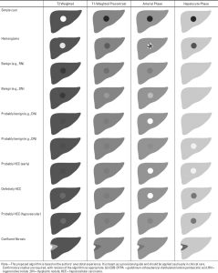
Expected appearance of different lesion types under different imaging phases using hepatobiliary contrast agents.
The article also contains numerous MRI images of different lesion types under different imaging phases, and is well worth a read. The reference for the article is:
Cruite, I., Schroeder, M., Merkle, E. M., & Sirlin, C. B. (2010). Gadoxetate Disodium–Enhanced MRI of the Liver: Part 2, Protocol Optimization and Lesion Appearance in the Cirrhotic Liver. In American Journal of Roentgenology (Vol. 195, Issue 1, pp. 29–41). American Roentgen Ray Society. https://doi.org/10.2214/ajr.10.4538

