ANIMAL TRANSPORT PRE-READING NOTES
when in doubt, there is probably a pressure gradient
KEY CONCEPTS
- Animals require oxygen intake and carbon dioxide expulsion to sustain cell respiration.
- Gas exchange organs maximize the rate of O2 and CO2 diffusion by
- large thin surface area
- steep partial-pressure gradient favouring O2 in and Co2 out.
- Blood is specialized tissues that transport gases, nutrients, wastes
- Hemoglobin = protein that carries oxygen
- Very good at take up oxygen at lungs and other gas exchange organs
- release oxygen at tissue
- Hemoglobin = protein that carries oxygen
- Circulatory systems use positive pressure generated by one or more hearts to transport stuff throughout body.
Major questions:
- How are oxygen and Co2 exchanged with the environment? (Oxygen and Carbon dioxide important bc associ with cell resp
- How are these gases along with nutrients, waste, etc transported throughout body?
8.1 Why is respiration and circulation necessary for animals?
8.2 Air and water as respiratory media
- What is the differences between total pressure and partial pressures of a gas mixture?
- Why are the partial pressures of gases such as oxygen and carbon dioxide important?
8.3 O
rgans of gas exchange
- What is the importance of the various parts of Fick’s equation
- Describe how fish gills generally work.
- Describe with detail how the fish gill is a countercurrent exchange mechanism. What is countercurrent exchanges?
- How do vertebrate lungs work?
- Must name the main structures of mammalian lung
- What are the mechanisms of ventilation of the mammalian lung?
- How do changes in pressure result in lung ventilation?
- Explain the concept of “dead space”
- How do bird lungs work?
ANSWERS
How do animals exchanges gases with the environment and transport substances within their bodies?
8.1 Brief overview of respiratory and circulatory systems.
- Important bc need to O2 to produce ATP. Making ATP makes Co2. You need to get rid of Co2 because it can become acid in your blood. Not good.
- What are the four steps of gas exchange between environment and mitochondria?
- Ventilation = air or water moves through specialized gas exchange organ (lungs, gills)
- Diffusion at respiratory surface
- Circulation = transported in circ system
- Into tissues where O2 low due to cell respiration
- Which system helps with ventilation and gas exchange with the environment?
- resp system
- which system help with transport in body and exchange into tissues
- circ sys
8.2 Air and water as respiratory media
- DIFFUSION!!!
- How much oxygen in environ vs tissue?
- oxy high in enivorn and low in tissue so oxygen wants to go from environ to tissue
- carbon dioxide in environment vs tissue
- carb dix lower in envir than in tissue cuz in tissue there is lots cuz produced from cell resp. so co 2 tend to go from tissue out to environment
What is the differences between total pressure and partial pressures of a gas mixture?
- partial pressure = pressure of a particular gas in a mixture of gases
- partial pressure is like percentage of that gas in a mixture but gas style
- Partial pressure = (fraction) of ( total pressure of mixture).
- fraction = what fraction that gas is of the total air
- atmospheric pressure = 760 mmHg
- note: our atmosphere is mostly N2 and oxygen with a little co2 and argon. we ignore nitrogen and argon bc not useful to us
Why are the partial pressures of gases such as oxygen and carbon dioxide important?
- oxygen and carbon dioxide diffuse between the environment and cells along their partial-pressure gradients
- move from high partial pressure to low partial pressure
Compare water breathers with air breathers
- water breathers face more challenge than air breather
- the oxygen content of water is less than the oxygen content of air
- so water breathers have to take in much more water than air breathers need to take in air
8.3 Organs of gas exchange
How do small animals that lack lungs or gill exchange gas?
- diffusion across body surface is rapid enough to fulfill gas exchange needs
- e.g. sponges, jellyfish
- but must live in wet environment
- skin must thin which is no protective, prone to water loss
so animals that are big, live in dry (e.g. land
- respiratory organs provide greater surface area for gas exchange, large enough to meet gas demands of body cells
- land animals – lungs inside body to minimize water loss
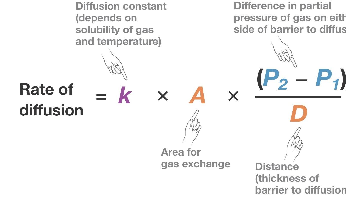
What is the importance of the various parts of Fick’s equation?
- Fick’s law of diffusion = rate of diffusion depends on 5 things
- solubility of gas in the aqueous film lining gas-exchange surface
- temperature
- surface area available for diffusion
- difference in partial pressures of gas across gas exchange surface
- thickness of the gas exchange surface
- So gas will diffuse quickly if very soluble, high temperature, lots of sruface area, big diff in partial pressures, thin surface
- Fick’s law states that all gases, including O2 and Co2, diffuse in the largest amounts when three conditions are met:
- A is large. – lots of surface area for exchange
- D is small – thin surface
- P2 – P1 is large. large partial pressure gradient -> maintained by efficient circ sys in contact with resp surface.
Describe how fish gills generally work. (not very important)
- gills = body surface or throat used for gas exchange
- large surface area for oxygen to diffuse across a thin surface
- Some gills contact water directly
- internal gills: water must be pushed over gills by cilia, limbs or other
- fish gills located on both sides of head,
- operculum = tissue cover gills
- pumping action of mouth and operculum creates pressure gradient -> cause water to move over gills (Obs: all of this plant/animal class is All about the gradient: pressure, solute, concentration etc. Always from high to low)
- ram ventilation = fast swimming with open mouth
- water flows in one direction over gills
- water passes through gill filaments -> gill lamellae -> capillaries – > gas exchange
Describe with detail how the fish gill is a countercurrent exchange mechanism. What is countercurrent exchanges?
- one way flow of water through gill lamellae
- flow of blood through capillaries in lamellae is OPPOSITE to flow of water
- counter current exchange = two fluids flowing in opposite direction
- lamellae always have oxy press gradient
- So water is always adjacent to capillary blood with LOWER oxygen content so large partial pressure gradient
- most of oxygen in water diffuses into blood
- If concurrent flow, then oxygen transfer stops where partial pressure gradient stop bc same partial pressure.
- counter current flow makes gills effcient at extract oxygenfrom water
- because difference in partial pressure of oxygen and carbon dioxide in water vs blod maintained over entire gas-exchange surface (lamellae)
- effect of counter current change = maximum P2 – p1 difference over entire gill surface
Must name the main structures of mammalian lung
- trachea – tube
- bronchi
- bronchioles
- lungs = organs of ventilation, foldings of throat
- alveoli – surface area: thin film, 1 layer epithel, extracellular matrix, wall of capillary
What are the mechanisms of ventilation of the mammalian lung? How do changes in pressure result in lung ventilation?
- actively contract muscles to pump air
- negative pressure ventilation – pressure gradient
- changes pressure in chest cavity
- inhalation = increase cavity volume by contract diaphragm, which decreases lung pressure
- diaphragm downward, ribs outward
- active
- exhalation = passive except exercise
- lung is elastic = return to original shape (normally collapsed shape)
- chest cavity volume decrease
Explain the concept of “dead space”
- dead space = portions of air passage that do not have respiratory surface e.g. trachea, bronchi
- = why only 2/3 of 450 ml in per breath partakes in gas exchange
- breathing = more efficient during exercise cuz chest cavity greater changes in volume
How do bird lungs work?
- one way airflow through avian lung
- posterior air sacs fill with outside air – inhalation
- lungs fill with air from posterior sacs – exhalation
- anterior air sacs fill with air from lungs – inhalation
- anterior air sacs empty – exhalation
- inhalation, air flows through trachea and enter two posterior sacs
- exhalation, air leaves posterior sacs, enters parabronchi
- inhalation 2 air into parabronchi in anterior lung, then to anterior air sacs
- exhalation 2 air out of anterior, thru trachea, out to atmosphere
- less dead space = dead space is restricted to short stretch of trachea between mouth and opening of anterior air sacs.
- gas exchange during both inhale and exhale
- blood circulate through bird lung in capillies that cross parabronchi perpendicularly. cross current is less efficeint than countercurrent (fish) but better than weblike capillaries in mammals
CIRCULATORY SYSTEM
- carries blood or hemolymph into close contact with every cell in body
- close enough that diffusion is efficient
(open and closed circ systems not discussed in class but expected to know the generally open and closed. details not needed)
open circ systems
- hemolymph direct contact tissue
- do not need to diffuse across wall fo blodo vessel
- heart and body movement
- hemolymph low pressure, low flow rate. ok for sedentary not needing much oxygen
- insects
closed circulatory systems
- blood flows in continuous circuit through body under pressure generated by heart
- blood confined in vessels
- high pressure -> high flow rate
- blood flow can be directed to specific areas when needed
IMPORTANT to know types of blood vessels, general characteristics. won’t be discussed in class but will tested on exams
arteries = thick wall, take blood away from heart at high pressure
-
- all have both muscle fibres and elastic fibres in walls
- elastic fibres dominate aorta all so can expand in response to high pressure
- when contraction of heart ends, diameter of aorta return to resting state. elastic response propels blood away from heart “secondary pumping
- maintains forward blood flow between contractions
- arterioles
- sphincters = muscle fibres around circumference of vessels
- control resistance to flow
- Constriction relaxation controlled by nervous system
- allows for nervous system to be able to control blood flow
- When sphincters relaxed, arteriole diameter increases, resistance to flow decreases
- when sphincters contracted, arteriole diameter decreases, resistance to flow increases, slow blood flow, can divert this blood floow to other tissues
- capillaries = one cell thick, low pressure
- one red blood cell at a time
- dense network throughout body
- exchange
- in some organs e.g. liver, capillaries have multiple openings so less barrier to diffusion
- veins
- all veins have some muscle fibres that contract in response to signals from nervous system, decreasing diameter and overall volume of vessels. Blood pressure in closed system is partially regulated by actively adjusting volume of blood within veins.
- low pressure, thinner walls
- larger interior diameter
- flow speeded by moving limbs
- one way valves – prev backflow
- muscle fibres contract when signals from nervous system say decreasing the diameter and volume of vessels
- blood pressure is regulated by actively adjusting the volume of blood within veins
- venules
Interstitial fluid. Focus on physiology of how lymph is formed.
- interstitial fluid = fliud that fills area between cells
- high pressure of closed circ systems + thin walsl of cappiles means that small but steady leake of plasma fliud from blood vessel
- why does interstitial fluid build up?
- there is outward-directed hydrostatic force in capillaries, created by the pressure on blood generated by heart
- inward directed osmotic force in capillaries, created by higher concentration of solute in blood plasma than in interstitial space
- at the end of capillary nearest to arteriole, hydrostatic force exceeds osmotic force so fluid moves out of capillary into interstitial space
- venuous end, osmotic force exceeds hydrostatic so fluid lost on the arteriole end gets reclaimed at venuous end of cpillary.
- but not all interstitial fliud reabsorbed by capillaries
What is the role of the lymphatic system?
- lymphatic system = branching tubules called lymphatic ducts or vessels
- lymphatic ducts
- permeate all tissues
- eventually join with one another
- largest lymphatic vessels return excess fliud to major veins entering the heart
- assuming that total solute concentration in plasma and interstitial fliud must be different enoguh to bring fliud into capillaries via osmosis
- fliud that leaks out of capillaries must have low OSMOLARITY
- capiliess must act as filters that retain large prrotines
- albumin = large and net negative prtoeins that can’t exit capillary
- keep solute concentration in blood high
- maintains strong osmotic gradient that brings fluid back to capillaries
How does the heart work?
Carefully read “The human heart” and “the cardiac cycle”
- pulmonary circulation = lower pressure, to and from lung
- systemic circulation = to and from body
What are the parts of the human heart?
- thin walled atrium receives blood
- thick walled ventricle pumps blood out
- artioventricular valves seperate atria from ventricles
- venae cavae (inferior, superior)
- pulmonary artery
- blood flows form artium to ventricle to arterty only one way
- one way valves seperate heart chembers and ffrom blood vessel
- heart murmur = back flow
- pulmonary veins
Trace the flow of blood through the chambers
- right atrium
- right ventricle
- lungs
- left atrium
- left ventricle
- body
What are the steps of the cardiac cycle – how do changes in pressure inside heart result in blood flow?
- systole = contraction phase of atria and ventricles
- diastole = relaxation
- cardiac cycle = sequence of contraction, relaxation , one diastole, one systole
- ventricular systole -> increases pressure in both ventricles, blood to pumonary artery and aorta,
- systolic blood pressure = measure at peak of ventricular ejection
- disatolic = low bp
electrical acitation of heart
- pacemaker cells initiate contractions of cardiac muscle cells
- pacemaker cells located in sinoatrial (SA) node
- SA node and muslce cells receive inputs from nervous systesm and chemcial messengers
- to regulate heart rate
- stregnth of ventricular contraction
- amoutn fo blood mvoing through cri c varies in repsones to electrical signa dn and hormoones
- to regulate heart rate
- an electrical impulse that stimulates contraction is genereated in SA nodea nd rapidly conducted through right and left atria
- signal spreads quickly from cell to cell because cardiac muscle form physical and electrical connections to each other (this is special and unique to cardiac muscle cells)
- all cardiac muscle cells branch to ocntact other cells
- intercalated cells =
- elf
- sa node initates signal
- sa node signa spread ove ratria. atria contract simulatnaeously and fill ventricles
- signal from atria conducted to AV node. AV nod delays and then pas to centricles. delay allow ventricles ot fill
- electrical impulse form va transmit through fibres in muscular wall seperated ventricles. both ventricles contract as atria relax.
- ventricles realax and cells recoves, restore electrical state prior to contraction
(see fig 8.29)
patterns in blood pressure and blood flow
- blood p in capillaries drops bc resistance increases. lots of fricton loss. seen in graph as lots of squiggles
- velocity of blood flow decreases in capillary
How bp and blood flow regulated?
- arteriole sphincter
- homeostatic control of bp
- sensors e.g. baroreceptors – dec in bp
- cardiac output increases
- artioles constrict to divert blood
- veins constrict
- integrator process info about change
- effects diminish impactof chage
- sensors e.g. baroreceptors – dec in bp
Stuff to remember:
- if atmos is 45, predicted partial pressure in water surface is 45 mmHg.
- oxygen concentration is water is lower than concentration in air
- cross sectional area of vessels increase, velocity of flow also increases. FALSE
- Left ventricle of heart fills ???????
 Follow