Organization of Tissues into Tissue Systems
| The tissues of the stem, as with the other organs, are organized into three tissue systems: dermal, vascular, and ground tissue systems.Below you can see how the tissue systems are arranged in the stem. |
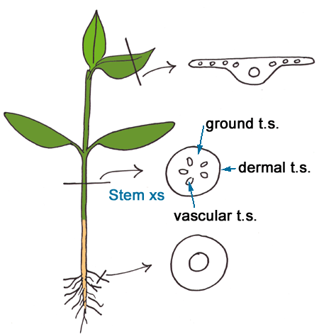 Diagram of a young plant, Illustrating the cross section of the stem |
Primary Growth in Stems
| The growth in length in plants is due to primary growth. Growth is the result of cell division and elongation. New cells are generated in the apical meristem. Coleus is commonly used for microscopic purposes (it is also a common house and garden plant). |
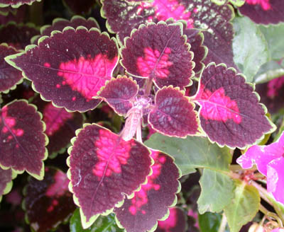 Coleus |
A longitudinal section through the shoot tip of Coleus demosntrates the arrangement of the growing tip of a plant. The apical meristem is indicated with an arrow. This is a region of active cell division. You can also see the two long structures on either side of it (leaf primordia). The leaf primordia are produced from cells of the apical meristem. Axillary buds (bud primordia) are generated in the axils of leaves. Each bud primordium has an apical meristem.
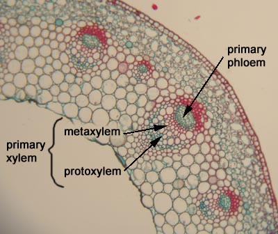
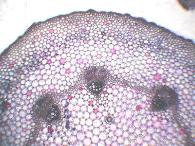
| The cross-section of Ranunculus (buttercup) shows the distribution of vascular bundles in the primray part of a stem. The tissue system is organized during primary growth. Some plants, such as buttercup, only have primary growth and are said to be herbaceous. The cortex and pith are regions of the ground tissue system |
 Ranunculus Stem Cross Section |
| Monocots, such as corn, do not produce secondary tissues. |
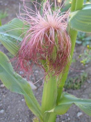 Corn Plant (in flower/fruit) |
| Corn can get to grow quite tall. Support is provided by the many vascular bundles that are scattered throughout the ground tissue system. Each bundle has fibres which contribute to the structural support of the stem. |
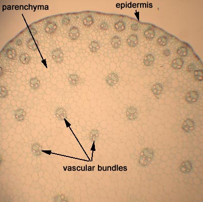 Corn Stem Cross Section |
Secondary Growth in Stems
| Secondary growth or lateral growth refers to growth in girth (width). This occurs in many plants to varying degrees. Some plants we call herbaceous, such as mints, actually have a limited amount of secondary growth. Other plants, such as conifers , have extensive secondary growth. |
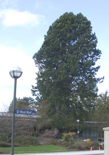 A Conifer Pinus ponderosa |
| Meristems, called lateral meristems, are responsible for lateral growth. Plants have two lateral meristems: vascular cambium and cork cambium. For such meristems to develop, cells able to divide must be present. Parenchyma cells are such cells.The figure to the right is a cross-section through the stem of Ricinus (castorbean). Primary growth has finished and secondary growth is about to commence. |
 Castorbean Stem Cross Section |
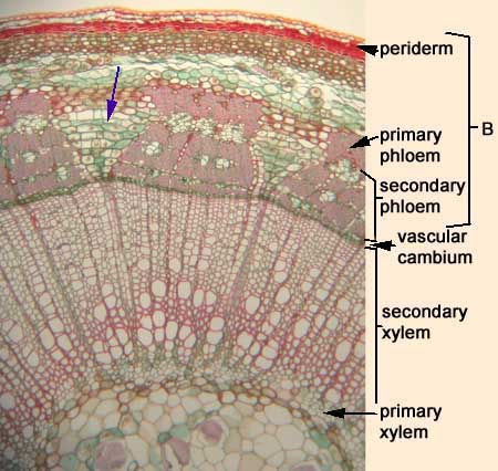
| After primary growth is complete, undifferentiated cells (parenchyma) between the primary vascular tissues starts to divide in two directions. These dividing cells are called the vascular cambium, indicated with arrow in the picture below. Vascular tissues are produced by the vascular cambium. Xylem is produced to the inside of the dividing cells (vascular cambium) and phloem is produced to the outside. Because the vascular tissues are derived during secondary growth they are referred to secondary xylem and secondary phloem. | 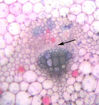 |
| Bidirectional cell divisions occur in the ground tissue (parenchyma) adjacent to the vascular cambium formed in the bundles, as indicated with an arrow in the picture below. The vascular cambium then becomes a complete ring. |
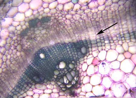 Bidirectional Cell Divisions of Vascular Cambium |
| The activity of the vascular cambium results in an increase in girth. A new layer of tissue, called periderm (cork), is generated to replace the single-layered epidermis. In the picture of geranium the secondary vascular tissues are labeled. You can also see the cork layer to the outside. The cells of both vascular and cork cambia divide to produce rows (files) of cells; this is evident in the resulting files of differentiated cells of the secondary xylem and the cork. |
 Geranium Stem Cross Section |
| The cork cambium differentiates from cells just inside of the epidermis to produce a cork layer. Cork cells are produced toward the outside whereas parenchyma cells (phelloderm) are producd to the inside. In most plants the phelloderm is liimited to only a few cells whereas the cork layer can be many cells thick. Periderm includes the cork cambium (phellogen), cork (phellem), and phelloderm. Unlike the vascular cambium, cork cambium has a limited lifespan. New cork cambia are generated to the inside of the old cork cambia. |
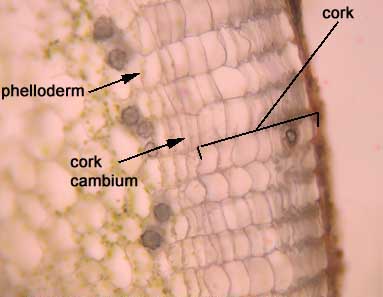 Close up of Cork Cambium |
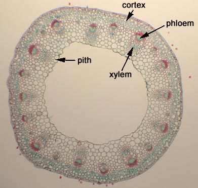
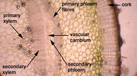
| The section in Figure 3(n) is through the stem of a linden tree (Tilia). Remember that phloem and xylem are complex tissues made up of many cell types. The purple arrow indicates a wide band of parenchyma of the phloem (phloem ray). Why is it not surprising that the primary xylem and primary phloem started out close to each other, yet in older plants they are far apart? |
 Cross-section through a Woody Stem of Tilia |
Back to Lab 2.