
SELAGINELLACEAE
Selaginella is probably one of the oldest of all the extant (living) generas of the vascular plants, second only to Lycopodium. There are approximately 700 species and several additional very closely related fossil forms included in this group. Selaginella is distributed from the tropics to the near subarctic latitudes. It is most common in areas of abundant rainfall and warm weather, but also occurs in British Columbia.
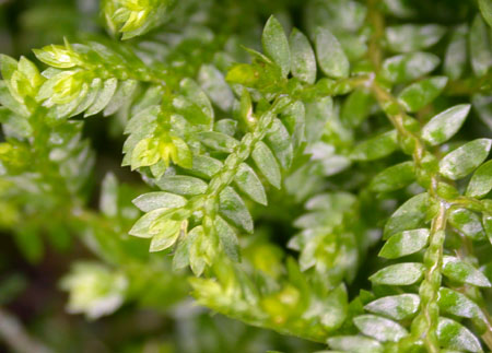
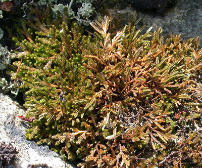
Most species grow in deep soil, but Selaginella wallacei, common on rock bluffs in southern parts of British Columbia (White Cliff Park), prefers shallow soil. The picture below was taken at Eagle Ridge above Horseshoe Bay. Lighthouse Park is another place to find this pretty little clubmoss.
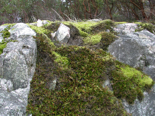
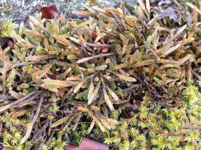
The picture below is of a patch of Selaginella. It is the darker green. The lighter green plant is an honest to goodness moss.
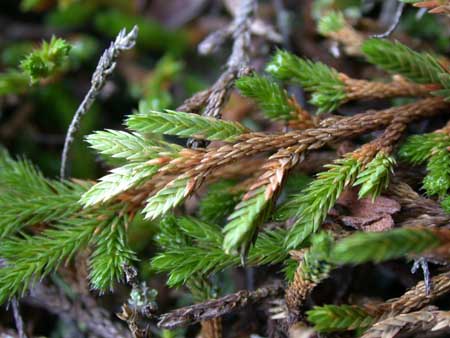
Selaginella vegetative structures
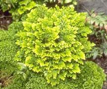
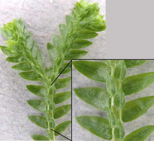
This is Selaginella krausiana a common potted houseplant.
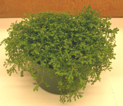
Its delicate leaves make it an attractive addition to anyone’s coffeetable.
You can see that all the leaves of this plant are not the same. They are different sizes and are thus said to be heterophyllous (heter = different, phyll = leaf).
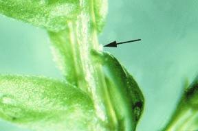
Selaginella can be differentiated superficially from Lycopodium by the presence of a tongue-like structure known as a ligule, which grows from the axil of vegetative and reproductive leaves. It is something you cannot see without a dissecting microscope.
When you pull back a leaf from the stem you can see the small ligule, a central and basally attached outgrowth of the leaf. The ligule may function as a secretory organ, secreting water and mucilage to keep the leaf moist.
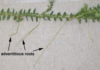
Roots are a little easier to see. They are adventitious and borne along the stem.
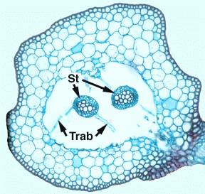
Here is a cross-section through the stem of Selaginella. Note that the stele (St) is suspended in a cavity (lacuna) by elongated cells of the endodermis, the trabeculae (Trab). The stele of Selaginella is protostelic.
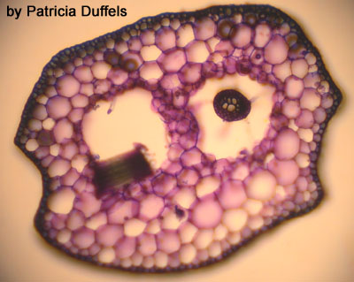
Here is a section showing you the same components as above. see if you can identify them.
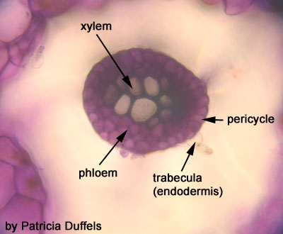
Here is a close-up of a stele.
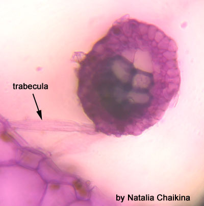
A trabecula:
Compare your observations with Raven, your lab manual, and the GLOSSARY OF STELE TYPES.
Selaginella reproductive structures
Differences between the members of the Selaginellales and Lycopodiales can be seen in the reproductive structures. In members of the Selaginellales the strobilus shows complete dimorphism in the sporangia. The male microsporangia produces many tetrad microspores in the axiles of the microsporophylls near the apex of the strobilus, whereas the female megasporangia produce four (rarely up to 12) large yellow megaspores in the axiles of the megasporophylls closer to the base of the strobilus. These spores will be shed and eventually develop into the male and female gametophytes respectively.
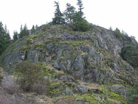
Selaginella wallacei grows on rocky cliff-faces like this one overlooking Fulford Harbour on Saltspring Island.

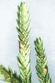
Here is a picture of Selaginella wallacei. You can see the vegetative form on the left and the reproductive on the right. The strobilus as a squared-off appearance.
You can see this feature a little better in this picture.
The sporangia-bearing microsporophylls are sporophylls which are associated with microsporangia.
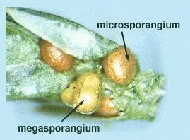
Parts of the sporophylls have been removed from this strobilus to expose the megasporangia and microsporangia.
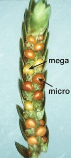
This shows you a close-up of a megasporangium and a microsporangium. Within the megasporangium there are four megaspores (only three are visible because the fourth is in the back). The microsporangia contains lots of microspores. The next image demonstrates this as well.
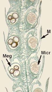
In this picture of a longitudinal section through the strobilus you can see that there is a different size of spores found within different sporangia. The microphylls which bear the sporangia with large spores (megasporangia = Meg) can be called megasporophylls and those sporangia with small spores (microsporangia = Micro) can be called microsporophylls.
Where do the microspores and megaspores occur in the life history of Selaginella, (see Raven 7th, p. 386-387; 8th, p. 410-411)? How is heterospory related to the sex of a gametophyte? Make sure you understand the difference between homospory (as in Lycopodium) and heterospory (as in Selaginella).