Presentation skills in science

Over the past few years, and particularly as I have begun to work on my thesis project, I have found myself listening to talks from other researchers. I have sat in on seminars from fellow biology students, mock thesis defenses for Masters and PhD candidates, and prominent researchers from Vancouver and across Canada. All of these talking points and Powerpoint slides have shown me that, without a doubt, presentation skills are key to one’s success in the sciences. However, it still amazes me how many people in the sciences do not seem to possess these fundamental skills.

I admit that I am a bit biased in this respect. I have worked as a campus tour guide for several years on campus, and have trained many new members of the team on how to deliver presentations effectively. I am comfortable speaking in front of a crowd, and recognize that not everyone else feels this way. Still, I think that developing presentation skills is often overlooked by students who are hoping to go into graduate school. Instead, undergraduate students focus on developing their study skills, or gaining hands-on experience in a lab. Are these things important for grad school? Absolutely, but if you are unable to stand in front of a room and tell people why your research matters, you will have a hard time getting that graduate degree.

Therefore, my advice to people who hope to continue in the sciences: practice speaking in public! Volunteer to present at journal club, apply to deliver a talk at a conference, or just present your research to your family and friends! The more you practice, the better you will be able to communicate why your research is important.
“So, what’s your thesis project?”

This is a question that I have been asked many times over the course of this year. I am currently in my final year of study, and am working on my honours thesis project at the BC Cancer Agency. Since I am frequently either on my way to or from the lab, my thesis often comes up in conversation with my friends who have little to no background in the sciences. I also had to field many questions at the Thanksgiving and Christmas dinner table from my family, which meant I needed to figure out how to explain my research.

My honours project is focused on two components of a protein complex called the MRN complex, which is involved in the Fanconi anemia DNA repair pathway. I am investigating whether these proteins, Mre11 and Nbs1, are able to reduce the level of R loops in human cancer cells and therefore promote genome stability. Explaining this to my relatives, some of whom did not complete high school, was a huge challenge over Christmas, but I eventually figured out a way to explain it. “Cancer is caused by mutations, and there are these structures in our DNA called R loops that make mutations more common,” I explained. “I’m studying a couple of molecules to see if they can fix the R loops and prevent cancer.” An oversimplification? Sure, but now my relatives and friends have a better grasp on what I am doing in the lab all day.

This has made me understand the importance of knowing how to simplify complex concepts for the general public in science. Granted, in my case this ability to explain my research has little effect on the state of scientific research as a whole, but the same skills must be used by scientists who are communicating with the public every day. As the political climate in the United States has shown, particularly with regards to climate change, scientists can often be seen as “elitist”, and research can be challenging to explain to people with little formal education. It is important to always be mindful of our audience in science, especially as research becomes increasingly specific and the public becomes increasingly hostile towards that which they do not understand.

Chromatin remodeling and bivalent histone modification in ESCs

  • Chromatin remodelling: any change or modification to the chromatin
    • In the paper, authors used term to describe ATP-dependent remodelling factors
  • Epigenetic modification of histones makes different transcription patterns possible with the same genome
    • Includes acetylation, methylation, ubiquitinylation, phosphorylation, sumoylation, ADP ribosylation, and many other modifications
  • Histone modifications can assist with activation and repression of transcription
    • H3K4me3: trimethylated lysine-4 of histone 3, activating histone mark
    • H3K27me3: trimethylated lysine-27 on histone 3, repressive histone mark
    • These two can be found in combination on certain promoters, called bivalent modifications
Bivalent modifications
  • HCNE: highly conserved non-coding elements, tend to be enrichev d with these bivalent marks
    • Upon depletion of PRC2 subunit, there was a loss of H3K27me3, which led to an upregulation of the genes
    • Under wild-type conditions, bivalent genes showed little to no activation, suggesting genes are poised for activation in mouse ESCs
  • In human ESCs and iPSCs, bivalent domains were often identified, on developmentally regulated genes
  • H3K4me3 and H3K27me3 were found together on regulators of development that were expressed at low levels
    • Also seen in other stem cell types, e.g. hematopoietic stem cells
    • Some promoters still had bivalent promoters, even when they were terminally differentiated, suggesting that maybe bivalent marks are present at a low level in all cell types
    • Trithorax group (TrxG) deposits the H3K4me3 mark
    • Polycomb group (PcG) proteins deposit the H3K27me3 (repressive) mark on histones
    • Bivalent domains are mainly associated with CpG islands in ESCs
  • H3K4me3 is deposited by the SETD1A, SETD1B, and MLL complexes
    • Globally, the mark is deposited by the SETE1A and SET1B complexes
    • MLL1 through MLL4 appear to carry out more specific functions
    • MLL2: main methyltransferase at bivalent promoters
    • MLL1/2: contain CXXC or zinc-finger CXXC (ZF-CXXC) domains, which specifically recognize unmethylated CpGs
  • H3K27me3 is deposited by the PRC2 complex at bivalent promoters
    • The core PRC2 complex is made up of EZH2 or EZH1, EED, SUZ12
    • EZH2: enhancer of zeste homologue 2, catalytic subunit of the PRC2, methyltransferase
    • H3K27 is recognized by chromodomain-containing proteins such as CBX
    • many developmentally regulated genes are marked by bivalent domains bound by PRC and PRC2, though some exclusively by PRC2
    • PRC2-specific bivalent domains are usually found on the promoters of genes that are not true developmentally active genes
  • key pluripotent genes are shown to interact with MLL and PRC proteins
    • Depletion of Oct4 in ESCs leads to selective depletion of H2K4me3 levels
    • When ESCs are undifferentiated, those deficient of PRC2 show aberrant differentiated potential
    • PcG proteins are therefore vital for embryonic stem cell differentiation
Chromatin remodelling and bivalency

Translating epigenetic research to the general public

Last week, a new study was published in the open-access online journal eLife, titled “Differential methylation between ethnic sub-groups reflect the effect of genetic ancestry and environmental exposures”. As frequently occurs with new research findings, a summary of the study was written on the popular science website, IFLScience.

The author of this article did a good job of summarizing a key concept that we have discussed in this course: how cells with the same genetic material can develop into different cell types with various functions. One of the ways in which this can happen, the author states, is through the addition of methyl groups to DNA, which can alter gene expression patterns. From there, he went on to explain how different environmental factors can alter the methylation patterns within the cell, using the example of smoking. This provided the key background information necessary to understand the results of the study, which showed that some differences in methylation were associated with ethnicity, rather than shared ancestry. As race/ethnicity are known to be social constructs, the authors concluded that a shared environment or culture could be at play in mediating these patterns.

However, there were some scientific missteps within this article. The most egregious error occurred when the author equated “epigenetics” with “methyl groups”, which is an oversimplification of the epigenetic controls over DNA. As we know, epigenetic alteration is not limited to DNA methylation. Many different covalent modifications can be added to nucleotides, as well as to the histone proteins contained within the nucleosome. Furthermore, there are epigenetic mechanisms such as miRNAs that do not involve the covalent modification of genetic elements at all. Granted, this is a complex field that could not be entirely explained in a short article, but the author could still have mentioned that there are mechanisms other than DNA methylation that are involved in epigenetic regulation of gene expression. This goes to show that as members of the scientific community, we should sometimes be cautious about news reports surrounding research studies, and look instead to the primary literature for the real answers.

Enhancer function: mechanistic and genome-wide insights

Plank, J.L., Dean, A. (2014). Enhancer function: mechanistic and genome-wide insights. Mol Cell. 55(1): 5-14

  • Enhancers: cis-acting regulatory elements that increase transcriptional output of target genes to affect cells in development
    • May reside far away from targets
  • Models proposed for interaction with promoters:
    1. Enhancer looping
    2. Linking via protein complexes
    3. Combination of the two models
  • Enhancer sequences contain binding sites for TFs, confers tissue specificity
    • Factors binding to enhancers and genes could stabilize chromatin loops
    • Tissue-specific proteins are critical to looping, identified in some model systems
      • e.g. beta globin locus, Th2 cytokine loci, human IFNy locus
  • Looping interactions of chromatin-binding proteins (e.g. CTCF) facilitate gene-enhancer contacts
  • Genome-wide studies help to place interactions in 3D context
    • Chromosome conformation capture technology: measures physical interaction frequencies between enhancers and targets
    • Refinements in 3C approach detects chromatin loops at multiple levels
    • Enhancer transcription into eRNA may function as part of gene activation, looping
    • Specific features of enhancers, targets, chromatin structures found

Long-Range Interactions with Target Genes

  • TFs bind enhancers in clusters
  • Exclude nucleosomes, contribute to the DNase I hypersensitivity
  • Certain loci show specific enhancers binding to proteins required for looping
  • Complex including GATA1 and cofactor FOC1 required for beta clobin LCR coding
  • Erythroid cell transcriptional activation requires enhancer action
    • Complex includes LBD1, TAL1, LMO2
    • Dimerization domain of LBD1 underlies enhancer-gene proximity
    • When linked to the beta globin promoter, was capable of driving loop formation and partial activation of transcription
    • Large cohort of erythroid genes are activated by the LBD1 complex
  • CTCF/cohesin binding elements and enhancers bind T-bet, a lineage-specific Th1 factor in the IFNy locus
    • shows proteins can participate directly in enhancer-gene looping
    • CTCF promotes a Th1 specific IFNy locus looped conformation
    • Need to join both CTCF and enhancer sites to activate IFNy transcription
    • CTCF: transcriptional repressor, creates boundaries between topologically associated domains in chromosomes, facilitates interactions b/w/ transcription regulation sequences
  • During cell division, enhancer-gene interactions are disassembled
    • FOXA1 (lineage factor for hepatoma cells), GATA1 (key erythroid cell lineage factor) remain associated with chromosomal sites containing enhancers

Enhancer Loops and Transcriptional Activation

  • Enhancers function primarily to promote increased transcriptional output
    • Interaction with promoters may involve transcriptional machinery components
    • Mediator occupies the enhancers of many ESC genes, pluripotency factors
    • Links promoters to Pol II at target promoters thru direct interaction with cohesin
  • Enhancers may be involved in the initiation of transcription
    • Some function to release Pol II in order to allow elongation
    • Loop to target promoters, permit activation of the P-TEFb complex
    • PTEF-b: complex required for the release of Pol II into the elongation stage
  • Study showed enhancer-promoter gene interactions enriched for cell-specific genes
    • Gene families regulated by common TF were overexpressed
    • Overrepresentation of multigene complexes, linked to Pol II foci
    • Pluripotency genes in ESCs were also connected within one hub
  • Shows enhancer looping key, maybe required for transcription activation
    • Formation of new enhancer loops precedes transcription at the beta-globin locus
    • Long-range enhancer-gene interactions may drive nuclear relocalization and clustering

Enhancer Loops within the Nucleus

  • Genomic context: long range interactions between loci predominantly within TADs (topologically associating domains)
    • TADs largely conserved across range of cell types in development
    • Borders enriched for CTCF sites (also found within TADs)
    • Long range interactions within the same TAD more common than between TADs
    • Intra-TAD contacts may be involved in cell-specific transcriptional activation
    • Intra-TAD more variable among different cell types
  • EPUs: enhancer-promoter units, clusters of coregulated enhancers and promoters
    • Super enhancers: large domains of up to 50 kb that contain clusters of individual enhancer elements, highly occupied by Mediator, pluripotency factors
    • Enhancers within these regions are associated with genes that encode cell identity regulators
    • Sub-TADs have also been identified, vary in tissue-specific manner
  • CTCF/cohesin and Mediator play roles at different levels of long-range interactions contributing to TAD or sub-TAD organization
    • HoxA genes and enhancers occupy same TAD but are grouped into specific topological domains in the limb buds (where the genes are active)
    • Enhancer-gene long range interactions are strengthened by but do not depend on enhancer activity
  • Are TADs functionally relevant for long-range enhancer activation of genes?
    • Deletion of a TAD border in Xist locus led to new ectopic contacts, long-range transcriptional misregulation
    • CTCF (not cohesin) knockdown in vitro led to increased inter-TAD interactions
    • Cohesin knockdown in post-mitotic thymocytes had no change in TAD organization
    • CTCF may help maintain TAD borders
  • Is an enhancer able to fucntion outside of its normal TAD context?
    • Ectopic B-globin LCR re-established contact with the gene and increased transcription
    • Inter-TAD interactions may help enhancer function
    • HoxD cluster: some genes switch enhancer contacts from one TAD to another as gene expression changes across locus, close to the TAD border
    • There may therefore be a flexibility to the border of TADs
  • Within TADs enhancers cluster together into topological domains, relevant gene targets, EPUs, sub-TADs
    • May all be different examples of same enhancer-dependent clustering phenomenon
  • Enhancers, promoters must scan a limited nuclear area in order to make contact
    • Interruption of contact sites within a cluster of coregulated genes affects transcription of other interacting genes
    • Consistent w/ model that enhancer-gene clustering within TADs, association of TADs of similar character serve to nucleate transcription factors
    • Emphasizes role of enhancers and looping in the spatial organization of transcription in the nucleus

Temporal and Spatial Regulation of Enhancers

  • Enhancers are progressively modified to activate transcriptional programs
    • Occurs specifically through acquisition of H3K27ac mark
    • “poised” enhancers in ESCs have p300, BRG1 occupancy, H3K4me1, low nucleosome density
    • Enhancers also have H3K27me3 mark, PRC2
  • Later in development loss of PRC2 and H3K27me3, acquisition of H3K27ac and ability to activate gene expression
  • “Inactive” state of ESC enhancers with high nucleosome density identified, have occupancy by ELL3
  • ELL3: polymerase II elongation factor, may mark enhancers for subsequent activation
    • As ESCs differentiate, enhancers of pluripotent genes inactivated
    • Some mechanisms proposed:
      1. L-SID1 (histone H3K4/9 demethylase) removes H3K4me1
      2. PRC2 deposits H3K27me2 mark at ESC enhancers before the H3K27ac mark
      3. OTX2 transcription factor pioneers new enhancers, allows Oct4 to move to new site and activate new targets
        • Permits cells to exit their naive state
        • Occurs during transition between naive and “primed” cells
  • Changes occurring genome wide within regulatory landscape in regulation
    • ~90,000 enhancers show specific tissue/stage-specific activity windows
    • DNase I hypersensitivity used as a proxy to measure enhancers, gave similar results
    • Enhancer usage varies among cell lineages, linked to lineage-determining TFs
  • Enhancers in differentiated cells capable of responding to external stimuli
    • TFs can mark “inactive” enhancers in unstimulated differentiated cells, required for later activation of targets
    • Unclear how latent enhancers are recognized and activated
    • Also unclear how enhancer modifications poise/activate enhancers
    • HoxD had some enhancer-gene contacts prior to gene activation, so cannot say that activation depends on the establishment of loops to target promoters
      • May be necessary but not sufficient?

DNA Methylation as a Modulator of Enhancer Activity

Long non-coding RNAs: lessons from genomic imprinting

Kanduri, C. (2016). Long non-coding RNAs: lessons from genomic imprinting. Biochimica and Biophysica Acta.

Introduction

  • In gametogenesis ~1% of protein-coding genes undergo genomic imprinting
    • >150 imprinted genes identified in mouse
    • Typically located in clusters that range from a few kB to 3.0 Mb
  • Long non-coding RNAs found in all imprinted clusters
    • Inverse expression pattern relative to protein-coding counterparts
    • Promoters map to differentially methylated regions (DMRs)
    • Deletion of the DMRs often leads to loss of imprinting
    • ICRs: imprinting control regions, 1-3 kb in size, most DMRs are ICRs
  • Human genome has more lncRNAs than protein-coding genes
    • Perform various functions in development, differentiation, disease
    • Multiple mechanisms both at transcriptional and post-transcriptional level
    • Most target chromatin modifying complexes e.g. PRC2, SW1/SNF, hnRNPK, G9a
    • Implicated in gene regulation at post-transcriptional level when they are localized to the cytoplasm

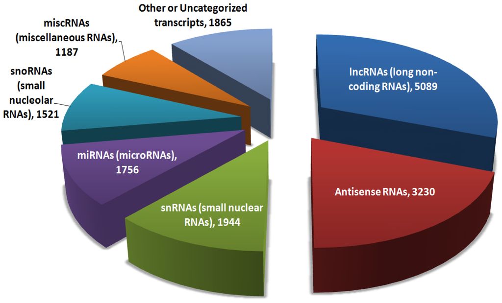
Da Sacco et al. (2012). IJMS. Pie chart of the major categories of >16,000 non-coding RNAs in the genome.

Intergenic lncRNAs in Genomic Imprinting

  • lncRNAs can be classified into 4 main categories: intergenic, antisense, intronic, and enhancer
    • All except intronic implicated in imprinting and parent-of-origin specific expression
  • H19: lncRNA (2.3 kb in length)
    • Maps to a well-investigated cluster on mouse chromosome 7, human chromosome 11
    • only expressed from the maternal allele, silenced on paternal
    • Paternal allele silenced via CpG methylation at the promoter
    • May have lineage-specific roles in the body
      • Deletion of H19 affected Igf2 in mesoderm but not endoderm
      • Deletion also has effects on growth, but is not embryonic lethal
    • Part of IGN (imprinted gene network) that includes 16 genes
    • Potentially controls growth via regulation of the IGN in trans
    • Interacts with MBD1, a methyl CpG-binding protein
      • Complex recruits H3K9 methyltransferase to DMRs of some members of the gene network
      • Establishes H3K9me3 marks to “fine tune” expression from both parental allelles
    • Expressed at high level in embryogenesis, downregulated after birth
      • Exception: remains highly expressed in muscle tissue
      • May promote myogenic differentiation?
    • Also shown to have oncogenic, tumour-suppressive properties
      • Overexpression of H19 is linked to metastasis
  • IPW: paternally expressed lncRNA, maps to an imprinting cluster on mouse chromosome 7, human chromosome 15
    • Deletion observed in 70% of cases of Prader-Willi syndrome patients
    • Mouse expression mainly restricted to the brain, but humans seen in all tissues
    • 5′ end contains tandem repeats
    • functional role of IPW cluster has not been investigated, but shown to interact with G9a methyltransferase
      • Targets IG-DMR to modify chromatin structure via H3K9
      • IG-DMR is a master controller of gene expression at the DLK-D103 imprinted cluster
      • First example of a lncRNA shown to promote chrosstalk between two imprinting clusters by altering ICR chromatin
  • MEG3: maternally expressed gene 3, imprinted lncRNA, maps to DLK-DI03 locus on human chromosome 14, mouse chromosome 12
    • IG-DMR controls maternal specific expression of MEG3
    • MEG3 expression is a marker of iPSCs with a fully pluripotent state
    • iPSCs without MEG3 expression are not viable to support embryonic development
    • MEG3 may promote interaction of PRC2 with JARID2
    • Complex of PRC2-JARID2 shown to contribute to ESC differentiation
      • MEG3 may therefore underlie the fully pluripotent state

Enhancer RNAs in Genomic Imprinting

  • Enhancers are responsible for spatio-temporal gene regulation
    • “landing site” for transcription factors, co-activator complexes
    • Can activate or increase transcription from distal promoters
  • Characteristics of enhancers include:
    • DNase I hypersensitivity
    • Post-translational histone modifications (especially H3K4me1/2, H3K27ac)
    • Bidirectional transcription
    • Transcripts generated are low copy number, non-polyadenylated
  • Enhancers promote target gene expression via recruitment or stabilization of basic transcription machinery binding
    • Establish higher-order chromatin contacts between enhancer and targets
    • Transcripts from IG-DMR control expression of maternally expressed transcripts at DLK1-Dio3 locus
    • Methylated on the paternal chromosome, unmethylated on the maternal
    • Unmethylated version is critical for expression of multiple lncRNAs including MEG3
      • Therefore acts as putative enhancer with enhancer-specific histone marks, encodes bidirectionally transcribed ncRNAs
  • Maternal chromosome bidirectional transcription in ESCs correlates with early replication, inner subnuclear positioning of Dlk1-Dio3 locus
    • IG-DMR transcripts may promote higher order chromatin
    • Enables early replication, subnuclear localization
    • Maintenance of expression of maternally expressed genes

H3K9me3-Dependent Chromatin: Barrier to Cell Fate Changes

Becker et al. (2016). H3K9me3-dependent chromatin: barrier to cell fate changes. Trends in Genetics. 32(1): 29-41.

Models of Developmental Gene Silencing

  • Genetic material in the nucleus divided into 2 categories:
    • Euchromatin: DNA with relatively low density, high gene transcription rates
    • Heterochromatin: regions of the chromosome that are compact, transcriptionally repressed
      • Can be constitutive (present in all cell types, phases of cell cycle) or facultative (repression temporally specific or cell-specific)
    •  Large proportion of genome has repeat-rich sequences
      • Risk to genome integrity due to possible recombination, duplication
      • Utility in keeping regions silent (constitutive heterochromatin)
      • Repeat-rich heterochromatin marked by H3K9 methylation (di- and tri-methylation)
      • Mammals: methylation catalyzed by 5 members of the SET-domain containing methyltransferase family
  • Heterochromatin protein 1: HP1, three isoforms in mammals
    • Can self-oligomerize, recruit repressive proteins to modify histones
    • Contributes to compaction and spread of heterochromatin
    • Binds H3K9me2/3 via its chromodomain
  • Methyltransferases that deposit H3K9me2/3 required to establish hypermethylation at CpGs, low-level histone actylation
    • Two characteristics of heterochromatin
  •  H3K27me3: methylation of lysine 27 at histone 3, catalyzed by PRC2 (Polycomb repressor complex)
    • Facilitates facultative silencing in cell-type specific repression
    • Especially present at lineage-specifying TF genes eg. Hox genes
    • H3K27me3 marked promoters are still able to be bound by general TFs, paused RNAP
  • H3K9me3 involved in cell type-specific regulation of facultative heterochromatin
    • in differentiated cells, form large contiguous domains called patches
    • Expand in both number, size during differentiation
    • Span numerous genes repressed in cell type-specific manner
    • These domains largely exclusive of H3K27me3
  • H3K9me3: repressive modification, also forms megabase-scale domains that include genes
    • called LOCKS (large organized chromatin K9 modifications)
    • Binding sites for the repressor protein CTCF detected at boundaries
    • Unsure if domains expand during differentiation
    • Important to silence lineage-inappropriate genes in differentiation

Heterochromatin: A Barrier to Cell Reprogramming

  • Hallmarks of cell identity erased during reprogramming to iPSCs
    • Requires the reprogramming TFs to bind their targets in DNA
    • Reactivation of pluripotency genes, suggest that accessing heterochromatin important to the process
    • Only <0.1% of cells are successfully reprogrammed
  • OSKM are the key reprogramming factors
    • All 4 open chromatin sites, but only OSK target sites containing nucleosomes w/o histone marks
    • This makes them pioneer factors
  • DBRs: differentially bound regions, megabase-scale chromatin regions in which none of the 4 factors can target DNA in fibroblasts
    • Same domains bound by OSKM in pluripotent cells
    • Overlap with domains enriched for H3K9me3 in fibroblasts but not ESCs
    • Knockdown of SUV39H112 increases Sox2, Oct4 binding
    • Encode diverse genes and elements, including TFs essential to pluripotency
    • Pluripotency genes seem to be more refractory to activation
    • Majority of genome regions found to have altered non-CpG methylation in iPSCs vs. ESCs are DBRs
    • Some H3K9me3 domains stay in iPSCs – indicate incomplete reversion to ESC state
    • H3K9me3 removal may help increase reprogramming efficiency
    • Knockdown of SUV39H1.H2 led to increased iPSC colony formation
    • Also seen with other H3K9 methyltransferases, unclear which is more responsible for stabilizing the differentiated state
  • Other factors/components of repressive chromatin acts outside DBRs
    • Demethylation of H3K9me3 needed for reprogramming via Utx
    • Repressive histone variant macroH2A inhibits reprogramming
    • H3K27me3 methyltransferase EzH2 needed for iPSC reprogramming
    • Need deposition of H3K27me3 and removal of H3K9me3 simultaneously
    • MBD3: component of the NuRD histone remodelling and deacetylase complex, mediator of gene silencing
      • Knockdown leads to improved iPSC programming
      • Stops reprogramming factor activity at the sites they already bind
      • May play a role in regulating H3K9me3 – hasn’t been explored

Paucity of Heterochromatin Defines Pluripotent State

  • Reduction of inaccessible H3K9me3-marked heterochromatin fundamental hallmark of the pluripotent state
    • Chromatin of pluripotent cells shows increased rate of exchange at chromosomal proteins e.g. linker histones, HP1
      • This indicates a dynamic and accessible state
  • Repetitive sequences: DNA sequences with high copy numbers, organized in adjacent near-identical units or dispersed throughout the genome
    • Includes retrotransposons, tandem repeats, satellite repeats, endogenous retroviruses
    • More common expression of these in ESCs, repressed in differentiated cells
    • Deletion of proteins that maintain chromosomal accessibility leads to impaired self-renewal of ESCs
    • Developmental plasticity of ESCs linked to chromatin accessibility
  • Partially reprogrammed cells have highly compartmentalized heterochromatin structures
    • Contain dense chromatin fibres similar to diffrentiated cells
    • DNA methylation, H3K9me3 at specific pluripotency loci
    • Erasure of H3K9me3 can allow them to become full iPSCs
  • SCNT: somatic cell nuclear transfer, uses factors of egg cytoplasm to restore pluripotency
    • H3K9me3 heterochromatin is a barrier to SCNT as well
    • RRRs – reprogramming resistant regions, silenced only in SCNT condition
    • Reducing H3K9me3 led to improved SCNT success
  • Heterochromatin, especially H3K9me3, presents a barrier to reprogramming, regardless of the cell conversion methodology

H3K9me3 as a Regulator of Cell Fate In Vivo

  • Patterns of H3K9me3 must be reorganized in cell fate transitions in development
    • Early embryo and terminal lineage maturation
    • TF networks ensure that H3K9me2/3 is regulator
    • Setdb1 occupies and represses genes that encode developmental regulators
      • Also acts as a corepressor of Oct4, suppressing trophoblast genes
  • Implantation is followed by a progressive silencing of Oct3/4 and other pluripotency genes (Nanog, Stella, Rex1)
    • Deposition of H3K9me2, DNA methylation dependent on GLP and G9a occurs
    • G9a prevents Oct3/4 reactivation when differentiated ESCs returned to pluripotent state
    • Mutations in GLP that disrupt its ability to recognize H3K9me1 led to decreased dimethylation and a delay in pluripotency silencing, abnormal embryonic development
    • Cross-talk exists between H3K9me3 and H3K27me3
    • A direct role for H3K9me2/3 has been proposed in developmental control of gene expression
      • Reduced H3K9me2 occurs at LADs (lamina-associated domains), coupled to relative depletion of H3K27me3
    • G9a and GLP-null embryos have early lethality
    • SETB1 homozygous inactivation also embryonic lethal
    • Distinct lethal phenotypes in each case, shows they have different developmental contributions
  • H3K9me3 contributes to lineage restriction in mature cell types
    • Shown by examining methylation status in Th1 vs. Th2 cells
    • Showed that H3K9me3/H3K27me3 have different roles in the two different lineages

Molecular Control of H3K9me3 Deposition

  • Additional factors needed to explain site selectivity of H3K9me3
    • Sequence-specific TFs have been shown to recruit the heterochromatin machinery
    • KRAB-ZNFs: Kruppel-associated box zinc finger proteins, important for establishment of heterochromatin and have mostly lineage-specific expression
    • Noncoding RNA can function as a binding platform for heterochromatin establishment at specific positions
      • In yeast, heterochromatin dependent on RNAi pathway components, requires transcription of a locus to be silenced
      • Not well understood in mammals yet but believe RNA is involved in the H3K9me3 establishment

Concluding Remarks

  • Large domains of H3K9me2/3 form in a cell-type specific fashion
  • Machinery responsible for this is still mainly mysterious
  • Need to understand initiation, delimitation of the H3K9me2/3 domains to develop more targeted ways to reduce H3K9me3-dependent heterochromatin
  • RNAi knockdown of all 5 methyltransferases for H3K9 was shown to increase reprogramming efficiency
  • Establishment of conditional knockouts of genes alone/in combination could provide insight
  • Future studies of lineage-specific H3K9me2/3 domains should look at whether they impair transdifferentiation/conversion

“If I were a developmental biologist” Assignment

My two questions that I would like to investigate as a developmental biologist are:

1. To what degree do parental epigenetic marks influence the epigenome of their developing offspring? Are epigenetic marks hereditary, or are they generated de novo in the embryo?

2. What are the physiological/cellular factors that influence the dynamics and position of methylation and histone modification during development?

Reflection:

I find it very interesting to go back to the very beginning of the course and see how much my thinking surrounding and understanding of developmental biology has changed!

We have definitely answered the first question over the course of the semester. The first wave of global demethylation occurs in the developing primordial germ cells (PGCs), and de novo methylation is carried out by DNMT3A and 3B, with the help of DNMT3L. DNMT1 is responsible for the maintenance of parental methylation, and does so by targeting hemimethylated strands of DNA. The second wave occurs during early embryogenesis and assists with the establishment of pluripotency in cells. In spite of this, the methylation state of imprinted genes is preserved throughout the process.

As for the second question, this is a huge question and not one that could be completely answered throughout the course. I actually think that a better question to ask would be the opposite: how do methylation and histones affect development? I have learned that the methylation state, chromatin conformation (euchromatin vs. heterochromatin), and histone modifications (repressive, activating, or bivalent) greatly alter the cells’ potential cell fates and levels of gene expression.

Some learning objectives I demonstrated with this assignment:

  • Approach questions, concepts, and facts with curiosity
  • Assess your own level of knowledge in a field or area of interest and identify “gaps” in your knowledge or skills
  • Identify and ask informative questions so as to “extract” useful information from other people’s knowledge and from experiments
  • Distinguish questions that can be investigated experimentally from questions that cannot

Annotated Bibliography

Chen, B., Chan, W. (2014). The de novo DNA methyltransferase DNMT3A in development and cancer. Epigenetics. 9(5): 669-677

This 2014 review by Chen and Chan compares and contrasts the role of DNMT3A in development and cancer. The structure, variants, localization, and expression patterns of the DNMT3A gene are described in detail. DNMT3A plays a critical role in primordial germ cells, helping to re-establish the parental imprints that are needed for meiosis, spermatogenesis, and oogenesis. Studies have shown that DNMT3A is also involved in the de novo methylation that takes place post-implantation, with Dnmt3a knockout murine ESCs failing to differentiate normally. In addition to the frequent mutation of DNMT3A in AML, DNMT3A is also mutated in hepatic cellular cancer (HCC), melanoma, and lung cancers.

Collins, C.T., Hess, J.L. (2016). Role of HOXA9 in leukemia: dysregulation, cofactors, and essential targets. Oncogene. 35(9):1090-8.

This review summarizes the role of the homeobox family protein HOXA9 in leukemia. HOXA9 is a transcription factor that plays several important roles in development, specifically in the expansion of hematopoietic stem cells (HSCs). The molecular mechanisms through which HOXA9 can induce leukemia are not yet understood, and it is hypothesized that several additional upstream genetic alterations leading to overexpression of HOXA9 have yet to be identified. Given that HOXA9 has also been found to be hypomethylated in DNMT3A-null cancers, this paper brought up the question of whether loss of DNMT3A and HOXA9 hypomethylation are connected to the gene’s overexpression and leukemogenic activity.

Estey, E. (2006). Acute myeloid leukemia. The Lancet. 368(9550):1894-1907.

This review in The Lancet describes the key clinical, molecular, and pathogenic features of acute myeloid leukemia. AML is a heterogeneous disorder affecting the hematopoietic stem cells, characterized by a differentiation block that leads to accumulation of immature “blasts” in the bone marrow. AML is the most common myeloid malignancy in adults, and is particularly common in adults over age 65. Many older adults develop the cancer as a consequence of cytotoxic therapy for another type of cancer. The cancer is called “acute” as it progresses fairly rapidly, with a five-year survival rate of less than 25%. Prognosis is even more dire for older adults, with elderly patients surviving for an average of a few months. This paper provided me with a background in the clinical features of the disease, and helped me to understand the relevance of my research question.

Figueroa, M.E., Lugthart, S., Li, Y., Erpelinck-Verschueren, C., Deng, X., Christos, P.J., et al. (2010). DNA methylation signatures identify biologically distinct subtypes in acute myeloid leukemia. Cancer Cell. 17:13-27.

DNA methylation patterns are known to be aberrant in cancers. In this study, the authors investigated whether DNA methylation could be used to classify distinct subtypes in acute myeloid leukemia (AML), which may be relevant for treatment. The methylation profiles of over 300 patients with AML were examined, and several different subgroups were identified on the basis of DNA methylation signatures. This paper demonstrates that DNA methylation patterns have the potential to be clinically significant, and supports the significance of my research question.

Kim, M.S., Kim, Y.R., Yoo, N.J., Lee, S.H. (2013). Mutational analysis of DNMT3A gene in acute leukemias and common solid cancers. APMIS. 121(2):85-94.

This study by Kim et al. examined mutations in DNMT3A in several common solid cancers and acute lymphoblastic leukemia (ALL) to see if the mutation was common in other malignancies in addition to AML. Using a single-strand conformation polymorphism assay, 916 cancers from >400 blood malignancies were examined, as were >500 solid tumours. DNMT3A mutations were identified in most of the cancer types that were analyzed, including gastric cancers, lung cancers, and lymphomas. Different types of alterations in DNMT3A were observed, including allelic loss, somatic mutations, and loss of expression. This suggests that DNMT3A likely plays a critical role in multiple cancer types, and that it may initiate tumorigenesis through a multitude of mechanisms.

Qu, Y. et al. (2014). Differential methylation in CN-AML preferentially targets non-CGI regions and is dictated by DNMT3A mutational status and associated with predominant hypomethylation of HOX genes. Epigenetics. 9(8):1108-1119.

This primary research study characterized the genome-wide methylation differences between cytogenically normal AML (CN-AML) cells and healthy control bone marrow cells. A key finding from this study was that homeodomain-containing (HOX) genes experienced the most significant changes in methylation, with hypomethylation occurring in genes including HOXA5 and HOXA9. While this study showed only a correlation between DNMT3A mutation and hypomethylation of homeobox genes, it provides an intriguing potential mechanism through which DNMT3A may initiate cancer.

Redecke, V., Wu, R., Zhou, J., Finkelstein, D., Chaturvedi, V., High, A.A., Hacker, H. (2013). Hematopoietic progenitor cell lines with myeloid and lymphoid potential. Nature Methods. 10:795-803.

This group reports the development of a novel hematopoietic stem cell-derived line that is capable of differentiating into both myeloid and lymphoid lineages, but not erythrocytes or megakaryocytes. The potential to differentiate into cells of the myeloid lineage is assessed through differentiation with GM-CSF and M-CSF, which promote differentiation into dendritic cells and granulocytes, and macrophages, respectively. The differentiated cells expressed the surface markers characteristic of the myeloid cell lineage, and had similar immune function and properties. I decided to use this cell line as my model system in my project, since I needed cells to be predisposed towards a myeloid lineage and did not want to use HSCs.

Shan, W., Ma, X. (2013). How to establish acute myeloid leukemia xenograft models using immunodeficient mice. Asian Pacific Journal of Cancer Prevention. 14(12):7057-7063.

This review from Shan and Ma examined the various mouse models that are used in xenograft experiments for acute myeloid leukemia (AML). Xenografts require immunodeficient mice, as the cells come from another species and therefore are prone to rejection from the host. The authors detail the various positive aspects and challenges involved in using different immunodeficient mice, such as NOD/SCID, NSG, and NOG mice. I ultimately decided to use the NSG strain, since the authors note that it has the highest engraftment success rate when modelling acute myeloid leukemia in vivo.

Shlush, L. I. et al. (2014). Identification of pre-leukaemic haematopoietic stem cells in acute leukaemia. Nature. 506: 328–333.

This study investigated whether a pre-leukemic state prior to blast development could be observed in AML samples, as this could help with early detection and treatment. Upon examination of the hematopoietic stem cells (HSCs) of patients with AML, a high allele frequency of DNMT3A mutations were observed. This occurred, surprisingly, in the absence of the NPM1 mutations that typically coincide with DNMT3A mutation in AML blasts. DNMT3A was therefore one of the first mutations to arise in the AML clones. The DNMT3A mutant cells were also found to differentiate into a variety of lineages, and could survive chemotherapy. The authors concluded that HSCs bearing DNMT3A mutations clonally expand into a population of pre-leukemic cells that ultimately evolve into AML.

Yang, L., Rau, R., Goodell, M.A. (2015). DNMT3A in hematological malignancies. Nature Reviews Cancer. 15:152-165.

This review summarizes the research connecting mutations in the DNA methyltransferase 3A (DNMT3A) enzyme to blood malignancies such as acute myeloid leukemia (AML). This paper provided crucial information surrounding the structure of the DNMT3A enzyme, its catalytic activity, and the role it plays during development. The authors also examine the research implicating DNMT3A mutations in various blood malignancies of the myeloid and leukoid lineage, and propose a model for leukemia development in which DNMT3A acts to transform cells into a “pre-leukemic” state, and subsequent mutations in key proteins such as NPM1 result in different types of blood malignancies. This model greatly informed my own hypothesis and predictions for my research project.