Methods
Research Procedures
Our research began, for purposes of identification, gaining an understanding of the physiology of Rhizobium and Arbuscular Mycorrhizae Fungi (AMF). Our investigation revealed the range of relationships with host species, environmental conditions attractive to the two microorganisms, and the implications inoculation has on crop production.
Field Procedures
Plant and soil samples were obtained from fields C1-2, D3-1, D4-1, and D7 at the UBC Farm. Samples were also taken from The Orchard Garden randomly throughout a plot with well established cover crop, as well from other random sites throughout the garden (SHOW MAP). Samples were carefully taken to ensure roots stayed intact; the samples were bagged, labelled with plot and plant type, and refrigerated when not in use. Species we sampled at UBC farm were crimson and white clover, fava bean, strawberry, hairy vetch and ryegrass. Species sampled in the Orchard Garden were fava bean, white and crimson clover, hairy vetch and apple.
Lab Procedures
Part 1:
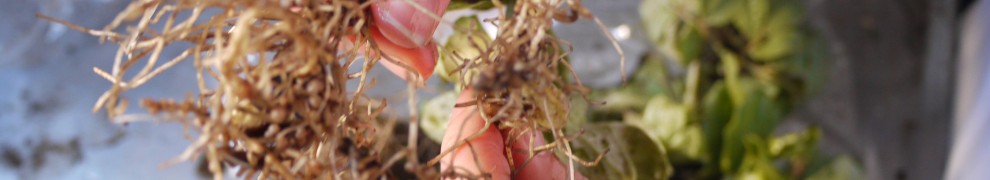
We started by gently rinsing the roots, using a bucket as a trap. Soaking loosened up large aggregates where many of the fine roots and much of the AMF reside.

Next we opened up the root mass with our fingers to reveal the nodules formed from Rhizobium infection.

We cut fine roots from each sample to use later in our search for AMF under the microscope in Part 2.
For each sample we recorded nodule width, range of widths, estimates of total nodule volume (mL) and total volume of roots(mL). See results.
Part 2:
We prepared the solutions and gathered the materials we needed to clean, stain and clear the fine root samples.
The clean roots were placed in individual 200 mL beakers and soaked with KOH solution. They were baked at 90 F for 1.5 hours. Once cooled KOH was drained (remained in the beaker) and the root samples were covered with the staining solution. Again the roots were baked for 1 hour. Once baked, the stain was recycled through a filter funnel and stored in a labelled bottle. Finally, we covered the samples with the destain solution and let stand while we cleaned up and prepared the microscopes.
The samples of fine roots were placed in a petri dish and examined for AMF under a dissecting microscope. Positive samples were clipped to sizes appropriate for slides. With needle and tweezers, roots were spread out on a slide. Destain was used as a mounting medium and the cover slip was pressed down to flatten the roots.
Dr. David MacArthur Guided us through multiple tutorials and revealed us the structures representative of AMF. Pictures of these structures were taken, and AMF counting allowed us to quantitatively determine the AMF presence.
Many samples reveal the presence of AMF, early stages of Rhizobium infection, as well as the presence many other microorganisms. The identification of microorganisms other than AMF is beyond the scope of this research.
Tentative counts of Mycorrhizae from the UBC farm and Orchard garden were performed and the results were recorded.