A comparison of 4 TELEs

I decided to summarize the 4 TELEs in table format. This is what I came up with:

Below is a link to a PDF in case the above does not read well.
Module B synthesis

Synthesis
All methods noted in the table are based on constructivist learning theories. All methods aim to achieve a deeper understanding of complex phenomena or concept in math and/or science. I found that the use of technology for presentation of information was the same in each TELE, however, the degree to which technology was used for other things differed. For example, in the Jasper series (Anchored instruction), technology use was limited to presentation of information, whereas in WISE (SKI), MyWorld (LfU), and Chemland (T-GEM), it was also used for data evaluation, visualization of ideas and collaboration. The TELEs also differed in the degree of guidance or scaffolding that was provided as well as the provider of that scaffolding. For Jasper, the purpose was for generative learning, and thus minimal scaffolding would be provided (though this can be adjusted dependent on the preference of the teacher and experience of the students). In contrast, with MyWorld, the teacher provides guidance at each step and also provides explanations of relationships that were uncovered by students during the knowledge construction observation stage.  In Jasper, MyWorld, and Chemland scaffolding was provided by the teacher. However in the WISE projects, much of the scaffolding was provided by technology in combination with the teacher particularly for those groups that were struggling.

Continue reading “A comparison of 4 TELEs”

T-GEM for cardiovascular physiology

A commonly cited conceptual challenge in medical education is cardiovascular physiology. This seems to be a consistent finding at different educational levels (Mikkila-Erdmann, 2012). If applying the T-GEM model (Khan, 2010)to teaching this, I would organize it as follows:

  1. I would provide some background content information, such as the definition of cardiac output, mean arterial pressure, heart rate, and systemic vascular resistance (total peripheral resistance)
  2. Have the students pair up and access the following simulator: https://ilearn.med.monash.edu.au/physiology/Cardiovascular/index.html 
    • on the Home tab, have the students look at the relationship between cardiac output, mean arterial pressure, and heart rate
    • have students generate a relationship between the above variables (by seeing what happens to these variables during different activities)
  3. Based on this relationship, have students predict what will happen to these variables with postural change
  4. Have students run the postural change simulation and evaluate the relationship
  5. Have students review their initial predictions and compare to the relationship that was observed through the simulation.
    • ask students to modify their initial predictions/relationships based on new data.


I think this method will offer students a chance to challenge their conceptual models by simulation and make modifications as needed. In trying to find software for TGEM in the medical context, I ran into a significant challenge. This is because a lot of medical simulations were for clinical skills or other technical skills improvement, and not for the purposes of understanding certain phenomena. But I do think this is a great way to learn and really understand concepts in a deeper more meaningful way (compared to the superficial rote memorization that is still common in medical education). I hope that simulations that examine concepts in my field become more readily available as time goes on.


References

Khan S. New Pedagogies on Teaching Science with Computer Simulations. J Sci Educ Technol. 2010;20(3):215-232. doi:10.1007/s10956-010-9247-2.


Mikkilä-Erdmann M, Södervik I, Vilppu H, Kääpä P, Olkinuora E. First-year medical students’ conceptual understanding of and resistance to conceptual change concerning the central cardiovascular system. Instr Sci. 2012;40(5):745-754. doi:10.1007/s11251-012-9212-y.

LfU – an application for pelvic anatomy?

According to Edelson (2001), LfU is based on 4 theories of learning, which are:
1) learning takes place through construction and modification of knowledge structures
2) knowledge construction is a goal-directed process, guided by a combination of conscious and unconscious understanding of goals
3) circumstances in which knowledge is constructed and subsequently used determine its accessibility for future use
4) knowledge must be constructed in a form that supports use before it is applied.

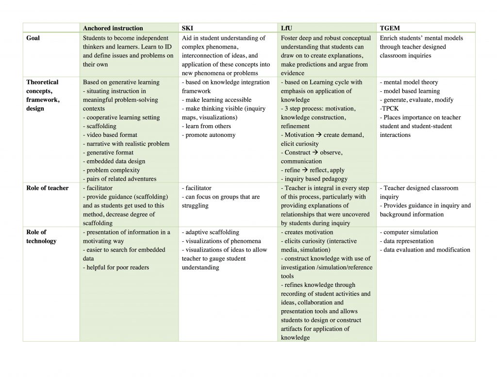
These principles underlie the LfU model, which is a three-step process involving
1) motivation
2) knowledge construction
3) knowledge refinement

In terms of technology, it plays many important roles within the LfU model. For example, it can play a role in eliciting curiosity (motivation), help students interactive with phenomena not possible in the real world which aids with knowledge construction and refinement.

So how would I apply it to a topic that I teach? The one thing that comes to mind is pelvic anatomy. Students tend to struggle with pelvic anatomy because it is quite complex and has multiple layers to it. I would start off by presenting students with a case of a patient who underwent surgery and shortly thereafter developed leg weakness and pain (this is actually one of the patient that presented to me early in my career). I would then have the students come up with theories based on their current knowledge level, regarding the cause of this pain/leg weakness. This is to draw their attention to their current level of knowledge and to have them recognize their limitations and thus increase motivation to learn. This would also create a context in their memory for integrating new knowledge (Edelson, 2001). Then I would use AnatomyTV, which is an interactive 3D anatomy resource (available through UBC at http://resources.library.ubc.ca/page.php?id=888) as my software of choice. This interactive resource that allows students to manipulate the body in 3D (select 3D Atlas —> Pelvis, then click female pelvis and perineum ~ tumble under 3D views from menu on the left to try it out!), which is really helpful, as most textbooks only present the learner with the upright position of the human body. This is not practical because in most gynecologic clinical practices, we examine patients and operate on them in the supine position, and thus knowledge of anatomy in this position is much more applicable. This program also allows the user to strip away all layers of the body and add them one by one, which allows students to understand how each layer relates to the other. I would also have student work in small group to promote interaction and discussion as this also aids in knowledge construction and refinement. Finally, I would have the students reflect on their initial theories, make any changes they feel are needed and present it as a group to the class and further apply their new knowledge to come up with a management plan for the patient. In this way, I believe I have applied the LfU principles to this topic.

References
1. Edelson DC. Learning‐for‐use: A framework for the design of technology‐supported inquiry activities. Journal of Research in Science Teaching. 2001;38(3):355-385. doi:10.1002/1098-2736

Spam prevention powered by Akismet