Category Archives: Daily Science

Science in your day-to-day life, not science published every day! News from recent published journal articles, and cool science facts and tricks!

Protein Showcase: Chaperon(ins) to the Rescue

The process of protein synthesis has probably been ingrained in your brain if you have taken any introductory biology or cell biology courses. It is so important that it is often referred to as the central dogma of molecular biology: DNA is transcribed to RNA, which is then translated to proteins.

Proteins essentially carry out the functions needed for the cells to remain alive. Need something made or broken down? Enzymes. Need something transported or moved? Carrier and intramembrane proteins. When it comes to proteins, shape equals function.

With their vast variety in function, the way a protein folds as it is made is closely monitored. Protein folding is faster than translation, thus the moment the N-terminal is exposed to the aqueous environment of the cytosol, the chain begins to fold due to intramolecular forces. As the chain continues to elongate, the protein can become kinetically trapped because the native state of a protein is only partially stable. Partially folded or misfolded states are problematic because they tend to aggregate due to their exposed hydrophobic surfaces (Hartl, F., et al. 2011).

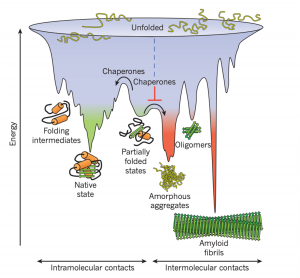
Figure 1: Competing reactions of protein folding and aggregation (Hartl, F., et al. 2011)

So, what to do with a misfolded protein? Chaperones and chaperonins are in charge or re-folding them. Also known as heat-shock proteins (hsp’s). These proteins seek and bind to exposed hydrophobic surfaces in newly translated proteins (Campbell, M., & Farrel, S. 2012).

Chaperones (Hsp70) bind and stabilize misfolded or partially folded proteins and prevent their aggregation as they are translated. ATP binding causes the chaperone to release the completed chain into the cytosol, allowing the protein to fold properly (Albert, B. 2015).

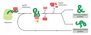
Figure 2: The hsp70 family of molecular chaperones (Albert, B. 2015)

Sometimes, even the chaperones are unsuccessful in helping the protein re-fold into its native state. In this case, additional chaperones or the more complex chaperonins (hsp60 and hsp10) provide additional help.

Chaperonins create a chamber for the proteins to re-fold post-translationally. First, the protein is captured though hydrophobic interactions with the entrance of the chamber (hsp60). The protein is then released into the interior of the chamber, which is lined with hydrophilic amino acids, and then it is sealed with a lid (hsp10). Here, the substrate can fold into its final conformation in isolation, where there are no other proteins that may aggregate. Finally, when ATP is hydrolyzed, the lid pops off, and the substrate protein is released from the chamber (Albert, B. 2015).

The best characterized chaperonin is the GroEL/GroES system in E. coli

Figure 3: Visualization of the GroEL/GroES chaperonins in E. coli (Rachel Davidovitz, 2014)

If the protein is still improperly folded, it is then targeted for degradation (ubiquitination, we’ll talk about it next time!)

References:

Albert, B, et al. (2015). Molecular Biology of the Cell (6th Edition). Garland Science, Taylor & Francis Group, LLC.

Campbell, M., & Farrel, S. (2012). Biochemistry (7th Edition). Brooks/Cole Cengage Learning.

Hartl, F. U., Bracher, A., & Hayer-Hartl, M. (2011). Molecular chaperones in protein folding and proteostasis. Nature, 475(7356), 324-332.doi:10.1038/nature10317

Rachel Davidovitz (2014, October 6). Active Cage Mechanism of Chaperonin-Assisted Protein Folding Demonstrated at Single-Molecule Level [Video]. https://www.youtube.com/watch?v=–NcNeLc1mo&ab_channel=RachelDavidowitz

Hydrogels as an Ophthalmic Drug Delivery System

If you’ve ever had some sort of eye surgery, you might have been prescribed an assortment of eyedrops with their own frequent dosing schedules. The reason for the frequent dosing is that topical drug delivery to the eye is quite difficult, and upon further evaluation this should make sense! The eye is a delicate structure that has many barriers to prevent the entry of foreign particles. Hydrogel technology has been heavily investigated as a sustained drug release vehicle, obviating the need for such frequent dosing.

barriers of the eye

Drug residence time delivered via eye drops can be cut short due to pre-corneal factors: tears, blinking, and drainage through the nasolacrimal duct. Some of you might have been told to pinch the bridge of your nose after administering eye drops, this is to block off the drug from draining through this duct into the nose. In addition, drug that does remain on the eye must penetrate through the thick multi-layered cornea to get to the deeper tissues of the eye. The conjunctiva at the surface of the eye is also highly vascularized, meaning the drug will also be absorbed into the bloodstream before penetrating through the eye.

Anatomy of the eye. Credits: American Academy of Ophthalmology

Drainage through the nasolacrimal duct and absorption through the highly vascularized conjunctiva may cause unwanted side effects, as the drug is being distributed to off-target tissues through the circulatory system. Ironically, drugs cannot be delivered via consumption or systemic injection as there are blood-ocular barriers (analogous to the blood-brain barrier) that prevent systemic distribution of drugs to ocular tissue.

hydrogels to the rescue!

Hydrogels are basically polymers (long chemical chains) composed 95% of water. The advantage of hydrogels is that they are viscous, meaning that they can stick onto the eye longer before being removed. They can also encapsulate drug molecules, and can release the desired drug at a certain rate, based on their initial preparation conditions. Currently, there are many types of hydrogels being investigated for there use in drug delivery. These can be broken down into synthetic polymers, which have the advantage of being easily tunable in mechanical properties and natural polymers, which have the advantage of being biocompatible to the eye. In-situ forming hydrogels have been an area of focus, as they can be administered as a liquid, but gels in response to a stimulus. For example a gel could be liquid at room temperature but turn into a gel at body temperature.

Despite being a potential solve to a long-standing problem, there is currently no FDA-approved hydrogel used in drug delivery. A lot more research in terms of in vitro, in vivo, and clinical studies are needed to evaluate the long-term efficacy and biocompatibility of these options. However, hydrogels have made their way into clinical use in other ophthalmic departments! An immediate one that comes to mind is the use of contact lenses which are practically just hydrogels. A lesser known use is in cataract surgery, where hydrogels have been used as ocular adhesives to seal any surgery-induced wounds.

References

Lynch, C.R.; Kondiah, P.P.D.; Choonara, Y.E.; du Toit, L.C.; Ally, N.; Pillay, V. Hydrogel Biomaterials for Application in Ocular Drug Delivery. Front. Bioeng. Biotechnol. 2020, 8, 228, doi:10.3389/fbioe.2020.00228.

Embarrassed of Asian Glow? Don’t Worry, The Future is Promising

Ever find yourself beet red after one small drink? You’re not alone! Over one-third of East Asians and eight percent of the world population experience this awkward phenomenon; however, a solution is in the works. Just last month, researchers from Weill Cornell Medical College have solved this problem by experimenting with targeted gene therapy on mice.

What does asian glow look like? A before and after comparison. (Credits: Wikimedia Commons)

The Dangers of Asian Glow

Apart from causing embarrassment, asian glow comes with more serious consequences than just flushing red. The red glow is caused by a deficiency in the ALDH2 enzyme, a key component in detoxifying alcohol. When you drink alcohol, your body converts the alcohol into acetaldehyde. Normally, acetaldehyde is further converted to a safer compound by ALDH2; however in individuals with asian glow, ALDH2 does not function, causing acetaldehyde to build up. Since acetaldehyde is a cancer-causing agent, its accumulation drastically increases the risk of developing esophageal cancer by six to ten folds.

Conversion of alcohol to acetate is stopped in people with asian glow. This leads to toxic buildup of acetaldehyde. (Credits: Me – created with Notability)

A Glowing Solution…

Matsumura’s team reasoned if a lack of ALDH2 enzyme was the problem, maybe they could simply add it back in.

“We hypothesized that a one-time administration of a […] virus […] expressing the human ALDH2 coding sequence […] would correct the deficiency”

They tested their idea on three strains of mice: mice with functional ALDH2, mice lacking ALDH2, and mice with a non-functional version of ALDH2. The latter two simulated the asian flush syndrome seen in humans. After introducing all the mice with the ALDH2 gene and feeding them alcohol, the researchers carefully monitored acetaldehyde levels in the blood.

Their hard-work paid off! In the two strains deficient for ALDH2 function, acetaldehyde levels and abnormal behavior associated with alcohol consumption were back to near-normal levels. Furthermore, they found that one dose was enough to confer persistent and long-term protection.

From Mice to Humans: A Complicated Decision

Matsumura’s team emphasize that a long-lasting treatment for ALDH2 deficiency currently does not exist. Although making the jump from mice to humans will be challenging, they assure that virus-mediated gene therapy shows the most promise in becoming an effective therapy. The million-dollar question is whether the risks of the glow outweigh the benefits of reduced alcohol consumption seen in affected individuals. To this Matsumura’s team say:

“the overall burden […] on human health, particularly […] cancer, supports […] gene therapy.”

What do you think?

The COSMIC Bubble Helmet: A Revolutionary Ventilation System

As the COVID-19 virus evolves into different strains so to has the methods of dealing with the consequences of this virus. In patients with severe COVID-19, they are plagued with acute respiratory distress disorder (ARDS). This is a condition where fluid fills the lungs and prevents air from reaching the necessary gas exchange compartments, similar to what occurs in drowning. These patients are subjected to invasive mechanical ventilation, where a tube is uncomfortably shoved down the patients throat to facilitate breathing.

a potential alternative?

To avoid ventilator-induced lung injury, non-invasive ventilation methods have shown to be well tolerated by patients. Instead of a tube, a tight-fitting nasal or facial mask is used and uses positive pressure to facilitate ventilation. In the context of COVID-19, the use of current non-invasive ventilation methods is not recommended due to virus aerosolization risks which puts healthcare staff in danger of contracting the virus.

Example of a non-invasive ventilation face mask.

A cunning solution

COSMIC Medical, a Vancouver-based organization has designed a “Bubble Helmet” that remains noninvasive but also reduces the aerosolization risks mentioned previously! Additionally, the materials needed to make the helmet are low cost and can be easily be retooled or adjusted by manufacturers due to its simplistic design.

Owing to its flexible design, patients freedom of mobility will not be hindered as they can lie face up or down comfortably. Furthermore, the helmet is made of transparent material, thus a patient’s vision will be unobstructed, further enhancing comfortability.

the next steps

There are still many hoops to jump through before this helmet can be offered to patients. These steps include obtaining regulatory approval and completing clinical trials. So far results have been obtained in an experimental setting, thus the effectiveness of the helmet must be measured in clinical settings as well. Currently the Bubble Helmet design is open source and available to all to peruse. Check out the open source paper!

Stress and Grey Hair: An Answer to a Biological Mystery

Everyone has heard that too much stress will cause grey hair. This is easily seen in former president of the United States, Barack Obama, whose hair could not escape the stress of the Oval Office! But what exactly links grey hair and stress? This year, researchers at Harvard University found that the nervous system eliminates pigment-regenerating stem cells responsible for coloring our hair!

Barack Obama’s hair color at the start of his presidency versus seven years after. Credits: DailyMail.com

THE ROOT OF THE PROBLEM

When you are stressed, your body responds in three distinct ways: the activation of your immune system, the activation of your sympathetic nervous system (SNS), and the release of cortisol, an energy-stimulating hormone. All these responses put your body into a “fight or flight” mode; increasing heart rate and blood pressure. The challenge for Zhang’s team was to sort through these three responses and determine which caused grey hair.

Zhang’s team tackled this problem by performing a series of experiments on black-furred mice. They first tested if immune system activation was the cause by seeing if the fur greyed under stress, even when the immune system was deactivated. They indeed found that stressed immune-deficient mice still greyed, indicating that stress causes greying, independent of an immune response.

They also ran similar experiments using mice mutated to not respond to cortisol or noradrenaline, a molecule involved in SNS activation. The idea being that if  a response was involved, stress should not cause the fur to grey if it was removed. In mice lacking response to cortisol, the fur still greyed; however, in mice lacking the response to noradrenaline, their fur remained black! This indicated that the SNS was the main driver in hair greying.

Figure 1. The results of the experiments described above are shown. Note that mice unable to respond to SNS activation do not grey under stress. “Control” refers to unmutated mice. Also note that a different type of control (non-stressed vs stressed) was ran in the immune-deficient case. (Sample size = 6 for each condition, standard error bars). Credits: Adapted from Zhang et al.’s data.

ZOOMING IN FURTHER…

With the culprit in hand, Zhang’s team didn’t just stop there! Through further experimentation, they illustrated that the SNS over-stimulates MeSC, the stem cells involved with hair pigmentation. During hair growth, these MeSC cells transform into pigment-producing cells and color the hair. Under stress, the SNS causes these MeSC cells to transform at an abnormally high rate, quickly depleting these cells and leading to grey hair.

THE REASON BEHIND THIS LINK?

In truth, the reason why this MeSC and SNS interaction exists is unclear. Zhang’s team suggests an evolutionary perspective. Since octopuses, a distant relative to mice and humans, can modify pigmentation of their skin using the SNS, they hypothesize that this interaction was simply conserved. Whatever the reasons may be, this just further shows that the mystery has yet to be completely solved!

Sugar Chemistry: A Pathway to Antibiotics

We’ve all heard it endlessly as kids. Don’t eat too much sugar, it’s bad for you. However, what if I told you that sugars aren’t all that bad and in fact, careful changes to its chemistry can lead to life-saving drugs, such as antibiotics! Just last year, researchers at the University of British Columbia, led by Stephen Withers, found a unique way to tinker with sugars’ chemical structures by using molecules in bacteria. The same bacteria found in our poop!

A view of E. Coli, the bacteria that was used by Wither’s and his team. Credits: The Philadelphia Inquirer

SWEET…BUT WHAT ARE THEY?

Before we go further, let’s start with a simple question: What exactly are sugars? Sugars are molecules shaped like hexagons which are often joined to other molecules known as “acceptors”. In a way we are kind of like sugars; we find someone we like, confess how we feel, and they accept our love! Right? Wrong. As we all know the last part rarely happens and this is the same in sugars, as chemists have yet to find easy ways to join the sugar and acceptors. Luckily for sugars (and unluckily for us), Mother Nature has come up with some solutions, using helper molecules known as enzymes.

Curious as to what type of sugars chemists work with? There’s no ambiguity here, chemists use the same molecules found in sugar cubes. Yes! The ones you put in your coffee. Credits: The Verge

A SOLUTION UNDER OUR NOSES…

Instead of struggling to find ways of joining sugars and acceptors, Wither’s team thought: Why not just use these enzymes? In other words, hijack Mother Nature. To make their idea a reality, they extracted sugar-specific enzymes from E. Coli, a bacterium that lives inside the human digestive tract. Their efforts gave them 175 sugar-specific enzymes, and from this they chose 8 enzymes that were most specific to the type of sugars and acceptors they were interested in.

“With the 8 enzymes in hand, Withers and his team could now easily make these sugar-acceptor linkages” is what I would like to report; however, things are never so simple. It turns out that the sugar-specific enzymes they got from E. Coli did the exact opposite of what they wanted. Instead of forming sugar-acceptor linkages, they were specialized in breaking them.

Unsurprisingly the savvy researchers expected this and already had a reliable strategy to reverse-engineer these enzymes from linkage breakers to linkage makers. You may be wondering how they re-purposed something to work completely opposite of what it was intended for. To reconcile this, think of this example: hammers. If you’re feeling angry one day you would likely use the hammer to smash things. However, if you’re feeling innovative one day, the hammer would help you build things by hammering in nails. These enzymes are similar; an enzyme that breaks sugar bonds differs very little from one that builds sugar bonds.

MORE THAN JUST A BOND…

Sugars go way beyond than just satisfying your sugar fix. They are molecules essential to the maintenance and regulation of not only your body, but in most living things! Because they are found everywhere, including infectious bacteria, sugar-based molecules serve as effective antibiotics, however making these drugs are difficult. Why? Well as mentioned before, chemists have trouble making these sugar-acceptor bonds; however, the research done by Wither’s team show that this will not remain the case. On a lighter note, they also created a sugar-based molecule that had nothing to do with health; detergent. This just further shows that these bonds are far-reaching and relevant in many contexts.

Story source

Armstrong, Z.; Liu, F.; Chen, H.-M.; Hallam, S. J.; Withers, S. G. Systematic Screening of Synthetic Gene-Encoded Enzymes for Synthesis of Modified Glycosides. ACS Catalysis 20199 (4), 3219–3227.

Remdesivir Authorized for Treatment of Severe COVID-19 Symptoms

There may be hope for people suffering severely from COVID-19. On July 28 2020, Health Canada has approved the use of Remdesivir for critically ill COVID-19 patients.

how does it work?

Remdesivir is an antiviral drug that acts as an inhibitor. Basically, the COVID-19 virus uses a protein complex called RdRp to replicate its genetic material and further infect the body. Since Remdesivir inhibits RdRp, the virus can no longer replicate and the infection is impeded.

proof that it works

A double-blind, randomized, placebo-controlled trial was conducted on 1063 patients suffering from COVID-19. The results of this trial showed that those who took the drug recovered 4 days faster than those who didn’t. They also found that lung infection in treated patients were significantly better than those who didn’t get the treatment.

who can use this drug

This drug isn’t for every COVID-19 case. You need to present severe symptoms such as pneumonia and require extra oxygen to help breathe (respiratory machine). The safety and effectiveness of the drug also needs to be further evaluated. To this end, Health Canada has authorized two clinical trials to gather more data.

Plants: Making Air Easier to Breathe

We’ve all heard on the news or learned in elementary schools about the mass deforestation going on all over the world. But have you ever stopped to wonder, why exactly is this bad? Why do we need plants and trees at all?

It turns out, plants are efficient in resupplying the air with oxygen, while removing carbon dioxide! The former, being essential to our survival, and the latter being a gas involved in global warming.

an inside look into plants

So how exactly do they do this? In turns out that inside the plants’ cells there are special machinery capable of splitting water (H2O). This machinery is called an electron transport chain (ETC).

Using sunlight, the ETC extracts energy from the water – leading to the generation of oxygen as a “waste” product. Ironically what’s considered waste for them is gold in terms of survival for us.

When it comes to removing carbon dioxide they have another set of machinery. For the plant, carbon dioxide is like food: they trap the carbon dioxide and convert them into carbohydrates and other nutrients.

rubisco – the single bad life-essential solution

A key piece of machinery in this conversion is RuBisCo – an enzyme. An enzyme is a molecule that speeds up biochemical reactions, and surprisingly RuBisCo is one of the least efficient in existence (think of RuBisCo as a bike and other enzymes as the newest Tesla).

So you might be thinking, if RuBisCo is such a bad enzyme, can’t scientists just make a better version of RuBisCo? This would increase crop yields, and be good for the environment! Well, scientists have tried and failed … it seems like this is the only bad solution to a complex problem. Along with the ability to split water at ease (which scientists also can’t do), this is why plants are biochemical miracles.

Giving COVID-19 What It Wants: A Potential Cure

COVID-19 needs no introduction, the familiar spiky ball has been tormenting us since the beginning of 2020. Consequently, researchers around the world have been working to find a vaccine and one potential solution seems rather odd. UBC researchers, led by Josef Penninger, have found that administering ACE2 decreases the virus’ infectiousness. The odd part? ACE2 is the same protein on lung cells exploited by COVID-19 to gain entry into these cells.

COVID-19 structure. Credits: Newscientist

COVID-19 structure. The red blobs coating the virus are Spike Glycoproteins, which facilitate infection of cells. Credits: Newscientist

Infecting the lung cells…

One of main targets of COVID-19 is the lungs. This is because the surface of the lung cells are coated in ACE2 proteins. On the surface of COVID-19 there are Spike Glycoproteins, which recognize and bind ACE2 proteins, facilitating infection of the lung cells (see our previous post for general information on COVID-19). Tinkering with this ACE2 – Spike Glycoprotein interaction is the goal of many developing vaccines and was also what Penninger’s team targeted.

satisfying the virus stops the infection!

The way Penninger’s team approached this problem was truly ingenious. Since the Spike Glycoprotein binds to ACE2 on the cells, why not just administer an outside source of ACE2, so the Spike Glycoprotein can bind to those instead? The administered ACE2 would effectively bind to all the Spike Glycoproteins on the virus, rendering it inactive and unable to target cellular ACE2.

They researchers tested this theory by infecting cell cultures with COVID-19. They showed that by incubating these cultures with hrsACE2 (genetically modified ACE2), the virus growth was inhibited.

To take it a step further, the researchers grew blood vessel and kidney organoids, which are models of these respective organs. Upon administering hrsACE2, infection and spread of COVID-19 in these organoids were significantly reduced. This demonstrated that hrsACE2 could inhibit infection in human organs!

Spike Glycoproteins on COVID-19 will bind to hrsACE2 instead of cellular ACE2 – inhibiting infection. Adapted: Penninger et al. (2020)

More work is still needed

Although the results are promising, Penninger’s team caution that there are still some kinks that need to be worked out:

The inhibition [by hrsACE2] is not complete […]. This may be due to […] other co-receptors/auxiliary proteins or even other mechanisms by which viruses can enter cells.

They also suggest that future studies should look at the systems that model the lung, as this organ is the primary infection target. With all this being said, Penninger’s research is still without doubt groundbreaking, and a big push forward into getting rid of this virus once and for all.

Journal Reference

Monteil, V., Kwon, H., Prado, P., Hagelkrüys, A., Wimmer, R. A., Stahl, M., . . . Penninger, J. M. (2020). Inhibition of SARS-CoV-2 infections in engineered human tissues using clinical-grade soluble human ACE2. Cell, 181(4), 905-913.e7. doi:10.1016/j.cell.2020.04.004

Everything You Need to Know About COVID-19

Who would have thought that the mere 27 cases of pneumonia in Wuhan, China would end up throwing the world into turmoil? On March 11, 2020, COVID- 19 was declared a pandemic by the World Health Organization, and since then, researchers worldwide have been trying to develop a vaccine. As of now, researchers have identified some parts of how the virus works, how it’s transmitted, and ways we can slow down its spread.

how did covid-19 arise?

COVID-19 is similar to most animal viruses in that it is an infectious genetic element packaged in a lipid membrane with associated membrane proteins. Upon sequencing of this element and comparisons to different coronavirus strains, researchers have determined the virus to originate from bats.

However, these comparisons also showed that humans did not contract the virus directly from bats. Instead, it is likely that the virus was transmitted through an intermediate host, pangolins.

What are pangolins? Credits: Shutterstock

Continue reading