Tag Archives: biochemistry

Protein Showcase: Chaperon(ins) to the Rescue

The process of protein synthesis has probably been ingrained in your brain if you have taken any introductory biology or cell biology courses. It is so important that it is often referred to as the central dogma of molecular biology: DNA is transcribed to RNA, which is then translated to proteins.

Proteins essentially carry out the functions needed for the cells to remain alive. Need something made or broken down? Enzymes. Need something transported or moved? Carrier and intramembrane proteins. When it comes to proteins, shape equals function.

With their vast variety in function, the way a protein folds as it is made is closely monitored. Protein folding is faster than translation, thus the moment the N-terminal is exposed to the aqueous environment of the cytosol, the chain begins to fold due to intramolecular forces. As the chain continues to elongate, the protein can become kinetically trapped because the native state of a protein is only partially stable. Partially folded or misfolded states are problematic because they tend to aggregate due to their exposed hydrophobic surfaces (Hartl, F., et al. 2011).
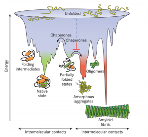
Figure 1: Competing reactions of protein folding and aggregation (Hartl, F., et al. 2011)

So, what to do with a misfolded protein? Chaperones and chaperonins are in charge or re-folding them. Also known as heat-shock proteins (hsp’s). These proteins seek and bind to exposed hydrophobic surfaces in newly translated proteins (Campbell, M., & Farrel, S. 2012).

Chaperones (Hsp70) bind and stabilize misfolded or partially folded proteins and prevent their aggregation as they are translated. ATP binding causes the chaperone to release the completed chain into the cytosol, allowing the protein to fold properly (Albert, B. 2015).
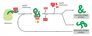
Figure 2: The hsp70 family of molecular chaperones (Albert, B. 2015)

Sometimes, even the chaperones are unsuccessful in helping the protein re-fold into its native state. In this case, additional chaperones or the more complex chaperonins (hsp60 and hsp10) provide additional help.

Chaperonins create a chamber for the proteins to re-fold post-translationally. First, the protein is captured though hydrophobic interactions with the entrance of the chamber (hsp60). The protein is then released into the interior of the chamber, which is lined with hydrophilic amino acids, and then it is sealed with a lid (hsp10). Here, the substrate can fold into its final conformation in isolation, where there are no other proteins that may aggregate. Finally, when ATP is hydrolyzed, the lid pops off, and the substrate protein is released from the chamber (Albert, B. 2015).

The best characterized chaperonin is the GroEL/GroES system in E. coli
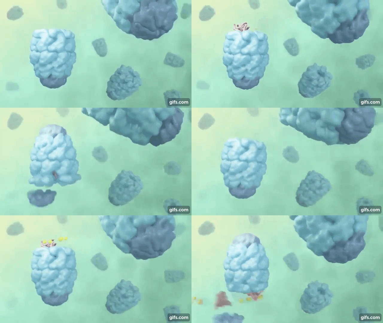
Figure 3: Visualization of the GroEL/GroES chaperonins in E. coli (Rachel Davidovitz, 2014)

If the protein is still improperly folded, it is then targeted for degradation (ubiquitination, we’ll talk about it next time!)

References:

Albert, B, et al. (2015). Molecular Biology of the Cell (6th Edition). Garland Science, Taylor & Francis Group, LLC.

Campbell, M., & Farrel, S. (2012). Biochemistry (7th Edition). Brooks/Cole Cengage Learning.

Hartl, F. U., Bracher, A., & Hayer-Hartl, M. (2011). Molecular chaperones in protein folding and proteostasis. Nature, 475(7356), 324-332.doi:10.1038/nature10317

Rachel Davidovitz (2014, October 6). Active Cage Mechanism of Chaperonin-Assisted Protein Folding Demonstrated at Single-Molecule Level [Video]. https://www.youtube.com/watch?v=–NcNeLc1mo&ab_channel=RachelDavidowitz

Arachidonic acid: a very important fatty acid

Arachidonic acid is a fatty acid (a carboxylic acid with a long carbon tail) with 20 carbons and 4 double bonds. Counting from the end without the carboxylic acid group, the first double bond appears at the 6th position from the end, making this an omega-6 fatty acid.

Chemical structure of arachidonic acid. Credits: Wikimedia (Public domain) https://commons.wikimedia.org/wiki/File:AAnumbering.png

Arachidonic acid is incorporated into phospholipids in cell membranes. In the process of some cell signalling events such as the inflammatory cascade, it is cleaved from phospholipids by phospholipase A2 (PLA2), after which it can be modified into many signalling molecules including the prostaglandins (PGs), thromboxanes (TXs), and leukotrienes (LTs). These are the most well-known and well-studied of the metabolites derived from arachidonic acid; however, there are also many other compounds including the endocannabinoids (ECs), and several less understood groups such as the eoxins (EXs)lipoxins (LXs), epoxyeicosatrienoic acids (EETs), hepoxilins (HXs), isoprostanes (IsoPs), and isofurans (IsoFs). Some of these compounds are quite recent discoveries and thus have little information available about them. Nevertheless, many of these compounds have biological activity associated with the inflammatory response, either with anti-inflammatory or pro-inflammatory effects. The latter molecules are still under active investigation in order to better understand the way they mediate actions in the body. [1]

The actions of all these molecules are too numerous to explain here. Some of the most well-known actions are those of the prostaglandins. If you have ever taken NSAIDs such as ibuprofen or naproxen, you will have affected this system. These drugs inhibit the cyclooxygenase enzyme, a critical enzyme in prostaglandin synthesis. Prostaglandins have diverse effects such as acting as pro-inflammatory mediators and regulating smooth muscle contraction and relaxation (such as PGE2), or causing vasodilation and preventing platelet aggregation (such as PGI2, also known as prostacyclin). Other molecules in the cyclooxygenase pathway, such as thromboxane, promote clotting and causing blood vessels to contract (such as TXA2). [2] The leukotrienes, synthesized from the lipooxygenase pathway, are inflammatory mediators. Some asthma medications, such as montelukast and zafirlukast, block the actions of the leukotriene LTD4 which can help with the bronchospasms in asthma. [3]

The endocannabinoid system is also a growing area of interest. The compound N-arachidonoylethanolamine, also known as anandamide, is probably the most well-known compound. The effects of this compound include pain sensation modulation, reward processes in the brain, and immune system modulation. [4]

This is only a small peek at the world of arachidonic acid metabolites: there remains much to be explained outside of this blog post, and much to be discovered!

Sources:

[1] Wang, B., Wu, L., Chen, J., Dong, L., Chen, C., Wen, Z., Hu, J., Fleming, I., & Wang, D. W. (2021). Metabolism pathways of arachidonic acids: Mechanisms and potential therapeutic targets. Signal Transduction and Targeted Therapy, 6(1), 1–30. https://doi.org/10.1038/s41392-020-00443-w
[2] Ricciotti, E., & FitzGerald, G. A. (2011). Prostaglandins and inflammation. Arteriosclerosis, Thrombosis, and Vascular Biology, 31(5), 986–1000. https://doi.org/10.1161/ATVBAHA.110.207449
[3] Dempsey, O. J. (2000). Leukotriene receptor antagonist therapy. Postgraduate Medical Journal, 76(902), 767–773. https://doi.org/10.1136/pmj.76.902.767
[4] Lu, H.-C., & Mackie, K. (2016). An Introduction to the Endogenous Cannabinoid System. Biological Psychiatry, 79(7), 516–525. https://doi.org/10.1016/j.biopsych.2015.07.028

BIOC 302: General Biochemistry

An interesting course where you dive into the biochemical pathways of lipids, proteins, and nucleic acids. BIOC 302 is a biochemistry course where you are assaulted with biochemical facts and information.

FORMAT OF THE COURSE

The lectures were live at 8AM…fortunately they were asynchronous and recorded due to the pandemic. Lectures were every MWF. There were also optional tutorials where the teaching assistants would go over a set of practice problems. The course was broken down into 3 broad sections. Biochemical processes and metabolic pathways of lipids, proteins, and nucleic acids and overview of DNA replication, transcription and translation. This course is super memory-heavy as you had to memorize most of the structures and names that appear in the slides as well as the complex metabolic pathways. What makes it even more difficult is that you also have to apply this memorized knowledge in different scenarios on the exams.

The assessments for this course consisted of one midterm and one final exam of equal weighting. The midterm tested the first half of the course only (lipids and proteins) while the final exam tested the latter half of the course only (nucleic acids, DNA replication, transcription, and translation). Both exams consisted of matching questions, structure recognition, multiple choice, and long answer questions adding up to 100 points.

GPA 🙂 OR 🙁

This course is very hard and you will need to put a lot of work in to be above the class average. It is imperative that you do not fall behind because then you will need to memorize more content, while trying to decipher what you are memorizing. The midterm exam was by far the hardest exam I’ve written this year (my 4th year); however, the final exam was much easier. The instructor is not shy about scaling, and usually scales the exams by 2-4% to reach an average above 70. The class average for my section was 74.

BIOC 302 Grade Distribtution. Credits: ubcgrades.com

verdict? to take or not to take

Despite being a very hard course, I would definitely take a course if you have a strong interest in biochemistry. This course is also a prerequisite for some professional schools in healthcare like dentistry. The assessments can be tough in terms of ambiguous questions in the exams, but if you’re not too worried about that then you’re good to go!

More steroids, plants, fungi

Steroids are not only relegated to the animal world; fungi and plants synthesize many steroids as well. One particular example of clinical relevance is ergosterol, found in the cell membranes of fungi where it serves a similar role to cholesterol in animal cell membranes. This can be exploited by antifungal medications: azole drugs such as clotrimazole and miconazole function by inhibiting ergosterol synthesis. Specifically, they inhibit the 14α-demethylase enzyme that converts lanosterol to ergosterol (note the similarities to the cholesterol pathway discussed in this previous post). [1]

Ergosterol can also be converted to ergocalciferol in a UV-light dependent reaction, similarly to the synthesis of Vitamin D3 in animals. In fact, ergocalciferol is also known as Vitamin D2, and like cholecalciferol, ergocalciferol can be hydroxylated twice to 1,25-dihydroxyergocalciferol or ercalcitriol, which binds to the Vitamin D receptor and causes its effects, although the binding of Vitamin D2 may not be as strong. [2]

There are diverse steroids made by plants, some of which have toxic effects. Of note are digoxin and digitoxin produced by the foxglove plant. These two chemicals consisted of a carbohydrate chain attached to a modified steroid, and they can be fatal if ingested. They inhibit the Na+/K+ ATPase responsible for establishing the electrochemical gradient within the cell, which is exploited for the use of digoxin as a drug for arrhythmias and heart failure due to the ability of the medication to increase the contractility of the heart when given at low doses. [3]

These are only some of the steroids occurring in plants and fungi. In the future, maybe more will be discovered with important biological activities!

Sources:
[1] Herrick, E. J., & Hashmi, M. F. (2021). Antifungal Ergosterol Synthesis Inhibitors. In StatPearls. StatPearls Publishing. http://www.ncbi.nlm.nih.gov/books/NBK551581/
[2] Houghton, L. A., & Vieth, R. (2006). The case against ergocalciferol (vitamin D2) as a vitamin supplement. The American Journal of Clinical Nutrition, 84(4), 694–697. https://doi.org/10.1093/ajcn/84.4.694
[3] Hauptman, P. J., & Kelly, R. A. (1999). Digitalis. Circulation, 99(9), 1265–1270. https://doi.org/10.1161/01.cir.99.9.1265

Steroids, salt, sugar, sex

Steroids are biologically active compounds composed of four fused rings. Although the word “steroid” is commonly associated with anabolic steroids and muscle growth, steroids are in fact a diverse group of compounds with varying effects on the human body.

The steroid cholesterol can be either synthesized via the mevalonate pathway or are obtained from the diet. The mevalonate pathway starts with acetyl-CoA, which is converted in a series of steps to isopentenyl pyrophosphate (IPP) and dimethylallyl pyrophosphate (DMAPP), which are the building blocks of isoprenoids, a diverse group of compounds that include steroids. The enzyme HMG-CoA reductase, which catalyzes the step converting HMG-CoA to mevalonate, is blocked by statins which are used for the treatment of high cholesterol levels. The IPP units are combined to form farnesyl pyrophosphate, which are then used to form squalene. From there, the squalene is cyclized to form lanosterol, which is then converted to cholesterol. Cholesterol is important for moderating cell membrane fluidity, and also participates in the formation of lipid rafts which are theorized to be involved in cell signalling. [1]

Cholesterol can then be converted into a variety of signalling molecules such as neurosteroids, vitamin D, glucocorticoids, mineralocorticoids, and sex steroids. Neurosteroids modulate complex activities in the brain, such as neural plasticity. They can act in an excitatory manner (such as dehydroepiandrosterone (DHEA), which modulates NMDA receptor activity) or inhibitory manner (such as pregnanolone, which modulates GABA A receptor activity). [2]

Vitamin D is involved in calcium homeostasis, increasing calcium absorption in the intestines and modulating bone remodulating. It is synthesized from cholesterol, including a step that involves UV radiation. It is then hydroxylated twice in order to be in the active form, 1,25-dihydroxycholecalciferol, also known as calcitriol, which binds to the vitamin D receptor to produce its effects. [3]

Glucocorticoids such as cortisol modulate metabolism and immune function. Cortisol promotes gluconeogenesis, which produces glucose, as well as promoting the breakdown of lipids and proteins. It also diminishes immune function by inhibiting the effects of various cytokines that promote inflammation and immune responses. [4]

Mineralocorticoids such as aldosterone helps to maintain blood pressure and electrolyte balance. Aldosterone acts in the kidneys to increase sodium reabsorption and potassium excretion, thus increasing sodium levels and decreasing potassium levels in the blood. Because of the sodium reabsorption, water is then retained, increasing blood volume and thus increasing blood pressure. Glucocorticoids and mineralocorticoids are both synthesized from cholesterol via progestogens in the adrenal cortex by 21-hydroxylase and 11β-hydroxylase. [5]

Sex steroids are classified as progestogens (such as progesterone), androgens (such as testosterone), or estrogens (such as estradiol). Estrogens are synthesized from androgens by the enzyme aromatase, while androgens are synthesized from progestogens by 17α-hydroxylase. Progestogens are synthesized by the conversion of cholesterol by cholesterol side-chain cleavage enzyme. Sex steroids regulate a variety of activities. Progesterone is important in the secretory phase of the uterus during the menstrual cycle, where it is produced by the corpus luteum to maintain the endometrial lining for implantation. Testosterone is important for sperm development, as well as increasing muscle growth and contributing to male secondary sex characteristics. Estradiol is responsible for inducing ovulation, bone maintenance, and female secondary sex characteristics. However, all sex steroids have diverse roles in people of all genders that are not described here. [6][7][8]

Steroids are a diverse group of compounds, and this is only the beginning. You can read about more steroids here!

Sources:
[1] Russell, D. W. (1992). Cholesterol biosynthesis and metabolism. Cardiovascular Drugs and Therapy, 6(2), 103–110. https://doi.org/10.1007/BF00054556
[2] Robel, P., & Baulieu, E. E. (1995). Neurosteroids: Biosynthesis and function. Critical Reviews in Neurobiology, 9(4), 383–394.
[3] Bikle, D. (2000). Vitamin D: Production, Metabolism, and Mechanisms of Action. In K. R. Feingold, B. Anawalt, A. Boyce, G. Chrousos, W. W. de Herder, K. Dhatariya, K. Dungan, A. Grossman, J. M. Hershman, J. Hofland, S. Kalra, G. Kaltsas, C. Koch, P. Kopp, M. Korbonits, C. S. Kovacs, W. Kuohung, B. Laferrère, E. A. McGee, … D. P. Wilson (Eds.), Endotext. MDText.com, Inc. http://www.ncbi.nlm.nih.gov/books/NBK278935/
[3] Arlt, W., & Stewart, P. M. (2005). Adrenal corticosteroid biosynthesis, metabolism, and action. Endocrinology and Metabolism Clinics of North America, 34(2), 293–313, viii. https://doi.org/10.1016/j.ecl.2005.01.002
[5] Connell, J. M., Fraser, R., & Davies, E. (2001). Disorders of mineralocorticoid synthesis. Best Practice & Research. Clinical Endocrinology & Metabolism, 15(1), 43–60. https://doi.org/10.1053/beem.2000.0118
[6] Aizawa, K., Iemitsu, M., Maeda, S., Jesmin, S., Otsuki, T., Mowa, C. N., Miyauchi, T., & Mesaki, N. (2007). Expression of steroidogenic enzymes and synthesis of sex steroid hormones from DHEA in skeletal muscle of rats. American Journal of Physiology. Endocrinology and Metabolism, 292(2), E577-584. https://doi.org/10.1152/ajpendo.00367.2006
[7] Penning, T. M. (2010). New Frontiers in Androgen Biosynthesis and Metabolism. Current Opinion in Endocrinology, Diabetes, and Obesity, 17(3), 233–239. https://doi.org/10.1097/MED.0b013e3283381a31
[8] Cui, J., Shen, Y., & Li, R. (2013). Estrogen synthesis and signaling pathways during ageing: From periphery to brain. Trends in Molecular Medicine, 19(3), 197–209. https://doi.org/10.1016/j.molmed.2012.12.007