Tag Archives: #biology

Protein Showcase: Chaperon(ins) to the Rescue

The process of protein synthesis has probably been ingrained in your brain if you have taken any introductory biology or cell biology courses. It is so important that it is often referred to as the central dogma of molecular biology: DNA is transcribed to RNA, which is then translated to proteins.

Proteins essentially carry out the functions needed for the cells to remain alive. Need something made or broken down? Enzymes. Need something transported or moved? Carrier and intramembrane proteins. When it comes to proteins, shape equals function.

With their vast variety in function, the way a protein folds as it is made is closely monitored. Protein folding is faster than translation, thus the moment the N-terminal is exposed to the aqueous environment of the cytosol, the chain begins to fold due to intramolecular forces. As the chain continues to elongate, the protein can become kinetically trapped because the native state of a protein is only partially stable. Partially folded or misfolded states are problematic because they tend to aggregate due to their exposed hydrophobic surfaces (Hartl, F., et al. 2011).
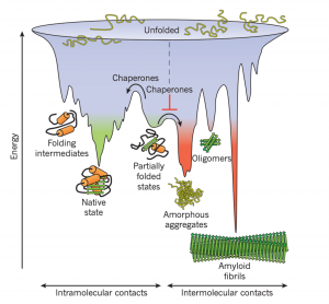
Figure 1: Competing reactions of protein folding and aggregation (Hartl, F., et al. 2011)

So, what to do with a misfolded protein? Chaperones and chaperonins are in charge or re-folding them. Also known as heat-shock proteins (hsp’s). These proteins seek and bind to exposed hydrophobic surfaces in newly translated proteins (Campbell, M., & Farrel, S. 2012).

Chaperones (Hsp70) bind and stabilize misfolded or partially folded proteins and prevent their aggregation as they are translated. ATP binding causes the chaperone to release the completed chain into the cytosol, allowing the protein to fold properly (Albert, B. 2015).
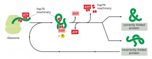
Figure 2: The hsp70 family of molecular chaperones (Albert, B. 2015)

Sometimes, even the chaperones are unsuccessful in helping the protein re-fold into its native state. In this case, additional chaperones or the more complex chaperonins (hsp60 and hsp10) provide additional help.

Chaperonins create a chamber for the proteins to re-fold post-translationally. First, the protein is captured though hydrophobic interactions with the entrance of the chamber (hsp60). The protein is then released into the interior of the chamber, which is lined with hydrophilic amino acids, and then it is sealed with a lid (hsp10). Here, the substrate can fold into its final conformation in isolation, where there are no other proteins that may aggregate. Finally, when ATP is hydrolyzed, the lid pops off, and the substrate protein is released from the chamber (Albert, B. 2015).

The best characterized chaperonin is the GroEL/GroES system in E. coli
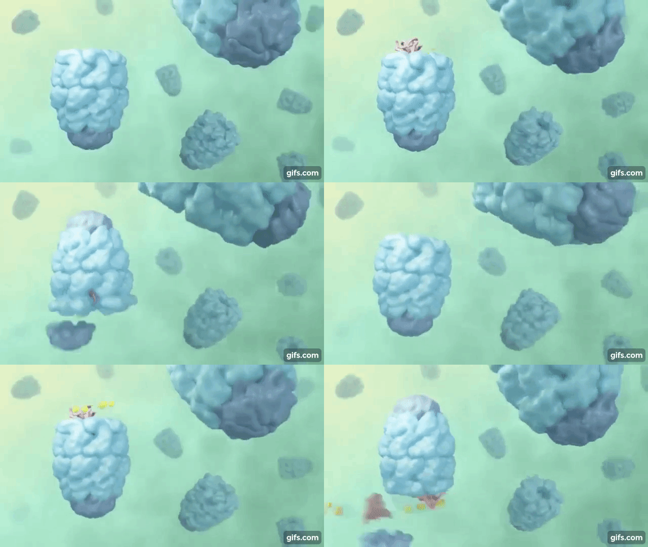
Figure 3: Visualization of the GroEL/GroES chaperonins in E. coli (Rachel Davidovitz, 2014)

If the protein is still improperly folded, it is then targeted for degradation (ubiquitination, we’ll talk about it next time!)

References:

Albert, B, et al. (2015). Molecular Biology of the Cell (6th Edition). Garland Science, Taylor & Francis Group, LLC.

Campbell, M., & Farrel, S. (2012). Biochemistry (7th Edition). Brooks/Cole Cengage Learning.

Hartl, F. U., Bracher, A., & Hayer-Hartl, M. (2011). Molecular chaperones in protein folding and proteostasis. Nature, 475(7356), 324-332.doi:10.1038/nature10317

Rachel Davidovitz (2014, October 6). Active Cage Mechanism of Chaperonin-Assisted Protein Folding Demonstrated at Single-Molecule Level [Video]. https://www.youtube.com/watch?v=–NcNeLc1mo&ab_channel=RachelDavidowitz

More steroids, plants, fungi

Steroids are not only relegated to the animal world; fungi and plants synthesize many steroids as well. One particular example of clinical relevance is ergosterol, found in the cell membranes of fungi where it serves a similar role to cholesterol in animal cell membranes. This can be exploited by antifungal medications: azole drugs such as clotrimazole and miconazole function by inhibiting ergosterol synthesis. Specifically, they inhibit the 14α-demethylase enzyme that converts lanosterol to ergosterol (note the similarities to the cholesterol pathway discussed in this previous post). [1]

Ergosterol can also be converted to ergocalciferol in a UV-light dependent reaction, similarly to the synthesis of Vitamin D3 in animals. In fact, ergocalciferol is also known as Vitamin D2, and like cholecalciferol, ergocalciferol can be hydroxylated twice to 1,25-dihydroxyergocalciferol or ercalcitriol, which binds to the Vitamin D receptor and causes its effects, although the binding of Vitamin D2 may not be as strong. [2]

There are diverse steroids made by plants, some of which have toxic effects. Of note are digoxin and digitoxin produced by the foxglove plant. These two chemicals consisted of a carbohydrate chain attached to a modified steroid, and they can be fatal if ingested. They inhibit the Na+/K+ ATPase responsible for establishing the electrochemical gradient within the cell, which is exploited for the use of digoxin as a drug for arrhythmias and heart failure due to the ability of the medication to increase the contractility of the heart when given at low doses. [3]

These are only some of the steroids occurring in plants and fungi. In the future, maybe more will be discovered with important biological activities!

Sources:
[1] Herrick, E. J., & Hashmi, M. F. (2021). Antifungal Ergosterol Synthesis Inhibitors. In StatPearls. StatPearls Publishing. http://www.ncbi.nlm.nih.gov/books/NBK551581/
[2] Houghton, L. A., & Vieth, R. (2006). The case against ergocalciferol (vitamin D2) as a vitamin supplement. The American Journal of Clinical Nutrition, 84(4), 694–697. https://doi.org/10.1093/ajcn/84.4.694
[3] Hauptman, P. J., & Kelly, R. A. (1999). Digitalis. Circulation, 99(9), 1265–1270. https://doi.org/10.1161/01.cir.99.9.1265

Steroids, salt, sugar, sex

Steroids are biologically active compounds composed of four fused rings. Although the word “steroid” is commonly associated with anabolic steroids and muscle growth, steroids are in fact a diverse group of compounds with varying effects on the human body.

The steroid cholesterol can be either synthesized via the mevalonate pathway or are obtained from the diet. The mevalonate pathway starts with acetyl-CoA, which is converted in a series of steps to isopentenyl pyrophosphate (IPP) and dimethylallyl pyrophosphate (DMAPP), which are the building blocks of isoprenoids, a diverse group of compounds that include steroids. The enzyme HMG-CoA reductase, which catalyzes the step converting HMG-CoA to mevalonate, is blocked by statins which are used for the treatment of high cholesterol levels. The IPP units are combined to form farnesyl pyrophosphate, which are then used to form squalene. From there, the squalene is cyclized to form lanosterol, which is then converted to cholesterol. Cholesterol is important for moderating cell membrane fluidity, and also participates in the formation of lipid rafts which are theorized to be involved in cell signalling. [1]

Cholesterol can then be converted into a variety of signalling molecules such as neurosteroids, vitamin D, glucocorticoids, mineralocorticoids, and sex steroids. Neurosteroids modulate complex activities in the brain, such as neural plasticity. They can act in an excitatory manner (such as dehydroepiandrosterone (DHEA), which modulates NMDA receptor activity) or inhibitory manner (such as pregnanolone, which modulates GABA A receptor activity). [2]

Vitamin D is involved in calcium homeostasis, increasing calcium absorption in the intestines and modulating bone remodulating. It is synthesized from cholesterol, including a step that involves UV radiation. It is then hydroxylated twice in order to be in the active form, 1,25-dihydroxycholecalciferol, also known as calcitriol, which binds to the vitamin D receptor to produce its effects. [3]

Glucocorticoids such as cortisol modulate metabolism and immune function. Cortisol promotes gluconeogenesis, which produces glucose, as well as promoting the breakdown of lipids and proteins. It also diminishes immune function by inhibiting the effects of various cytokines that promote inflammation and immune responses. [4]

Mineralocorticoids such as aldosterone helps to maintain blood pressure and electrolyte balance. Aldosterone acts in the kidneys to increase sodium reabsorption and potassium excretion, thus increasing sodium levels and decreasing potassium levels in the blood. Because of the sodium reabsorption, water is then retained, increasing blood volume and thus increasing blood pressure. Glucocorticoids and mineralocorticoids are both synthesized from cholesterol via progestogens in the adrenal cortex by 21-hydroxylase and 11β-hydroxylase. [5]

Sex steroids are classified as progestogens (such as progesterone), androgens (such as testosterone), or estrogens (such as estradiol). Estrogens are synthesized from androgens by the enzyme aromatase, while androgens are synthesized from progestogens by 17α-hydroxylase. Progestogens are synthesized by the conversion of cholesterol by cholesterol side-chain cleavage enzyme. Sex steroids regulate a variety of activities. Progesterone is important in the secretory phase of the uterus during the menstrual cycle, where it is produced by the corpus luteum to maintain the endometrial lining for implantation. Testosterone is important for sperm development, as well as increasing muscle growth and contributing to male secondary sex characteristics. Estradiol is responsible for inducing ovulation, bone maintenance, and female secondary sex characteristics. However, all sex steroids have diverse roles in people of all genders that are not described here. [6][7][8]

Steroids are a diverse group of compounds, and this is only the beginning. You can read about more steroids here!

Sources:
[1] Russell, D. W. (1992). Cholesterol biosynthesis and metabolism. Cardiovascular Drugs and Therapy, 6(2), 103–110. https://doi.org/10.1007/BF00054556
[2] Robel, P., & Baulieu, E. E. (1995). Neurosteroids: Biosynthesis and function. Critical Reviews in Neurobiology, 9(4), 383–394.
[3] Bikle, D. (2000). Vitamin D: Production, Metabolism, and Mechanisms of Action. In K. R. Feingold, B. Anawalt, A. Boyce, G. Chrousos, W. W. de Herder, K. Dhatariya, K. Dungan, A. Grossman, J. M. Hershman, J. Hofland, S. Kalra, G. Kaltsas, C. Koch, P. Kopp, M. Korbonits, C. S. Kovacs, W. Kuohung, B. Laferrère, E. A. McGee, … D. P. Wilson (Eds.), Endotext. MDText.com, Inc. http://www.ncbi.nlm.nih.gov/books/NBK278935/
[3] Arlt, W., & Stewart, P. M. (2005). Adrenal corticosteroid biosynthesis, metabolism, and action. Endocrinology and Metabolism Clinics of North America, 34(2), 293–313, viii. https://doi.org/10.1016/j.ecl.2005.01.002
[5] Connell, J. M., Fraser, R., & Davies, E. (2001). Disorders of mineralocorticoid synthesis. Best Practice & Research. Clinical Endocrinology & Metabolism, 15(1), 43–60. https://doi.org/10.1053/beem.2000.0118
[6] Aizawa, K., Iemitsu, M., Maeda, S., Jesmin, S., Otsuki, T., Mowa, C. N., Miyauchi, T., & Mesaki, N. (2007). Expression of steroidogenic enzymes and synthesis of sex steroid hormones from DHEA in skeletal muscle of rats. American Journal of Physiology. Endocrinology and Metabolism, 292(2), E577-584. https://doi.org/10.1152/ajpendo.00367.2006
[7] Penning, T. M. (2010). New Frontiers in Androgen Biosynthesis and Metabolism. Current Opinion in Endocrinology, Diabetes, and Obesity, 17(3), 233–239. https://doi.org/10.1097/MED.0b013e3283381a31
[8] Cui, J., Shen, Y., & Li, R. (2013). Estrogen synthesis and signaling pathways during ageing: From periphery to brain. Trends in Molecular Medicine, 19(3), 197–209. https://doi.org/10.1016/j.molmed.2012.12.007

Plants: Making Air Easier to Breathe

We’ve all heard on the news or learned in elementary schools about the mass deforestation going on all over the world. But have you ever stopped to wonder, why exactly is this bad? Why do we need plants and trees at all?

It turns out, plants are efficient in resupplying the air with oxygen, while removing carbon dioxide! The former, being essential to our survival, and the latter being a gas involved in global warming.

an inside look into plants

So how exactly do they do this? In turns out that inside the plants’ cells there are special machinery capable of splitting water (H2O). This machinery is called an electron transport chain (ETC).

Using sunlight, the ETC extracts energy from the water – leading to the generation of oxygen as a “waste” product. Ironically what’s considered waste for them is gold in terms of survival for us.

When it comes to removing carbon dioxide they have another set of machinery. For the plant, carbon dioxide is like food: they trap the carbon dioxide and convert them into carbohydrates and other nutrients.

rubisco – the single bad life-essential solution

A key piece of machinery in this conversion is RuBisCo – an enzyme. An enzyme is a molecule that speeds up biochemical reactions, and surprisingly RuBisCo is one of the least efficient in existence (think of RuBisCo as a bike and other enzymes as the newest Tesla).

So you might be thinking, if RuBisCo is such a bad enzyme, can’t scientists just make a better version of RuBisCo? This would increase crop yields, and be good for the environment! Well, scientists have tried and failed … it seems like this is the only bad solution to a complex problem. Along with the ability to split water at ease (which scientists also can’t do), this is why plants are biochemical miracles.

BIOL 340: Introductory Cell Biology Laboratory (Review)

Endless reports and hours in the lab; the bane of all Biology major’s existence: BIOL 340. As its name suggests, BIOL 340 is an intense lab that teaches different cell biology techniques, from fluorescence microscopy to SDS-PAGE.

format of the course

BIOL 340 is a 3 hour weekly lab course with a separate 1 hour lecture portion. However don’t be fooled, often most of the class were unable to finish on time and labs ended up being on average 4-4.5 hours long. Since each lab featured a new lab technique, pre-readings were very dense. There were also in-class pre-reading quizzes, so memorizing every little detail was crucial (imagine spending hours reading about the different steps in SDS-PAGE, but then being asked what SDS stands for…).

These labs were usually done in a group of four of five (random partners), and I was lucky to be in a good group. Each group was given a different mutant yeast strain to practice on using the lab technique of the week. In the later parts of the course, we had to run an independent study on this same yeast strain, which was by far the most stressful portion of the course. Continue reading

BIOL 300: Fundamentals of Biostatistics (Review)

We all know statistics courses can be relatively dry, but BIOL 300 spices things up with interesting biological examples! BIOL 300 is an introductory level statistics course at UBC, which fulfills the statistics requirement for life science majors.

format of the course

The format of this course is usually what you see in most science courses. What makes this course unique is the lab component, where you learn computer coding. Wait … computer coding in a statistics course? It turns out that most statistical analyses are tedious to do by hand, so instead we learned how to automate these calculations. In class, we learned about the different statistical approaches for different contexts, while in lab we learned how to actually run these analyses. Continue reading