Singing to Treat Parkinson’s Disease: It Works!

Do you know someone that has Parkinson’s Disease? Did you know that there are more than 10 million people around the world that suffer from this disease, but there is still no cure?

A recent study has discovered what could be a therapy for Parkinson’s Disease, but it is quite unconventional! Keep reading to find out more.

To provide some background information, Parkinson’s Disease (PD) is a “neurodegenerative disorder”, which means that it causes serious damage to the nerves of the brain. Symptoms of the disease include hand tremors, extreme difficulty walking as well as mental problems such as hallucinations or delusional thoughts. The disease typically affects people over the age of 50.

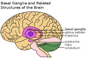
Diagram of the “Basal Ganglia”, which is the part of the brain affected by Parkinson’s Disease. Source: Wikimedia Commons. This image is part of the public domain.

Now, the good news! A group of researchers at Iowa State University have been investigating the effects of singing therapy on a group of PD patients, and found that the participants’ heart rate, blood pressure, as well as self-reported anxiety and sadness decreased over the 2.4-year-long treatment period. These results show that the overall health and well-being of the participants improved as a result of the therapy!

What is “singing therapy”, you ask? Essentially, the treatment is weekly or bi-weekly, and consists of sessions in which the PD patients undergo vocal exercises and sing well-known songs as a group. Researchers found that the PD patients were able to breathe more effectively after the therapy!

You might be wondering how singing could possibly affect such a complicated disease, but as lead investigator Elizabeth Stegemoller describes it,

“We’re not trying to make them better singers, but to help them strengthen the muscles that control swallowing and respiratory function. We work on proper breath support, posture and how we use the muscles involved with the vocal cords, which requires them to intricately coordinate good, strong muscle activity.”

Essentially, singing therapy is beneficial to PD patients because it helps them strengthen the muscles used for swallowing and breath control, which are tasks that become difficult with the onset of the disease.

In addition to these benefits, the study found that the patients experienced improvements in their tremors and walking.

A depiction of a PD patient drawn by neurologist Sir William Richard Gowers in 1886. This image is currently used by the medical community as a reference for the symptoms of PD. Source: Wikimedia Commons. This image is part of the public domain.

Looking to the future, researchers are hopeful that singing therapy will be implemented as a clinical treatment for PD patients, as it is cheap, extremely low-risk, and lots of fun!

YouTube Preview Image

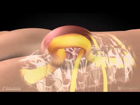
Watch this YouTube video (published by Iowa State University and available as part of the public domain) to find out more about the Parkinson’s Disease research being conducted by Elizabeth Stegemoller at Iowa State University.

Maya Liepert

The Venom that Can Cure You

Imagine hearing the words from your doctor, “You have cancer,” then feeling relived that it wasn’t anything fatal. Yes, scientists in Africa have discovered that the venom from Polybia paulista (South American Wasp) can kill cancer.

The polybia paulista (South American Wasp) is found native to Brazil, Paraguay, and Argentina.

The university of Brazil have begun experimenting its effects on mice with areas of tumour and seeing hopeful results. After stings from the wasps, the size of  tumours of the mice visibly shrunk significantly. This is cause by the venom having a protein Polybia-MP1 that causes the rupture on the parts of the cell membrane with these fatty component, phosphatidylserine (PS) and phosphatidylethanolamine (PE). Cancerous cell, unlike normal cells, have these parts on the outer membrane, making them more susceptible. The venom will actively damage these parts of the cells and cause the cell to degrade.

The process in which the wasp venom kills the tumorous cells.

Reachers are excited in the discovery of such medication. There exists a saying in Chinese “以毒攻毒” meaning using poison to attack poison.  The venom used does have its drawbacks. Normal cells are still susceptible to the the degradation and prolonged exposure can kill the healthy cells as well. Prolonged exposure to these venom will cause the patient to die. Researchers are still studying ways to implement this technique without harming the patient.

Dr Paul Beales, from the University of Leeds, stated that drugs that attacked the lipid structure of the cancerous cell’s  membrane are a revolutionary type of drug. “This could be useful in developing new combination therapies,” as he was keen on using multiple ways to deal with the tumour at the same time to increase the efficiency and to decease the time the patient is in contact with venom.

“This early stage research increases our understanding of how the venom of the Brazilian wasp can kill cancer cells in the laboratory.” said Dr Aine McCarthy, the science information officer for Cancer Research UK. She along with many others are excited to hear and is looking forward to do more tests on this subject before it is available to treat patients.

– Steven Li

A Setback in Animal Conservation Efforts: Reversal of Rhino and Tiger Trade Ban in China

Can you imagine a world without animals?! What about a world without rhinoceroses or tigers? Sadly enough, there has been a large decline in the abundance of these animals within the past few generations.

An image showing two South China Tigers sitting in an enclosed conservation area. Image Credit: Amur Tiger Programme, under Creative Commons License.

For example, the South China Tiger population, pictured above, is currently listed as critically endangered, since thousands of them have been killed from hunting practices. Another species in China that has a population directly threatened by hunters are the rhinos. Because poaching is persistent and there has been a gradual loss of habitat, there are very few rhinos that can survive outside of national parks.

The reason that animals, such as tigers and rhinos, are commonly sought after in China is because of their medicinal properties. The rhino horn is used to treat fevers, gout and even food poisoning! Tiger bones, on other hand, are used to boost health and oddly enough, they are believed to enhance male strength. Although there is a lack of evidence to support these “healing” powers, there is still a strong demand for rhino horns and tiger bones, which threatens these species’ chances of survival.

An image showing two rhinos after hunters have removed their horns, leaving them severely injured. Image Credit: Wikimedia Commons


As soon as Chinese government officials realized how urgent it was to protect these animals, they enforced a ban on the trade of rhino horns and tiger bones in 1993. This ban was a relatively effective measure aimed towards wildlife conservation. Although there was a subtle black market trade that persisted, it was definitely a step in the right direction towards saving these beautiful animals!

Despite China’s initial efforts to protect the endangered rhino and tiger species, they have recently decided to lift the ban on the trade of their body parts. This measure has spurred dissent amongst environmentalists and conservation groups, since poachers and hunters can now legally trade these items. Since rhino and tiger populations are already dwindling in numbers, the expansion of the wildlife trade market is a serious setback to prior conservation efforts. The urgency to continue to protect these endangered species is emphasized in the video below.

YouTube Preview Image

A video highlighting the criticism behind the removal of this ban.

Video Credit: Youtube, South China Morning Post, published under the Creative Commons License.


The removal of the wildlife trade ban will only encourage hunters/poachers to continue their practices… So therefore, it is crucial that we pursue any measure that might prevent these species from going extinct!


Lexynn Kwan

Killing the Ghosts: Beating Drug-Resistant Cancerous Cells

Rapid expansion of molecular biologists’ knowledge of how cancerous cells with damaged DNA sustain a long lifespan has helped us find new aggressive ways to wipe out cancer cells such as more effective chemotherapy. One key obstacle to tackle is the fact that cancerous cells quickly become resilient to DNA-harming medications. This leads to many chemotherapy failures.

A new research just appeared in Nature Structural and Molecular Biology  introduced an innovative method of dealing with death-evading cancerous cells by stripping these cells of their drug-resistance developing capabilities, more vulnerable to DNA damaging drugs hence.

Pancreatic cancer cells deficient in the expression of the human gene known as Schlafen 11 and resistant to chemotherapy (left panels) were re-sensitized to chemotherapeutic treatment (middle and right panels) by inhibiting the expression of the transfer RNA known as tRNA-Leu-TAA through specially designed antisense oligonucleotides. [Manqing Li, Michael David Lab, UC San Diego] (Genetic Engineering & Biotechnology News (GEN))

Built upon their prior works on HIV immunology, Professor Micheal David and Emeritus Professor Jean Wang from University of California (San Diego) say that a protein called Schlafen 11 stops the normal functions of the two vital proteins of cancerous cells, ATM and ATR. They explain that DNA damaging drugs activate Schlafen 11 which leads to cancerous cells death, and those cancerous cells which do not express Schlafen 11 simply survive the chemotherapy.  This study has a great potential for applications in immunology and virology, including HIV therapies, due to molecular mode of action of Schlafen 11.

David’s Lab further found out that, similar to the transfer RNA molecules of Schlafen 11, transfer RNA molecules of several gene families involved in DNA repair systems are encoded by transfer RNA gene rich in leucine amino acid. This provides a clue for making drug-resistant cancerous cells sensitive anew by attacking the transfer RNA molecules of DNA repair genes.

The findings show that disruption of normal functions of both ATR and transfer RNA could kill the cancerous cells combined with chemotherapy even though this technique could compromise whole DNA repair system. The paper also shows the role of cellular adjustments made in levels of transfer RNAs in survival or death of a cell with damaged-DNA for the first time.

by: Jamaledin Adel

https://www.nature.com/articles/s41594-018-0142-5?_ga=2.70392784.117150651.1540857600-2063335005.1540857600


The Americanization of Gut Bacteria in Immigrants

When immigrating to America everything becomes westernized, including the bacteria in your gut.

The green highlights the large intestines where the majority of the gut bacteria lies. Image credit: Mikael Haggstrom

Previous studies showed that living in the United states increase the risk of obesity and chronic disease among immigrants to the US relative to those who stayed in their country of birth. Researchers from the University of Minnesota, Dan Knights and Pajau Vangay, thinks that this increase in obesity and chronic diseases may be caused by the decrease in biodiversity in the gut of immigrants.

People in developing countries have more diverse microbiomes, while people living in industrialized countries like the United States have lower microbiome diversity. However the study found that when moving from a developing country to an industrialized nation would actually cause the biodiversity of the gut bacteria to decrease.

The Effect of Immigration on Gut Bacteria

The study focused on the Hmong and the Karen who are the most at risk for Obesity in Asian populations in Minnesota. The study studied the immigrants of different residency length and second generation immigrants and compared the biodiversity of the gut bacteria in these populations.

When immigrants move to the United States, their gut microbiome rapidly Americanizes within 6 to 9 months and becomes less diverse. The gut is invaded by with a rise in the population of bacteria of the genus Bacteroides which is associated with an American diet, while replacing the hey began to replace those of the genus Prevotella from the immigrants home countries diet.

When studying immigrants who have been in the US for years, they found the bacteria in the gut has become less diverse also the American genus Bacteroides have become more prevalent. The change of Bacteria is a long process and it starts immediately but it continues for many decades after and  those who have immigrated longer much more at risk for obesity and other chronic diseases.

As Immigrants reside in the US longer, the chances of obesity increases. Image Credit: Pharos

Bacteria Diversity Change with Long Term effects

The researchers also observed the second generation immigrant who have very similar diet as their parents with ten times the amount of rice as the average American. However even with a different diet the diversity of gut microbiome in these second generation immigrants very closely resemble the average American. Meaning the decrease in gut bacteria diversity does not only compound over time but across generations.

Know this decrease in gut bacteria diversity increases obesity and chronic illnesses, maybe it is worthwhile to find how to maintain or increase gut bacteria diversity to maybe help fight obesity and chronic illnesses.

Virtual Reality as Therapy for Mental Disorders

While many of us have heard of virtual reality (VR), we are likely less familiar with its applications. While VR can be an immersive gaming experience, it can also be used to confront your biggest psychological fears. In fact, some psychologists currently use this technology as treatment for patients with psychotic disorders and paranoid ideations. People with paranoid ideations believe they are being harassed and are suspicious of other people’s motives. As of 2013, more than 6.7 million people in Canada were living with mental conditions and Canadians with bipolar disorder and Schizophrenia, which both involve paranoia, each made up 1% of the population.

What is virtual reality?

Virtual reality creates the illusion of reality within different scenarios. The VR program is set up through a computer system, to which headphones and glasses are often attached. The recreated sights and sounds allow total immersion into the environment being experienced.

Samsung Gear VR virtual reality glasses

Specialized glasses for virtual reality
Image Source: flickr
Photographer: jocelynbird


Virtual reality as therapy

People with psychotic disorders often have similar responses to social interactions. Specifically, public settings and social activities tend to be avoided and time spent alone is higher than for an average person. One approach to treating patients and helping them reduce their social anxieties is through exposure. While therapeutic exposure-treatments are widely used, they present limitations for psychologists. Specifically, the therapist does not have control over the environment in which the patient is placed, which includes other peoples’ responses. Another difficulty is the willingness of a patient to undergo exposure to their fears in the real world. Virtual reality allows therapists to use exposure-treatments in a more controlled setting and therefore overcoming these limitations.

Virtual reality is used to treat paranoia
Posted By: Department of Psychiatry, University of Oxford


The effects of VR-assisted therapy were further explored in a study published in March 2018, which was conducted at seven Dutch mental health centres. The study consisted of outpatients between the ages of 18-65, who had been diagnosed with psychotic disorders and paranoid ideation, being randomly assigned to one of two groups: a control group (traditional therapy treatment) or VR-CBT (traditional therapy treatment with addition of VR). Patients within the VR-CBT group attended 16, one-hour VR-therapy sessions and were subjected to experiences such as busy streets, buses, cafés, and supermarkets. As mentioned earlier, the advantage to this treatment is the range of control that the therapist obtains. In this study, the number of human avatars in proximity to the patient, their sex and ethnicity and their responses to the patient, such as eye contact or hostile behaviour were all controlled by the therapist. The study found that momentary paranoid ideation and momentary anxiety were both decreased in patients. Their trigger-response behaviour was also reduced in the VR group compared to the control.

Supermarket virtual reality for paranoia patients
Posted By: Science News


Virtual reality can be used as a powerful tool in the treatment of mental illnesses. By exposing patients to their psychological fears in a controlled environment, we can decrease the anxiety and paranoia they would otherwise experience during real-life social activities.

-Shadi Elmi

Stroke and Motor Rehabilitation

Stroke is the most common neurological disorder and is the primary cause of physical disability. After cancer and coronary artery disease, stroke is the third leading cause of death in the United States, with an incidence of at least 100 per 100,000 people. In Canada, there are more than 50,000 strokes a year. Every seven minutes, a Canadian dies of heart disease or stroke.

What is a Stroke

Stroke is also called the cerebrovascular disease which means it happens when blood flow to a certain brain area is cut off. Without the oxygen in the blood, brain cells start dying within minutes.

Here is a video understanding some basic concepts of stroke by an animated explanation.

YouTube Preview Image

Video Credit: HealthSketch

Types

There are two main types of stroke:

The first type is called Ischemic Stroke. Due to lack of blood flow caused by a blood clot or fatty deposits lodging within a brain artery. Approximately 85% – 90%  of all stroke is due to ischemia.

The second type of stoke is the Hemorrhagic stroke, which is due to bleeding from a ruptured artery. It’s less common than an ischemic stroke, but it can be more serious. It can be a blood vessel in the brain balloons up and bursts, or a weakened one leaks.

The difference between the ischemic stroke and hemorrhagic stroke

Image Credit: FlintRehab

Causes

A common cause of ischemic stroke is the hardening of the arteries which is caused by plaques. Those plaques are mostly made of fat, cholesterol, and other substances build up in the arteries, leaving less space for blood flow. Therefore, a blood clot may stay in this narrow space and cause an ischemic stroke.

Risk factors that can be controlled include high blood pressure, high cholesterol, diabetes, blood vessel disease, heart disease, smoking, obesity, high alcohol consumption, poor diet, etc.

symptoms

The F.A.S.T test helps spot symptoms of stroke:

Related image

F.A.S.T Test

Image Credit: sleepapnea.org

motor rehabilitation

At least 70% of people will survive a stroke, but they may have profound motor deficits. Most people lose their motor functions of the other side of the body (different from the side where stroke happens) after stroke. Thus, functional recovery through active rehabilitation training is essential to achieve increased independence and quality of life.

Muscle weaknesses are pervasive after a stroke. This can interfere with walking and other daily motor activities. Physical rehabilitation is an effective way to regain strength, balance, and coordination; whereas occupational therapy is essential for fine motor skills, such as using a knife and fork, writing and buttoning a shirt.

Stroke rehabilitation works the best after 24 hours to 48 hours of stroke occur. Most people make the biggest recovery during the first three months after stroke. By continuing exercising and practicing new skills help part of your brain take over new functions.

Here is a video talking about how Steve recovered his motor ability from rehabilitative technology after a stroke.

YouTube Preview Image
Video Credit: Mary Free Bed Rehabilitation Hospital


Elaine Yuling Chen


Slow and Steady: New Research Claims That Removing The Appendix Decreases Risk of Parkinson’s Disease

Many people have a friend or family member who is afflicted by Parkinson’s Disease, a long-term degenerative disease affecting the brain’s central nervous system. Seemingly unrelated, many people also have removed their appendix, a small finger-like length of intestine that is widely removed due to appendicitis. Perhaps somewhat unexpectedly, a recent study analyzing more than 1 million people in Sweden has concluded that an appendix removal done decades ago may reduce the chances of developing Parkinson’s Disease (PD).

Scientists speculate that the removal of the appendix affects the development of an abnormally formed protein, named α-synuclein (alpha-synuclein), which is present in PD patients.

What Is Parkinson’s Disease?

Parkinson’s Disease is a genetic neurological (brain) disorder that can have significant impact on the physical and mental state of patients, with more than 55,000 Canadians diagnosed with PD. The onset of PD is slow, often alongside symptoms such as hand tremors and body stiffness.

Parkinson’s encompasses a wide subset of symptoms and related complications: restricting the movement of a patient gradually, eventually initiating other symptoms such as difficulty speaking, loss of coordination, and development of depression.

Prevalence of Parkinson’s disease in household population, by age group and sex, population aged 45 or older, Canada excluding territories, 2010/2011 Source: StatCan

Presence of α-synuclein In PD Patients

A protein called α-synuclein, in its misfolded (mutated) form, is fairly abundant in the appendix of both healthy people and PD patients. The mutated version of α-synuclein has the same structure as the version of α-synuclein that is found in the brains of patients afflicted by PD, which scientists have suggested causes PD by damaging the brain cells which control bodily movement.

Highly magnified image of mutated α-synuclein.
Source: Van Andel Research Institute

α-synuclein tends to travel from the appendix, leading scientists to investigate whether the protein present in the appendix is the same as the protein in the brain of PD patients.

Study on More than 1 Million Swedes Found…

Knowing this information about α-synuclein, Viviane Labrie (the first author of the study) and her team at the Van Andel Research Institute in Michigan analyzed 1.6 million Swedish medical records to find that appendix removal showed almost a 20% decrease in chances of developing PD.

The data analysis also showed that an appendix removal also delays the diagnosis of PD for the people in the data set who did go on to develop PD for more than 3 years.

Why Is This Important? 

Parkinson’s Disease is not only a devastating and debilitating disorder, but also it is increasingly common as the general population begins to age. In recent years, PD is affecting more than 60,000 people each year in the United States. As PD is the second most common neurodegenerative (brain disorder) disease in Canada, it is with hope that these findings will inspire investigations into the pursuit of a treatment for Parkinson’s Disease.

See below for a video posted by Parkinson Society of BC, showcasing a young patient and her goals despite her disorder.

An instalment of Parkinson Society of BC’s This Is Parkinson’s Disease campaign, sharing Hilary’s story: a real-life British Columbian living with PD.
Source: Parkinson Society of BC

-Allison Chiang

How much do you know about Spina Bifida? A Neurodevelopment Disorder

What is Spina Bifida?

I believe some of you may have heard about neurodevelopment disorder like hydrocephaly. What about the disorder strong related to hydrocephaly – Spina Bifida. Literally speaking, Spina Bifida means the spine split into two. It happens within the first four weeks the fetus in the uterus. Due to the spinal column is not fully closed, the spinal cord inside is not covered and even exposed and protruded out of the spinal column. There are about 120 children born with Spina Bifida each year in Canada, and 90% of them can survive and become adults.

Image result for spina bifida

Image Credit: Mayo Clinic


YouTube Preview Image
Video Credit: The Spina Bifida Resource Network


Spina Bifida has more than one type. What do they look like?

There are two main types of SB, Spina Bifida Occulta (SBO) and Spina Bifida Myelomeningocele (SBM).

SBO is the mildest case which does not show any symptoms of the condition. The defect is hidden under the skin, and the spinal nerves are not involved in the defect. This type of defect may be noticed early in life if there is an abnormal tuft or clump of hair, or small dimple or birthmark on the skin at the site where the spine is not formed correctly.

However, SBM, also known as open SB, is the most severe form which needs to be treated with surgical treatment as early as possible. The membrane and spinal nerves push through the opening between spinal column gap at birth, forming a sac on the baby’s back. In this situation, the spinal cord is exposed, causing partial or complete paralysis of the body.

Image result for spina bifida

Image Credit: familydoctor.org

Causes & Risk Factors

The specific reason for spina bifida is still not clear nowadays. It shows to result from a combination of genetic and environmental risk factors, such as family history of neural tube defects and folate deficiency during pregnancy.

Then how should we prevent this disorder? Scientists find out that folate plays a significant role in SB prevention. Low levels of the vitamin folic acid during pregnancy are linked to  SB.

Related image

Image Credit: Babyment

how to treat the disorder?

Treatments for SB also depends on the type of SB. Children with SBO typically do not have any symptoms or complications, only routine pediatric care is needed. However, the child with SBM needs to be treated with surgery. The research finds out that fetal surgery can be an effective management strategy for SBM patients, resulting in a significant benefit to these patients, typically in improving motor function.

Another study investigates the neurodevelopment outcomes of preschool children with SBM treated with myelomeningocele closure. They found children who have undergone fetal myelomeningocele closure have average preschool neurodevelopment scores.

– Elaine Yuling Chen

Spam prevention powered by Akismet