If anyone has siblings, or even a twin, could you imagine being physically joined together with them? And no, I don’t mean metaphorically. Could you imagine being conjoined and having to share the same body with them? Conjoined Twins, or sometimes known as Siamese Twins, may have been something you’ve seen on TV, like in Grey’s Anatomy or American Horror Story.
What are Conjoined Twins?
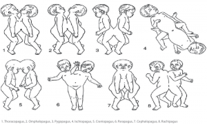
Conjoined twins are twins that are born with physically connected bodies. Every case of conjoined twins are different, as they could be joined at different parts of the body. For instance, they could share only a small amount of tissue, and each pair could have their own set of organs and structures they need to essentially stay alive if they are able to be separated.
Others, the connection can become quite complex. The twins could share a vital organ, like the heart, or digestive, genital, and urinary systems. They could also share a huge portion of the body, like sharing the entire lower half of the body, or be conjoined at the head where they share parts of the brain and skull.
Approximately 75% of conjoined twins are joined at least partially in the chest, or share organs.
The Success Rate
The progression of advances in imaging studies, surgical techniques, and anesthesia, the rate of successful separation of conjoined twins have increased more now than in the past.
However, at birth, ~40% of conjoined twins are not alive.
~35% die within a day after birth because their organs are not able to support them both.

Source: Research Gate
The succession of separating conjoined twins depends on so many factors:
- Where the twins are connected
- Which structures they share
and this can get quite complicated
This could result in both twins surviving, but because of a serious birth defect, there is a risk of one or both twins dying.
In the cases the structures and organs they share may be too high of a risk, or not able to perform the separation. Lots of conjoined twins live happy and healthy lives by remaining connected.
Causes of Conjoined Twins
Identical twins are called monozygotic twins, and this occurs when a single fertilized egg splits and develops into two individuals. 8-12 days after conception, the embryonic layers split, forming into specific organs and structures.
When the embryo splits later than that time period; usually between 13-15 days after conception, the separation stops before the process is completed, and the twins result in conjoinment.
An alternate theory is that two separate embryos somehow fuse together in early development, but what might cause either of those theories is unknown.
A Case of a Successful Separation

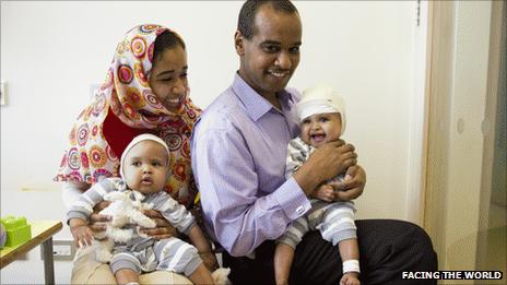
Conjoined Rital and Ritag Gaboura Source: NewScientist
Rital and Ritag Gaboura were joined at the head – and it took a team of more than 15 surgeons and doctors at Great Ormond Street Hospital in London to completely separate the conjoined twins.
Magnetic resonance imaging (MRI) and X-ray computed tomography (CT) was done to show how their skulls were fused. The Gaboura twins turned out to be craniopagus twins – meaning they shared a skull but not a brain. This is extremely rare, and occurs once out of every 2.5 million births.
If they shared the same brain, the surgery would have been too risky to perform.
The surgeons needed to study the twin’s skull and brain to meticulously strategize the network of delicate arteries and veins that transported blood to and from the brain. Most craniopagus twins do not share arteries that feed the brain, but their veins are tangled together, and some can run from one twin’s brain to the other.
When separating the veins, if one twin ends up with too many veins, it would cause the heart to collapse.
Leaving the other twin with not enough veins, the brain would swell up with blood.
Studying their blood flow was done by injecting dyes into the twins’ arteries and tracing the blood’s in and out pathway of the brain with a series of X-rays.
A media production company was involved, using the images to make a CGI video of the blood rushing in and out of the brain.
The surgery was chosen to be done in stages, so the skull and veins could recover between each operation.
The two major systems of veins drain blood from the brain:
- A superficial system woven into outer layers of dura mater (envelope of protective tissue surrounding the brain)
- Deep system in internal brain structures
One twin would be given most of the superficial system, and the other most of the deep system, and hopefully the brains would adapt to the loss by expanding small collateral blood vessels that could connect the two systems.
To divide each vein, the surgeons opened a window in the twins’ skulls and operated on the veins in that little window. A craniotome, which is a high speed drill was used to drill through the skull to reach the blood vessels, and essentially it would push the brain out of the way so only the bone would be cut.
The veins were sliced through, and tied off with ligatures and dissolvable sutures.
When it was possible, the shared veins were split at a junction so each twin could receive one part of the brain.
The final hurdle of full separation was that craniopagus twins do not have enough bone and skin between them to form a complete skull and scalp when it was time to sew everything up.
The problem was solved by putting four silicone balloons filled with saline solution under the scalps, between the bone and skin. The balloons would progressively stretch the skin of the scalp.

Inflatable balloons stretch the skin. Source: https://www.bbc.com/news/health-14969823
The skull bone has three main layers:
- Tough outer layer
- Spongy middle layer
- Tough inner layer
By driving chisels between each layer, the surgeons were able to split the bone and lift up a section so it would double the original area of the skull.
Rital and Ritag were able to recover after their separation, and the surgery was deemed a success as they were separated, and each child was getting the blood it needs and draining blood from the brain in the correct ways.

Source: BBCNews


