
Nuclear medicine is a branch of health care that never seems to stop growing. For decades, we have used forms of radiation to help detect illnesses in our body. Recently at the University of British Columbia (UBC), research involving the radioactive element Gallium-68 has shown promising results for nuclear medicine. When injected into a patient, Gallium-68 can be detected by medical scanners, helping to monitor blood flow as it is traced through the body. But if this isotope is radioactive (known as a radioisotope), then couldn’t this treat lead to cancers, or even mutations? To understand these misconceptions of modern nuclear medicine, it is helpful to know how these medical scanners function.
https://www.youtube.com/watch?v=1flwtte9fTg&feature=youtu.be
One of the most common types of medical scanners used today is Positron Emission Tomography, or PET. This scanner works by monitoring a radioactive dye that is injected into a patient’s bloodstream. Because isotopes like Gallium-68 have different numbers of protons and neutrons, they eventually ‘decay’ to another element. During this radioactive decay, isotopes release gamma radiation that PET scans can detect, creating a full 3-dimensional image of where the isotopes collectively decay in your body. This image can display areas of altered blood flow, such as cancerous tumours, or plaques in Alzheimer’s disease. Single-photon emission computed tomography (SPECT) is another form of medical scanning, providing a less-expensive and poorer resolution than PET.
Audio clip: Adobe Flash Player (version 9 or above) is required to play this audio clip. Download the latest version here. You also need to have JavaScript enabled in your browser.
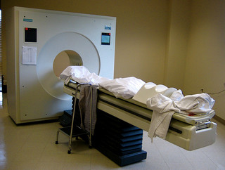
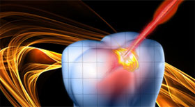
Despite using radioisotopes, PET scanning is less harmful than one may think. Reference: Flickr (Creative commons)
However, we have commonly heard of how deadly radioactive elements can be, such as with Uranium and its involvements in Chernobyl and nuclear weaponry. The fact is, the radioisotopes used in medical scanning, such as Technetium-99m in SPECT, release about as much radiation as other medical imaging tests like x-rays– a cornerstone of medical imaging. With the Gallium-68 radioisotope currently studied at UBC, the particle was found to be quite stable and left the bodies of tested animals quickly. As it also has a half-life of about one hour, the results ensure less gamma rays are emitted in the body during PET scans. This is contrasted with other media-frenzied radioisotopes like Uranium-235, which has a half-life of over 700 million years and is capable of nuclear fission: a chain reaction that is potentially deadly.
Because both Uranium-235 and Gallium-68 are radioisotopes, it is understandable that the general public would assume they share similar effects with the body and assert these ideals to nuclear medicine. However, the quick decays of isotopes used in PET and SPECT, along with their relative stability, should leave little to be worried about their emitted radiation.
References
Acyclic Chelate with Ideal Properties for 68Ga PET Imaging Agent Elaboration. Eszter Boros, Cara L. Ferreira, Jacqueline F. Cawthray, Eric W. Price, Brian O. Patrick, Dennis W. Wester, Michael J. Adam, and Chris Orvig. Journal of the American Chemical Society 2010 132 (44), 15726-1573
Triumf Article on the effects of radiation in Scanning: http://legacyweb.triumf.ca/welcome/petscan.html
HowStuffWorks Article on harmful effects of Uranium-235: http://science.howstuffworks.com/nuclear-power1.htm


