
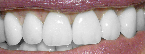
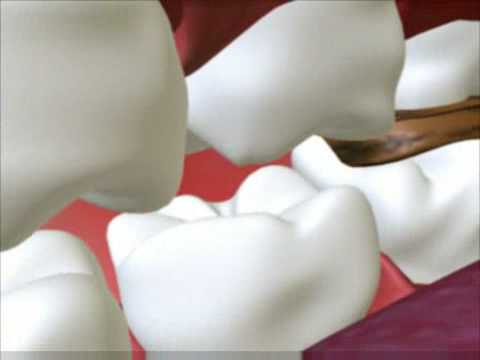
Teeth photo from Flickr by drburtoni
Last year, my wisdom teeth were hidden beneath my gums, seemingly content and not disturbing anyone. One day, I discovered that one wisdom tooth, in the lower left corner of my mouth, started to peek over my gum line. Curious, I started asking my friends about their experiences in getting wisdom teeth removed. Although everyone seemed to have a similar procedure, they all went through varying degrees of pain, swelling and functionality afterwards.
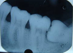
Impacted lower wisdom tooth from Flickr by Dr Parveen Chopra
The medical term for wisdom teeth that have not yet erupted (or become visible in the mouth,) is “third molars.” Some people don’t have wisdom teeth at all. Others have up to four (or even more) wisdom teeth. However, most people do need to have them removed as their jaws are not spacious enough to hold all 32 teeth. Wisdom teeth therefore, often become impacted, meaning they are blocked by other teeth or stuck in the gum tissue for various reasons. They can even move teeth around or crowd teeth, causing pain. Wisdom teeth usually come out between the ages of 16-25. They are called wisdom teeth because they come in later in life, when you are presumed to be “wiser” than when you were a child.
There has been much discussion amongst dental professionals of whether or not to remove wisdom teeth that are not symptomatically problematic. Some even deem intervention a “questionable practice.” (Intervention meaning the removal of wisdom teeth before they are problematic for the patient.) There are well defined guidelines for when impacted third molars should be removed, being when infections, cysts, tumors, or the destruction of adjacent teeth or bones occur. However, sometimes wisdom teeth do not enter into the mouth at all. In other words, it is difficult to predict whether third molars will become impacted in the future.
Risks of intervention include nerve injury, excessive bleeding, and infection. There are also several much rarer, but more serious complications that may come up after or during the procedure including the infection of a vital organ, injury to adjacent teeth and/ or joints, or even dysesthesia, painful or weird sensation caused by faulty central nervous system function.
The benefits of removing unerupted asymptomatic teeth include a gain of arch space, prevention of infections, cysts, or tumors, and the promotion of gum health. In addition, all studies done have pointed out that the younger the patient is, (optimally in their late teens to early twenties,) the less chance there is of encountering infection or disease after extraction.
In the end, it is up to your dentist to gage if it is necessary to extract wisdom teeth. But it is equally important for you, the patient, to inform yourself and decide what is best for you.
References:
http://scienceline.org/2007/02/ask-cooper-wisdomteeth/
CAPUZZI, P., L. MONTEBUGNOLI, and MA VACCARO. “Extraction of Impacted 3rd Molars – a Longitudinal Prospective-Study on Factors that Affect Postoperative Recovery.” Oral Surgery Oral Medicine Oral Pathology Oral Radiology and Endodontics 77.4 (1994): 341-3.
http://www.colgate.com/app/CP/US/EN/OC/Information/Articles/Oral-and-Dental-Health-at-Any-Age/Teenagers/Wisdom-Teeth/article/Should-You-Have-Your-Wisdom-Teeth-Removed.cvsp
Mercier, P., and D. Precious. “Risks and Benefits of Removal of Impacted Third Molars: A Critical Review of the Literature.” International journal of oral and maxillofacial surgery 21.1 (1992): 17-27.
http://www.thebrainhealth.com/what-does-dysesthesia-mean.html
http://www.aaoms.org/wisdom_teeth.php


