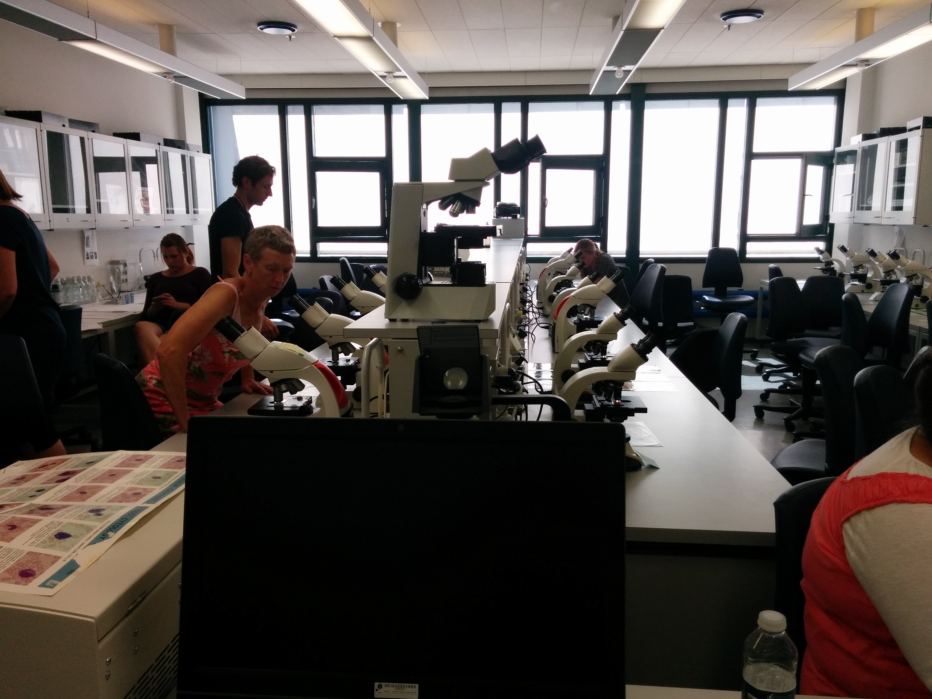
“You can’t learn microscopy in 3 hours, or even a month, or a year.. but we’ll see how you guys do” – Dan Meyrowitsch
Who remembers taking 1st and 2nd year general biology.. when you have to go to the lab to look at slides and its super boring usually because really.. who knows what you are supposed to see, and then you have to draw it and its always a blob.. or a dot with blobs around it. Well today we had a 3 hour lecture in the lab at Panum (the medical and science building at the University of Copenhagen), and it was nothing like any microscopy I have ever done before. Of course we were looking at malaria, schistosomiasis, leishmaniasis, worms, parasites, mosquitoes, etc. If it has something to do with disease, virus, bacteria, parasites found in developing countries – we looked at it. I have plenty of photos for you by the way!
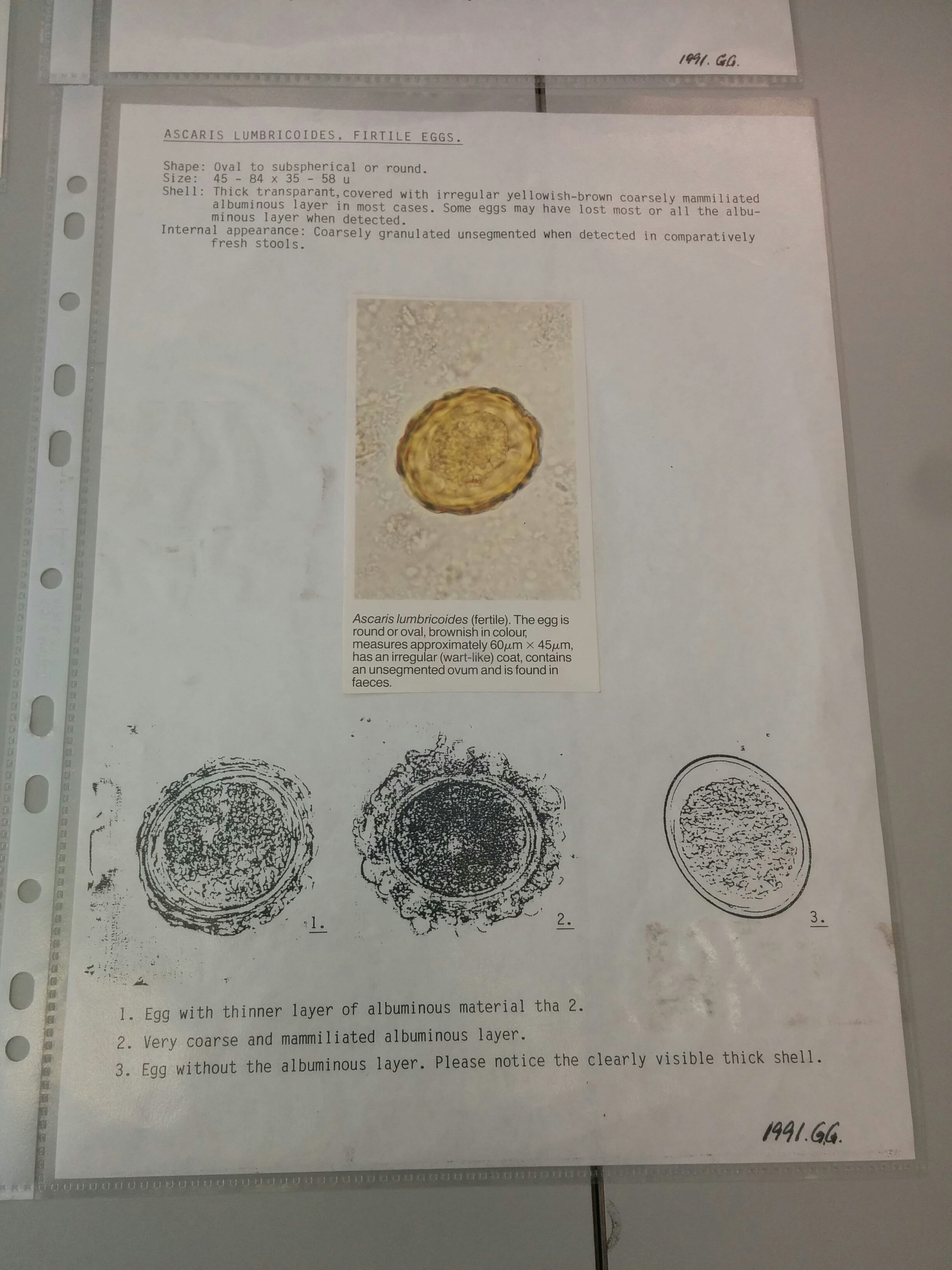
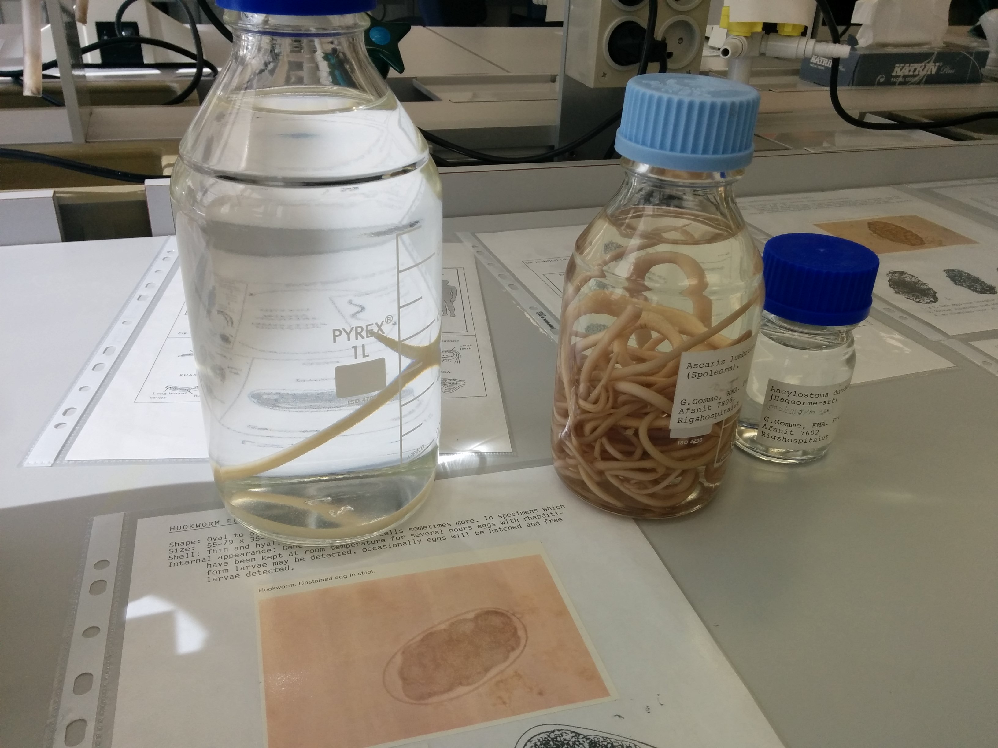
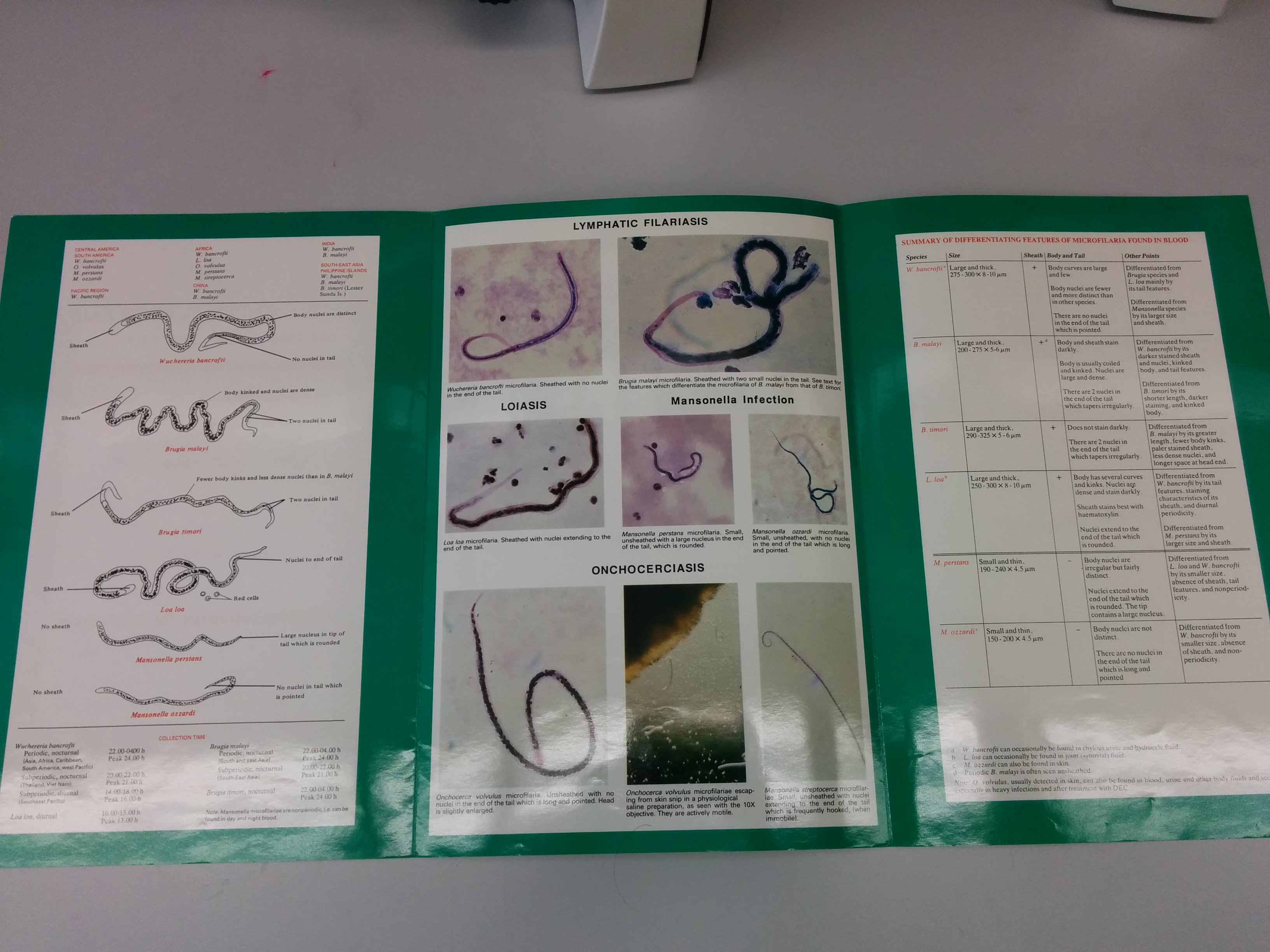
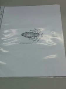
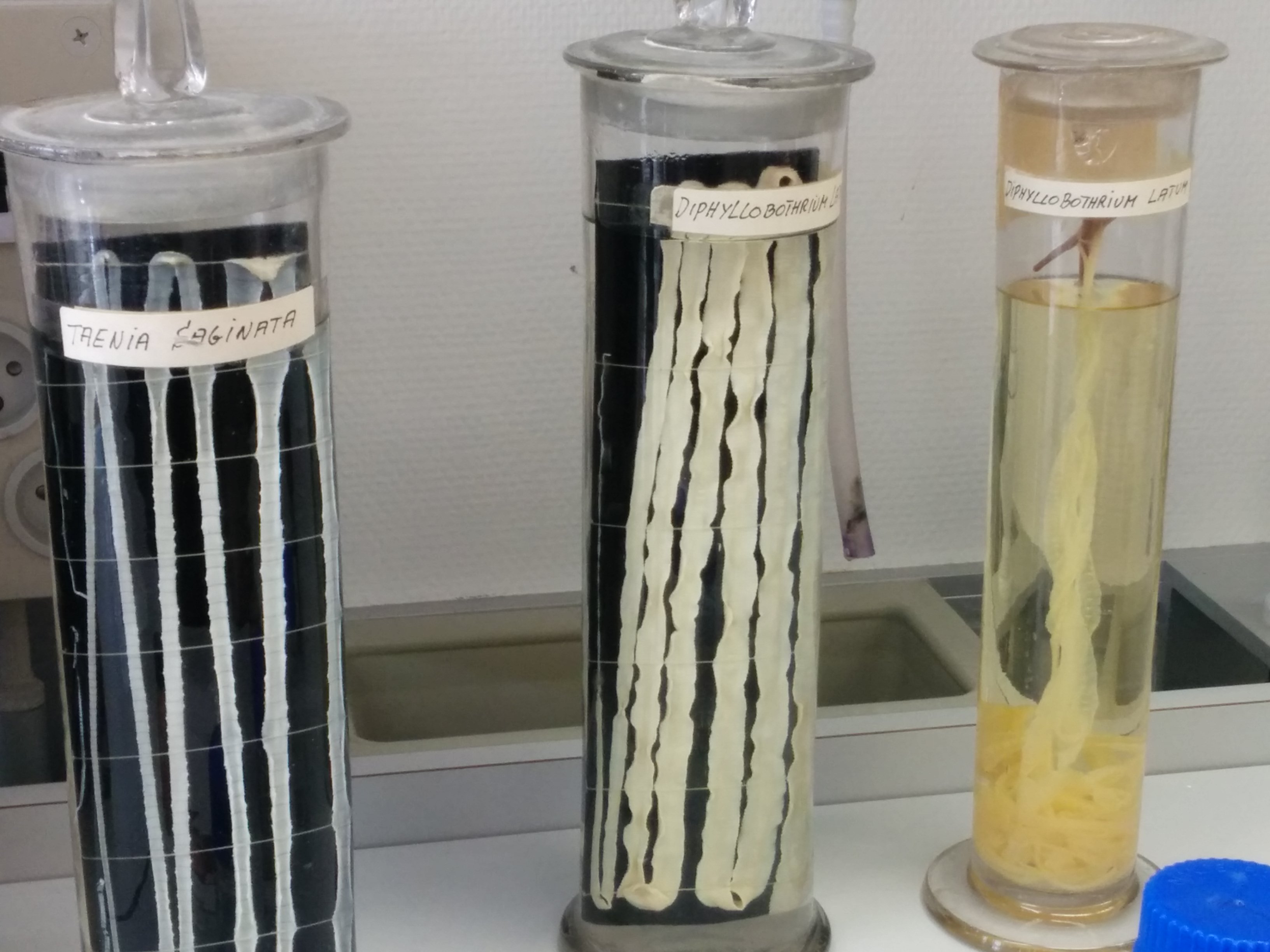
We were split into smaller groups to fit into each lab room. I started in the room full of eggs, worms, crabs, lice, TB and a few other things. We got a brief description of what each one was, and were sent off to explore. I started with the eggs.. hmm some interesting looking blobs! Each microscope was set up with a description of what you were looking for along with a picture and some interesting facts, I liked that part. I moved on. To the worms… seriously.. ew. The jar below with like 20-30 worms in it.. (brace yourself) those all came from one person! and that person, poor soul, had to poop them out after taking ‘de-worming’ medication. All I have to say to that is.. NO THANK YOU! but the slides were pretty cool, most of them were labelled male and female and really, there aren’t too many differences unless you know what you are looking for. Typically the males have a curl in their tail (also seen in the jar worms), and the females you can actually see the eggs inside of her, little dots inside near one of the ends. After the worms, I went over to the next row and that was almost worse. I had lice when I was a kid, who hasn’t – it’s a very common thing, but of course you never really know what a lice looks like because they are just soooooo small! Well I assure you, you probably don’t want to know what they look like. They literally look like little bugs and these ones in the slides, you could see their little claws clinging to hair. After the lice, crabs, I got to look at crabs and I don’t mean the ones you find on the beach. These guys are creepy looking little things resembling a crab. They also had their little claws hooked on to pubic hairs inside the slides and again I have to say NO THANK YOU!
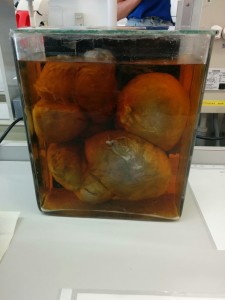
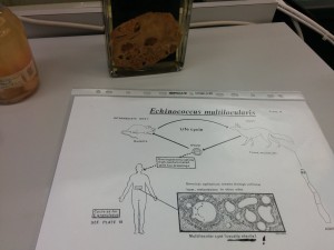
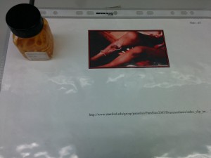
Although not part of the lab there were jars with tape worms in them, cysts caused by parasites and worms found in a foxes liver (which are infectious to humans as well) and it is kind of hard to see but a worm that actually just travels around under a thin layer of skin. The pictures below are these. and yes those big ball things are cysts that were cut out of a human.
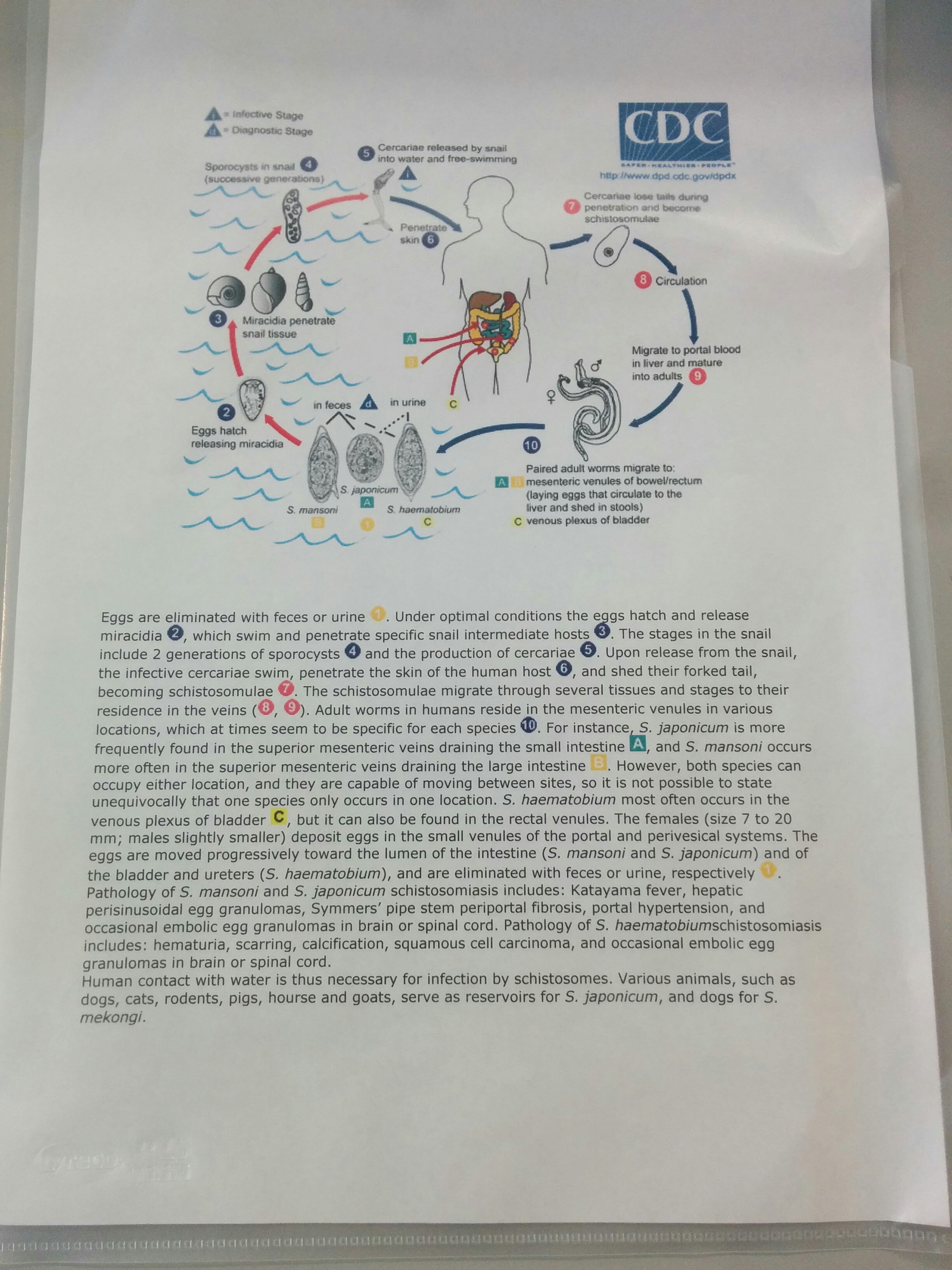
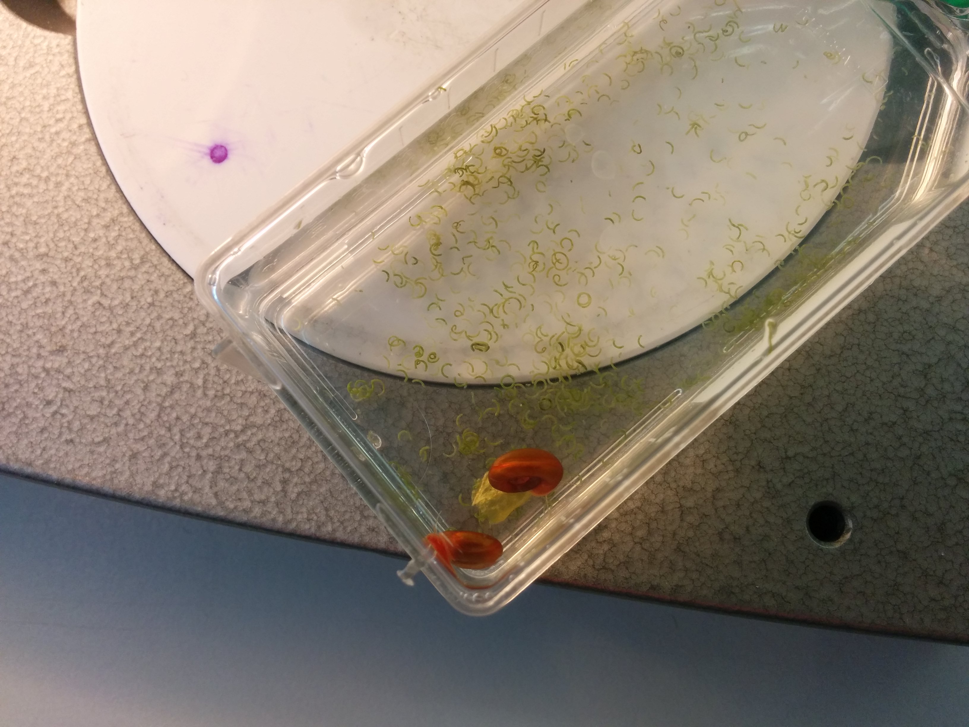
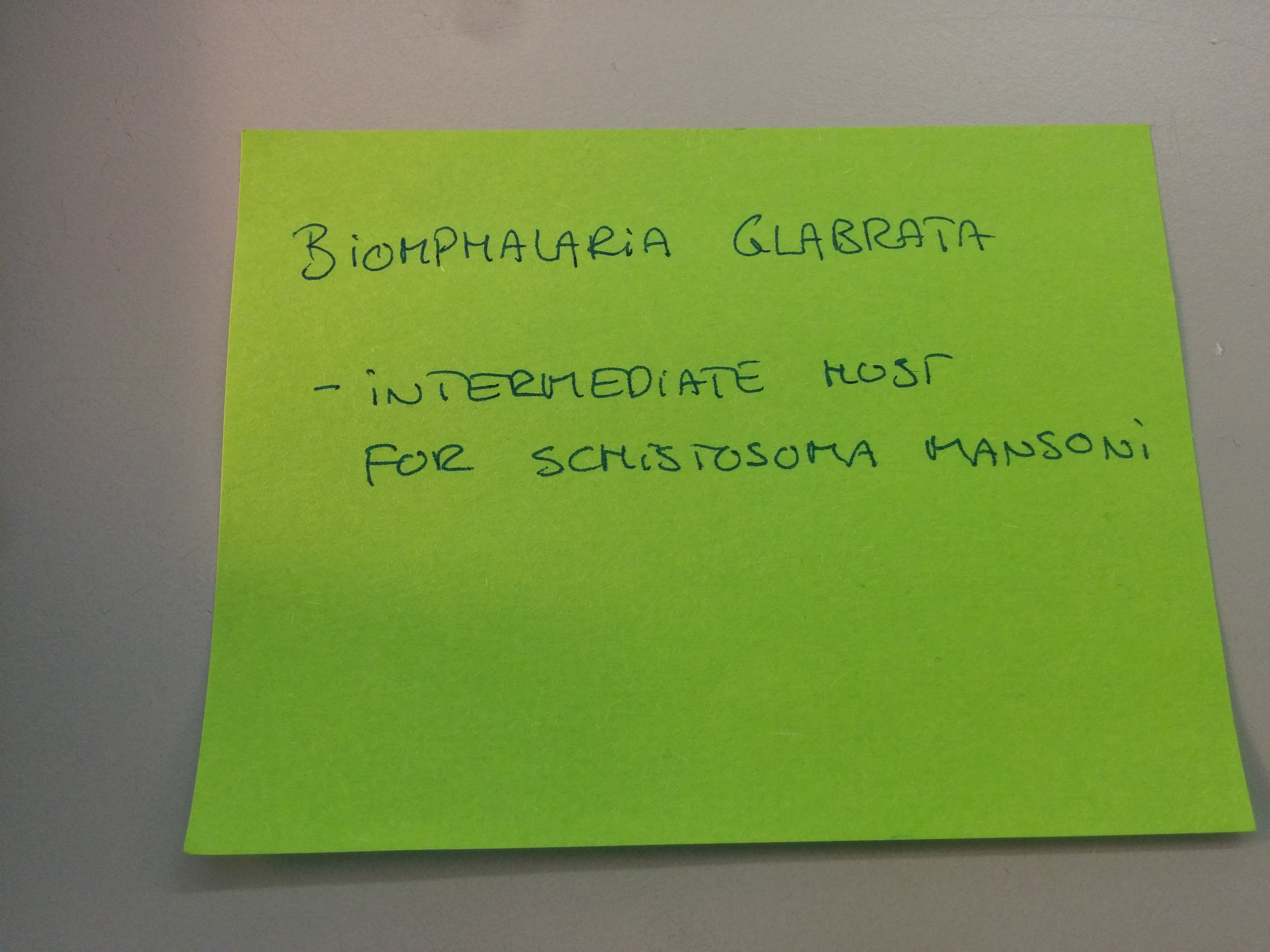
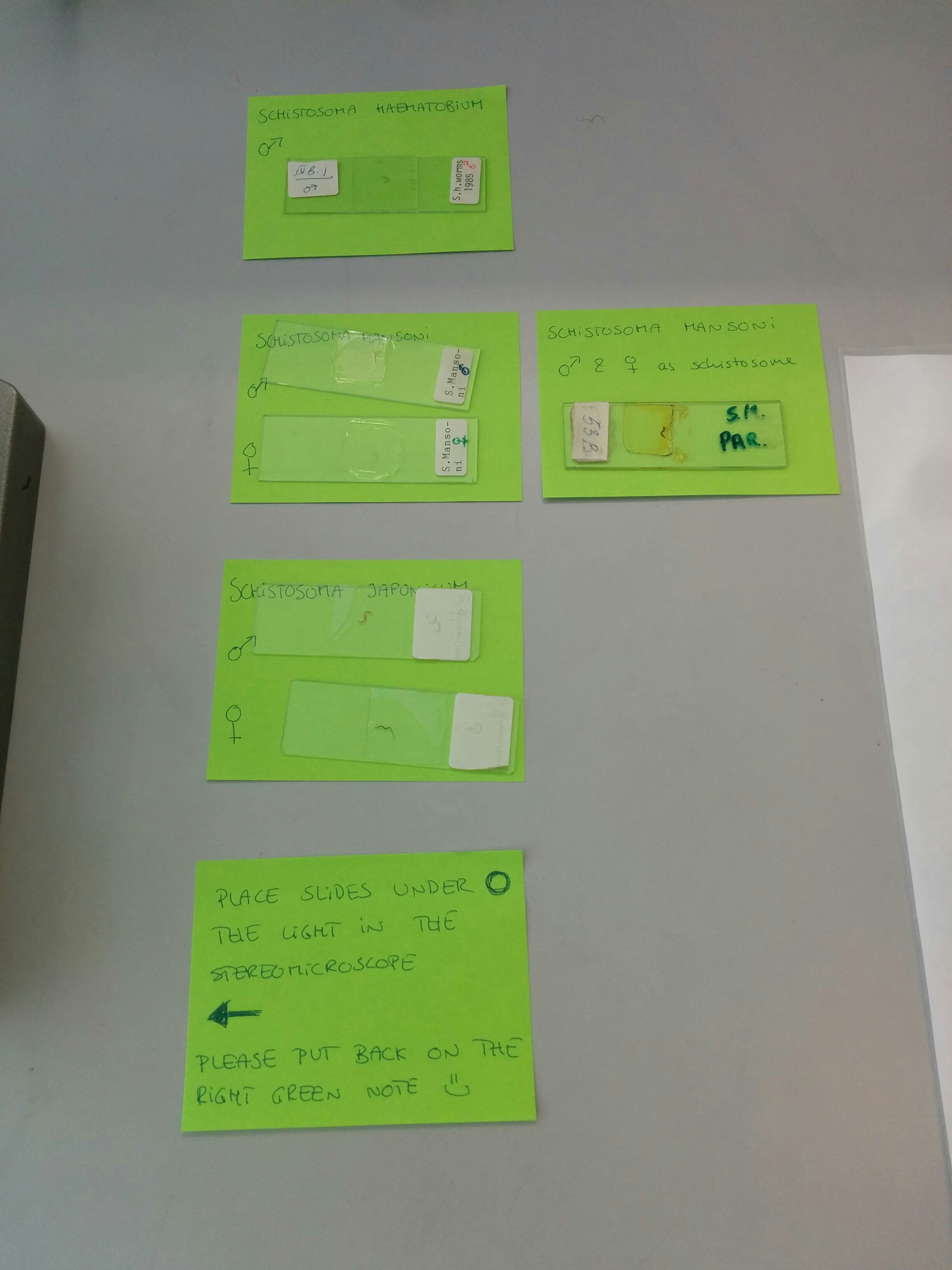
The second portion was about schistosomiasis. We learnt and got to look at the fresh water snails that are the intermediate host before the larvae infect a human (seen below). We got to make fecal slides (with peanut butter don’t worry) and look at fake urine samples. The urine samples were mostly just to be able to differentiate between color, how to use dip sticks and how to test for schistosomiasis. The slides included a real fecal sample, a few urine filter samples, a look at the snails (below) and many slides on the different forms, ages and eggs of the disease. The last photo there had many slides, a female and male from each form of the disease (dependent on geographical area) and one slide that was actually a female and male together just after they had bonded.. because really all they do is once they are inside, they travel to the liver and toward the bladder or intestines and meet a male or female and make babies.. and that’s all they do, so that was fairly interesting to see.
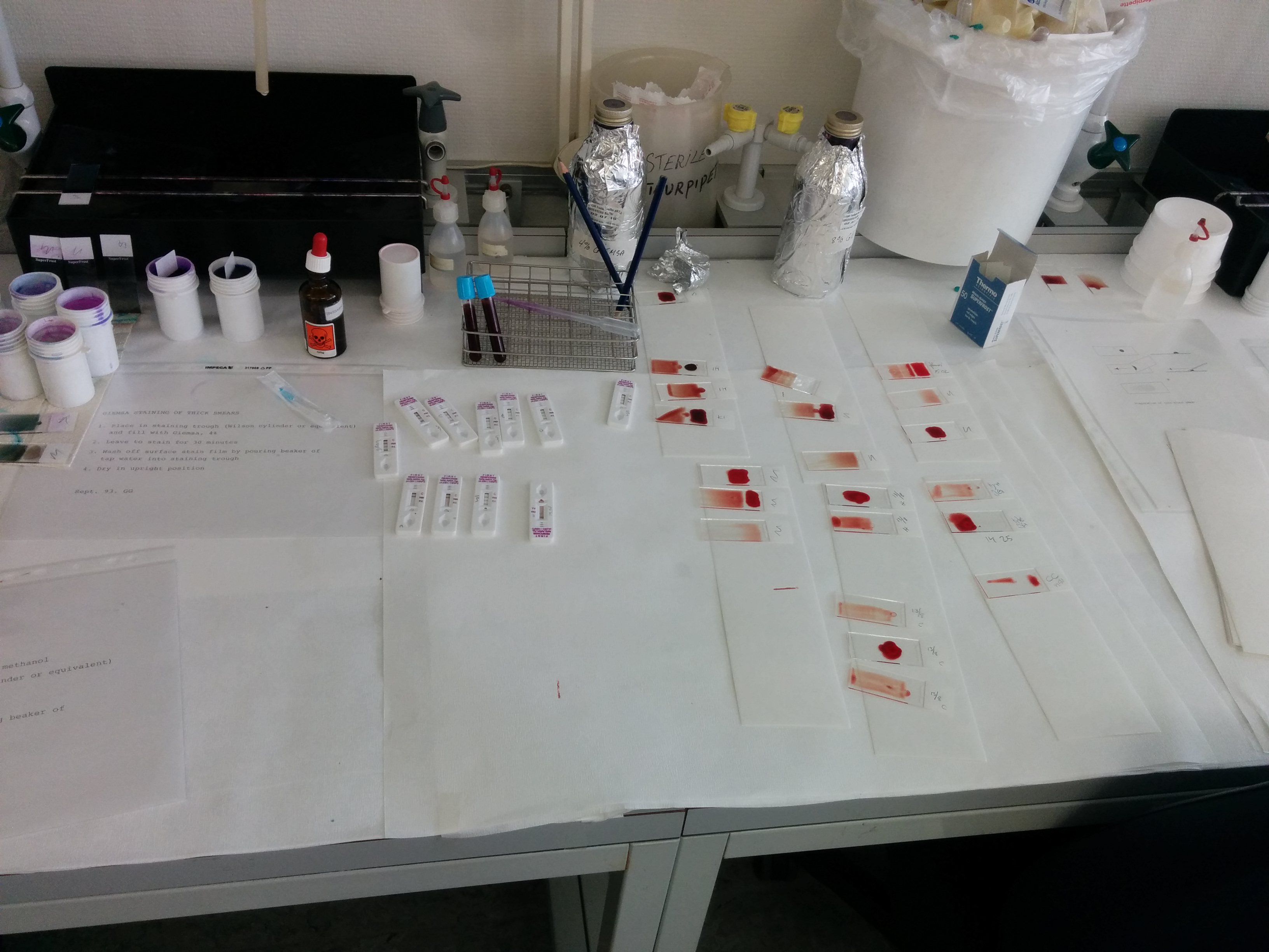
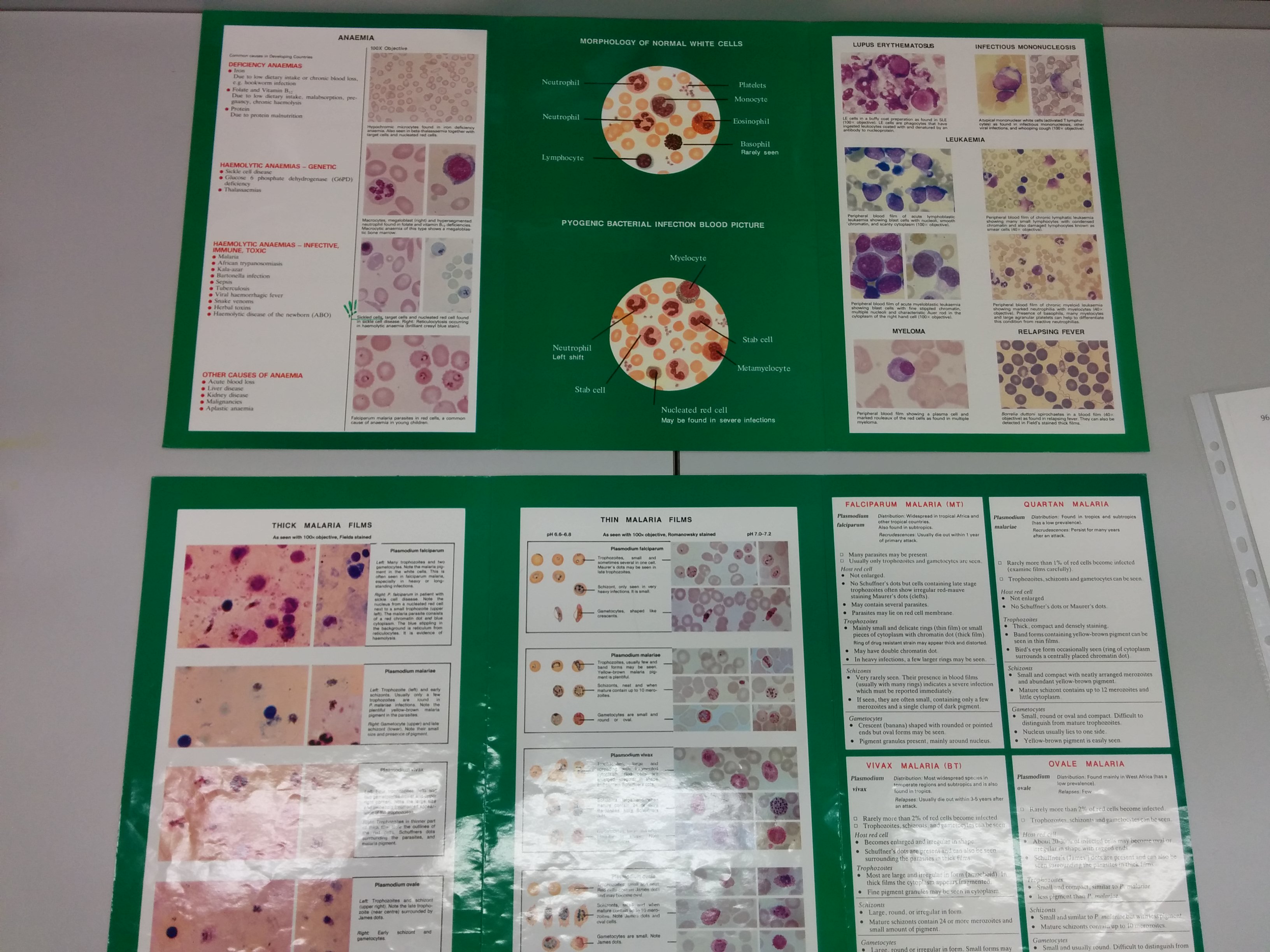
The last section was on malaria! We learnt how to do blood smears, thick and thin. We were taught and got to make our own rapid tests for malaria (mine was negative.. woo!). It was very interesting and fairly fun being able to stab people in the finger to get blood for the tests. We got to use infected blood to make the smears (cautiously wearing gloves of course). We made double slides – cost effective in countries with little resources – so half the slide is a thin blood smear and half the slide is a thick. So once you have made your thin smear, you fix it with chemicals and put the stain on. Each process takes about 30 minutes so it took a while, but it was actually very fun! After the stain was set you let it dry (another 20 mins or so – time for a coffee) and then we got to look at them in the microscope. There it was.. malaria! aka.. purple blobs.
Although I may never look into another microscope or use any of this, it was actually very interesting and who knows.. if I do end up going to a developing country to work in my field (Kinesiology) somehow, I may be asked to do stuff like this – they are severely understaffed in every region so you never know, if you have hands, a brain and can follow instructions, you’ll probably get used.