
One of the most fulfilling experiences in my undergraduate career thus far is my involvement in research at BC Children’s Hospital with neurologist Dr. Vann Chau. We are looking at how seizures affect brain maturation in children who have congenital heart disease (CHD). This is when a baby is born with abnormalities in their heart structure or function, which leads to problems with blood circulation. One example is “atrial septal defect” (see image below)
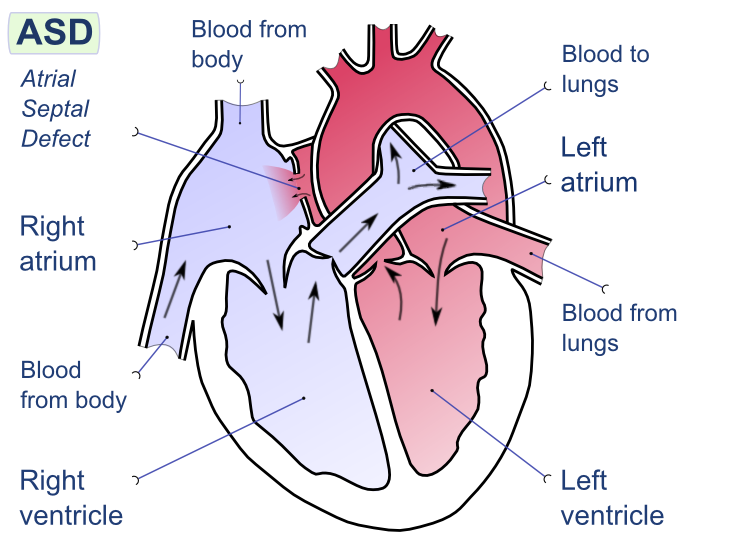
Atrial septal defect is where the septum separating the right from the left atria is missing (Image via Wikipedia)
After open-heart surgery, 15% of infants will have seizures— the most common neurological complication. Previous research using animal models has found that seizures can induce lasting brain injuries (Holmes, 2002). However, it is not known whether this is true in human newborns.

Cardiac surgery operating room at BC Children’s Hospital (With permission from Dr. Kenneth Poskitt)
Seizures are treated using anti-epileptic drugs which decreases excitation in the brain. However, there is a high risk for adverse effects when treating a newborn with anti-epileptic drugs. Thus it is not clear to doctors just how aggressive they should be with seizure treatment since it is unknown how seizures affect brain development in the first place.
This is where our study comes in. We are examining whether cases of CHD with seizures, compared to those without, is associated with disturbed brain development. If we do find that seizures are associated with alterations in brain development, then this would be compelling evidence that improved management of seizures may lead to better neurological outcomes.
So how exactly are we assessing brain development? We are using neuroimaging methods which I will write about for the remainder of this post!
(1) Standard Magnetic Resonance Imaging (sMRI)
This method allows us to detect stroke and white matter (nerve fiber) injury by looking at differences in the alignment of protons in the brain. Below is an example of a stroke injury.

sMRI showing a stroke injury in the upper left region of the brain (With permission from Dr. Kenneth Poskitt)
(2) Diffusion Tensor Imaging (DTI)
This is a specialized application of MRI which uses properties of water diffusion to visualize neuronal tracts (bundles of nerve fibers) in the brain. Below is an example of a DTI image.

DTI of the human brain. To the left of this image is the frontal part of your brain. (Image via Wikipedia)
(3) Magnetic Resonance Spectroscopy (MRS)
This method looks at differences in resonant frequencies to determine the concentrations of key metabolites in selected regions of the brain. Below is an example of a MRS spectra.

MRS spectra showing different peaks for each metabolite which appears at a known frequency (Image via Wikipedia)
Written by Vivian Kwan

