A “Nutty” Remedy for your Peanut Allergy?


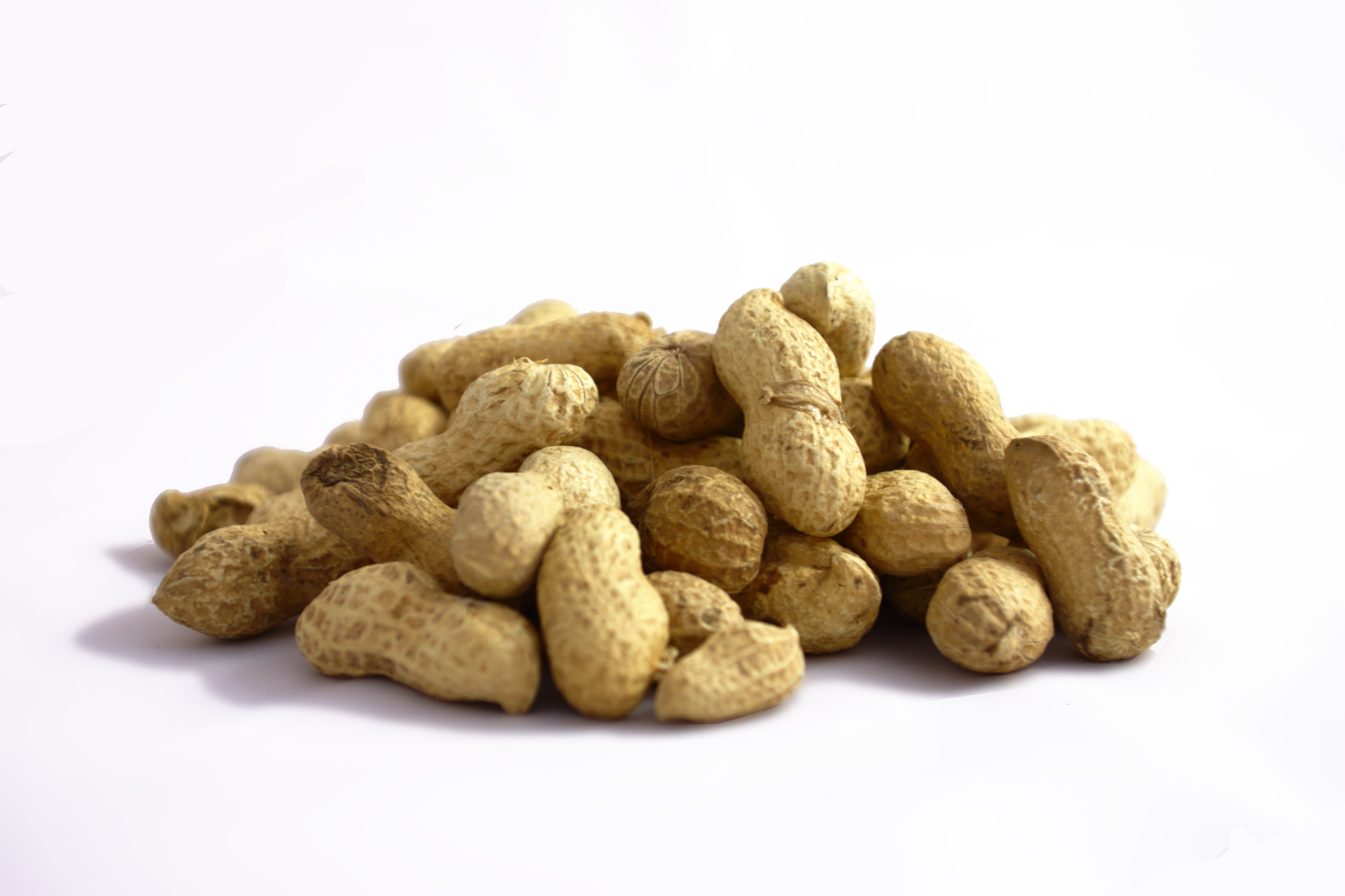
Image from Aktron on Wikimedia Commons

If you are allergic to peanuts, you must know how annoying and potentially deadly an allergic reaction is. It is the most common and severe food allergy, as it  affects one in fifty children. It causes problem with breathing and can even induce anaphylactic shock upon ingestion in severe cases. Peanut allergies can even go as far as making an impact on one’s social life as well when all action has to be taken to avoid making contact with peanuts at all cost. Needles to say, a peanut allergy is a big inconvenient. However, it seems as though scientists might have found a cure!

In an attempt to find a cure for the peanut allergy in children, scientists conducted a study where small increments of peanuts were exposed to children’s diet. They first started with peanut proteins equivalent to 1/70 of a peanut, then slowly increased the amount. After a few months, 88% of the participants built the tolerance to eat 5 peanuts a day, and 58% were able to eat as much as 10 peanuts. The experiment was carried out in two six-month periods; in the first six months, the children were given a placebo. Actual peanuts were prescribed in the second six months. No peanut tolerance was observed when the children were given the placebo, so the results in the end were definitely not due to the placebo effect. This study was recently published and the scientists hope that one day this will become a treatment for peanut allergies.

This is a video the details the overall experiment:

YouTube Preview Image


The peanut treatments were conducted in a controlled environment in case of the occurrence of an allergic reaction. This should not be tried at home. This study is still in it’s early stages and can not be considered a cure just yet. However, the results are significant and are a beacon of light for those who have severe allergies. If a cure for peanut allergy is possible, then perhaps a remedy for other allergies might someday be a reality as well. Hopefully, in the near future, allergies will no longer exist as a limit to people’s everyday activities.

By: Kimberley Xiao


References:

http://www.dailymail.co.uk/health/article-2548416/Have-scientists-way-cure-peanut-allergies-children.html

http://www.popsci.com/article/science/potential-cure-peanut-allergy-successful-test?dom=tw&src=SOC

http://www.thelancet.com/journals/lancet/article/PIIS0140-6736(13)62301-6/abstract


A ROCKY START: James Hutton and the Age of the Earth

It’s easy to mistake rocks as static objects.  You could stare at a rock for a few hours, and generally, it will do nothing interesting.

But the truth is, rocks are anything but static; it’s just that their dynamics are hidden by the long spans of time required for them to transform.  They erode, they shift, they change, and ultimately, they tell stories.  Rocks are the record keeepers of the Earth.  As we look downwards, through layer after dusty layer of sediment, we can, in a sense, travel backwards through time.

Rocks provide an extremely revealing way of examining the history of our planet, but it took until the late 18th century for scientists to fully grasp this concept.

In 1785, Scotland was at the forefront of Western science and philosophy, during a period of time later dubbed the Scottish Enlightment.  Despite recent advances in naturalism, chemistry, and medical science, people still thought that the earth 6000 years old, an estimate derived from the book of Genesis in the Bible.  Most scientists of the time agreed with this.  Even Isaac Newton (who died in 1727) accepted the idea of a young Earth.

But not everyone was convinced.  One man, James Hutton, had a very different idea.

James Hutton was a Scottish farmer, born in Edinburgh.  He had a degree in medicine, but by all accounts, never practiced medicine.   Hutton was an amiable and insatiably curious man, and initially applied his mind to developing and optimizing new farming techniques. At the same time, he had a much more ambitious pet-project on his mind – developing a geological theory of the Earth.

Hutton’s theory was based on observations, and asserted that rocks are constantly being formed, shifted and eroded; Hutton further concluded that these natural process likely behave in the same way now as they did thousands, even millions of years ago.

One of the primary pieces of evidence that Hutton used to support his theory is a rock on the east coast of Scotland called Siccar Point.   Siccar Point has an unusual structure – it is made up of two distinct layers of different types of sedimentary rocks (Devonian red sandstone, and Silurian greywacke) that contact each-other at a definitive angle.

Hutton concluded that Siccar point could only have been formed by a long sequence of sedimentation (formation of sandstone from small particles), folding and uplift (the buckling and lifting of rock masses over time) and erosion (the breakdown of rock surfaces by weathering), requiring extremely vast amounts of time – amounts far exceeding the mere 6000 year timeline proposed by biblical scholars.

Today, scientists have a variety of tools at their disposal for determining the age of the Earth, including the radiometric dating of fossils.  Although Hutton had no access to these types of techniques, he was still able to conclude that the 6000 year idea was incorrect using observations of modern sediments, and deductive reasoning.  It is a powerful example of one person’s curiosity and logic overcoming centuries of well-entrenched religious and scientific dogma.


Text and illustrations by Sam MacKinnon, 2014


REFERENCES:

Carruthers, M. W.  (2014).  Hutton’s Unconformity.  Natural History.  108(5): 86.

Repcheck, J.  (2003).  The Man Who Found Time:  James Hutton and the Discovery of Earth’s Antiquity.  Boulder, Colorado: Perseus (Basic Books). 


Sleep? Who has time for that?

Image: Clipart

For university students, balancing studies with time spent with your significant other can be fairly difficult. However, it is not the most prohibitive task they endure. Balancing school work with your sleep can be one of the most difficult things, as a student, to manage. It is suffice to say that through the progression from elementary to secondary school, and now to university, a vast majority of students can attest to the decrease in the amount of sleep they get each night. Most students’ can admit to falling asleep during lectures or even dozing off while studying. The matter in question is; how much of an effect does this lack of sleep have on grades? Studies show that the less sleep students get, the more their academic performance suffers.

One of the more recurring opinions regarding the lack of sleep university students get is, “I just don’t have the time”. Taking into account that most students wake up early for lectures, commute to and from campus, stay up late doing assignments and studying for a number of exams is reason enough to blame time constraints. Add in a part time job to pay for their tuition, along with a modest social life, these claims undoubtedly justify that they simply don’t have enough time to get the sleep required to sustain quality academic performance.

According to Medical News Today , only 30% of students get the required 8 hours of sleep a night, 20% of students pull an all-nighter once a month, 30% stay up past 3:00am and 12% of these students miss class three or more times a month. The main cause for students staying up late is the stress of having to do well in school. This stress can then affect their sleep, more so than relying on drugs such as  alcohol and caffeine.

YouTube Preview Image

Youtube video from a BBC article explaining how lack of sleep impairs learning.

Ultimately, it comes down to a student’s preparedness regarding how much sleep they’ll get in a given night. As outlined in this article from the UBC website, students must rid themselves of the notion that less than eight hours of sleep is sufficient enough to perform well in school. Focusing on getting a healthy sleep and to keep the mind fresh should be a primary concern for all students. Developing  a consistent and healthy sleep schedule, mapping out your day, while avoiding drugs like caffeine and alcohol, should help with falling asleep comfortably and lessen any chance of facing academic repercussions.

YouTube Preview Image

Youtube video from Watch Well Cast explaining ways to sleep better.

– Inderbir Bhullar

Snow Science!

Photo taken from snowcrystals.com

We are all taught at a very young age that every snowflake, much like a zebra or a fingerprint, is unique. However, despite being told that no snowflake is like another, we are never told why. In order to determine why each snowflake is unique, let’s look at snowflakes with more detail, and let’s discuss how a drop of water is transformed into a snowflake in the first place.

Through the observation of close-up photographs, it can be seen that snowflakes come in many different shapes and sizes; needles, columns, dendrites (the “perfect” snowflake) are among the many forms that they can take. The key factors that determine the final geometry of a snowflake are the temperature and relative humidity of the air. Data compiled by the Alaska Satellite Foundation plots just how a snowflake’s form varies due to these factors.

Now that we are beginning to understand some factors determining the variation of snowflakes, we can start to explore the formation of the crystal itself. The first ingredient to a snowflake is its nucleus. The snowflake nucleus is essentially a dust or pollen particle, or ice crystal that resides high in the atmosphere.

Geometric patterning of snowflake formation due to variance in temperature and humidity.
Photo from http://www.thenakedscientists.com

As it passes through a nimbus cloud, water molecules begin to aggregate onto the nucleus and form the familiar hexagonal shape of a snowflake. As the young snowflake descends, water molecules continue to condense onto the flake, creating the branches.


The following video from the popular YouTube channel “It’s Okay To Be Smart” illustrates the geometric reasoning behind the hexagonal shape of a snowflake on a molecular level.

YouTube Preview Image

Link: https://www.youtube.com/watch?v=fUot7XSX8uA

It is evident that there is a great deal of structure and order in a snowflake, but there is also a random, almost chaotic, influence in its design. The understanding that scientists have gained from snowflakes has been a valuable asset in many studies. For example, this study published by the American Physical Society (AMS) in 2004 compares the growth of a macromolecule by sedimentation with the formation of a snowflake.

So, next time you are out for a peaceful walk in the snow and a snowflake lands on your hand, you can truly appreciate what goes into its formation.

By: Kia Sanjabi

References

  1. Westbrook, C. D., Ball, R. C., Field, P. R., & Heymsfield, A. J. (2004). Theory of growth by differential sedimentation, with application to snowflake formation. Physical Review E, 70(2), 021403.
  2. Zentile, C. (2007). The Science of Snowflakes, Are no two ice-crystals alike? Retrieved 01/27, 2014, from http://www.thenakedscientists.com/HTML/articles/article/science-of-snowflakes/

Using nano-particles to protect teeth against bacterial damage.

Teeth are a very important part of our bodies. They play a significant role in the primary breakdown of food. But if not properly treated several complications can arise and cause deterioration or severe damage to our teeth. To protect our teeth from damage it is recommended to brush our teeth twice a day and floss on a regular basis. But sometimes despite taking precautions problems can arise, and dental problems often begin with plaque forming on the surface of teeth.  Biofilm formation also known as dental plaque has been identified to be the cause of many dental diseases such as dental caries, gingivitis, periodontitis, cavities etc.

http://thumbs.dreamstime.com/x/cartoon-tooth-dental-cavity-3234654.jpg

As a solution to protecting our teeth against damage scientists have discovered that coating the teeth with a layer of silver nano-particles, prevents biofilm formation on dentine surface as well as inhibits bacterial growth in the surrounding media.

Several different metal nano particles were compared in this study and various experiments were conducted to find the best metal nano particles. The results of the experiments suggested that silver nano particles are the most effective against pathogens as the silver coatings are found to be most susceptible to bacterial adhesion on the dentin surface.

YouTube Preview Image

Video Showing Nano particles in action

Furthermore it was discovered that silver nano-particle coatings do not affect the color of the dentine, unlike other metal coatings, which cause the dentine to appear discolored. Silver nano-coatings are also found to maintain their integrity (shape and chemical makeup) over time. Preventing the need for frequent re-coatings over a short period of time.

http://www.scielo.br/img/revistas/bdj/v20n4/v20a03f01.jpg

Dentin in teeth has several openings of dentinal tubules on its surface.  These openings are what that allows for dental plaque to form easily and bacteria can stick/adhere to the surface easily. Coating the dentin surface with silver nano particles fills these dentinal tubules,  which in turn prevents plaque formation.

Dentin Surface after the coating has been applied.

 http://www.jdentlasers.org/articles/2012/6/2/images/JDentLasers_2012_6_2_51_106653_u4.jpg

Thus the silver nano particle coating is the best way to protect our teeth. Not only is it safe, but it also does not cause implications such as changes in teeth color and nor does the coating have to be frequently reapplied. Lastly, to ensure that our teeth last us a lifetime it would be great to invest in such a procedure.

References:

Besinis A, De Peralta T, Handy RD. Inhibition of biofilm formation and antibacterial properties of a silver nano-coating on human dentine. Nanotoxicology. 2014, 8, 745-754.

By : Nitish Khosla

Cloning Organs for Transplant?

Although there have been great improvements in medical technology and an obvious increase in the number of donors over the past years, the supply of organs available for transplant is far lower than what we need. An average of 18 people die each day from the lack of available organs. More than 4,300 Canadians are currently waiting for an organ transplant.

We often hear about cloning plants and animals but have you ever imagined that you could clone your organs and store it somewhere safe in case you need a heart or liver transplant? Organ transplants itself are difficult to carry out because firstly, it is hard to find a donor, and secondly, there is no guarantee that your body will accept the new organ due to the rejection of tissues. However, if we could clone organs and produce an endless supply of transplants, people will no longer have to fear not being able get a donor.

So how does cloning organs work?

Figure. 1 Stem Cell Process

First, let’s talk about cloning itself. The most common method of cloning is somatic cell nuclear transfer (SCNT). SCNT involves removing the nucleus from a host’s egg. With the lone nucleus and an empty egg cell, the nucleus can then be fused with the DNA from the organism that is to be cloned. The cell is then incubated; and within a few weeks, the cells will multiply and form a blastocyst (early stage embryo) with almost identical DNA to the original organism. Scientists could theoretically clone organs with SCNT through this process and extracting the stem cells from the blastocyst could produce the desired organ. Nevertheless, coaxing the framework for the stems cells to grow in will require further research.

The video below will further discuss the cloning of organs:

The most obvious benefit of cloning organs is that it is an easy replacement of internal organs and tissues for patients in need of transplants rather than having to wait for suitable organ donors and since the cloned organ contains patient’s own cells and tissue, it will lessen the chance of rejection. Conversely, the biggest issue that arises from this topic is whether it is ethical to kill an embryo in order to produce its stem cells which that can be further cultivated into an organ for transplant purposes.  According to McGill’s Journal of Medicine, a new technique device shows that the manipulation of the cells can be done without killing the embryo during the process. If that’s the case, there will be nothing morally compelled and the process of organ cloning would be ethical to carry out.

While further research and engineering still needs to be made for cloned organs to work on the human body, it is almost certain that most body parts will eventually be replaceable and we will have an unlimited supply of organs for transplant.

– Rubina Lo

References:

http://www.ncbi.nlm.nih.gov/pmc/articles/PMC2323472/

http://learn.genetics.utah.edu/content/cloning/whatiscloning/

Common Misconceptions of Delayed Onset Muscle Soreness (DOMS)

Delay Onset Muscle Soreness
Source


A common barrier while attempting to persevere through an intensive long-term exercise program is having to deal with muscle soreness afterwards, which is also known as delayed onset muscle soreness (DOMS). Unlike the acute soreness that develops during the actual activity, delayed soreness becomes prominent approximately 24 hours after exercising, and is positively correlated to the duration and intensity level exerted. Although various studies have been performed regarding DOMS, the ultimate cause of the symptom is still unknown. However, there has not been any report of DOMS posing as a permanent threat to the body.

Most people unfamiliar with DOMS are often deceived into believing that there is a correlation between pain and injury, where continued exercise while experiencing DOMS leads to further injuries. As a result, many simply stop their exercise routines and turn to treatments such as taking a hot shower or using heat pads to alleviate the pain, which may take up to a week after the initial symptom. However, by that time, your muscles will have decreased its ability to adapt to the intensity level of your routine, and that hour of intensive exercise you performed earlier becomes a waste.

The main cause of DOMS still remains unknown, but many studies have investigated the mechanisms involved and suggested possible explanations. First, studies performed by Armstrong demonstrate that high intensity exercise result in greater metabolic waste, such as lactic acid, which may influence the calcium concentration gradient within the muscle tissue and stimulate neuron activity to increase pain sensation (1). Hough suggests that DOMS is related to the rate and force of muscle contraction during strenuous exercise, which leads to structural damage (2). Supporting Hough’s findings, Kumazawa et al. also suggest that elevated temperature plays a role in damaging muscle structure and promotes necrosis of muscle fibres and connective tissues (3). In brief, there is no main cause of DOMS. It is most likely caused by a combination of various factors, which leads to the difficulty in developing a treatment that can efficiently eliminate DOMS.

Compression to help reduce Delay Onset Muscle Soreness Source

There are various treatments for DOMS such as having a proper cool-down period after exercising, taking a hot shower, or wearing compression sleeves. However, continuing to exercise while experiencing DOMS is actually the only proven way to effectively eliminate DOMS. According to Armstrong, one the reasons why continued exercise can reduce DOMS is its ability to decrease the rate of muscle fibre necrosis (1). There has also been evidence for the reduction of exercise plasma enzymes, which indicates that continued exercising can reduce muscle fibre injuries (4). Lastly, lysosomal enzyme levels have been shown to decrease while exercising with DOMS, resulting in the reduction of the rate of muscle cell death (5). Therefore, although it may be difficult to motivate yourself while experiencing DOMS, the most effective way to treat DOMS is actually to continue exercising until your muscles adapt to it.

-Bailey Lei

References

1. Armstrong, R.B. Mechanisms of exercise-induced delayed onset muscular soreness: a brief review. Med. Sci. Sports Exerc. 16(6): 529-538, 1984.

2. Hough, T. Ergographic studies in neuro-muscular fatigue. Am. J. Physiol. 5:240-266, 1901.

3. Kumazawa, T. and K. Mitzumura. Thin-fibre receptors responding to mechanical, chemical, and thermal stimulation in the skeletal muscle of the dog. J. Physiol. (Lond.) 273: 179-194, 1977.

4. Schwane, J.A. and R.B. Armstrong. Effect of training on skeletal muscle injury from downhill running in rats. J. Appl. Physiol. 55: 969-975, 1983.

5. Vihko, V., A. Salaminen, and J. Rantamaki. Exhaustive exercise, endurance training, and acid hydrolase activity in skeletal muscle. J. Appl. Physio. 47: 43-50, 1979.