Many small molecules are crucial to life—not only are they used for life-saving drugs, but they also regulate important biological processes within our bodies. In order to understand these small molecules better, scientists have developed various methods to probe what atoms they are made up of, and how these atoms are connected.
Yet one of the newest and most promising of these methods is not so new at all—in fact, it has been a mainstream tool in the field of structural biology for years.
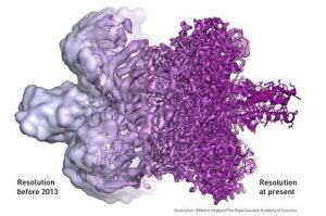
The imaging resolution of biological molecules before and after cyro-EM. Source: Martin Högbom/The Royal Swedish Academy of Sciences.
In 2017, the scientists Jacques Dubochet, Joachim Frank, and Richard Henderson received the Nobel Prize in Chemistry for the development of a powerful microscopic technique known as “cryo-EM”—so named because any samples used must first be cooled to very low, or cryogenic, temperatures. According to the Royal Swedish Academy of Sciences, cryo-EM has “moved biochemistry into a new era” by producing clear, high-resolution structures of biological molecules.
Now, cryo-EM may be poised to shake up the world of organic chemistry as well. In October 2018, researchers from UCLA and Caltech teamed up to show that cryo-EM can be used to determine the structure of small, organic compounds. According to Hosea Nelson from UCLA, their study in ACS Central Science proves that cryo-EM is a “game-changer” for organic chemists who want to perform “rapid unambiguous structure determination” on unknown compounds.
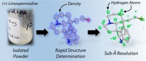
Source: ACS Central Science (Jones et al. 2018)
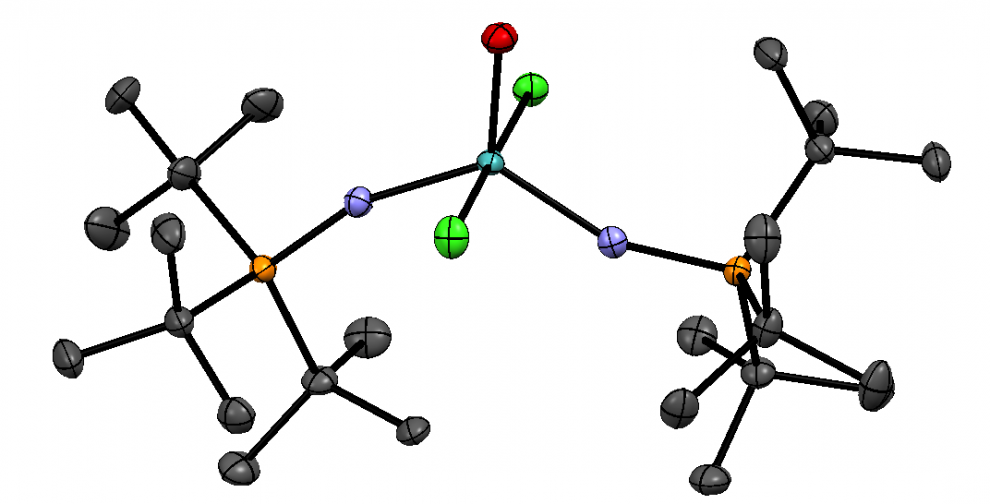
The researchers used cryo-EM to determine the atomic structure of twelve compounds and four powder mixtures that could not be imaged by X-ray crystallography. In cryo-EM, electron beams are directed at the sample, and a structure can be obtained from how these electrons diffract. Within minutes, the correct structures were revealed.
According to Robert Grubbs, a 2005 Nobel Laureate in Chemistry who is unaffiliated with this study, this technique “promises to revolutionize organic chemistry”. Compared to X-ray crystallography, cryo-EM offers benefits in terms of “minimal sample preparation and experiment time”. Furthermore, only crystals of a single compound can be imaged by X-ray crystallography, while cryo-EM can be used on mixtures or very small amounts of powders.
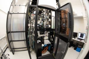
A Krios electron microscope at the Stanford-SLAC cryo-EM facility. Source: SLAC on Flickr.
One limitation of this technique is that “cryo-EM microscopes are very expensive, and not many institutions have them right now,” says the Salk Institute geneticist Dmitry Lyumkis. Another is that even if the samples being analyzed do not have to be crystals, they still have to be crystalline.
However, the UCLA/Caltech researchers remain optimistic about the potential of cryo-EM to become widely used in organic chemistry. According to Brian Stoltz, a Caltech professor and one of the project co-leads, “so many ideas are flying around”.
My opinion is that as more institutions gain access to expensive but useful cryo-EM technology, cryo-EM could play an important role in accelerating drug discovery or the characterization of natural products. For example, many compounds from marine sponges have anticancer potential but are structurally complex. As Stoltz said, “it will be exhilarating to see where this will lead.”
— Jessica Li
References
- Cressley, D.; Callaway, E. Cryo-electron microscopy wins chemistry Nobel. https://www.nature.com/news/cryo-electron-microscopy-wins-chemistry-nobel-1.22738 (accessed Jan 22, 2019).
- Thermo Fisher Scientific. The History of Cryo-EM. https://www.fei.com/life-sciences/history-of-cryo-em/ (accessed Jan 22, 2019).
- Royal Swedish Academy of Sciences. Press release: The Nobel Prize in Chemistry 2017. https://www.nobelprize.org/prizes/chemistry/2017/summary/ (accessed Jan 22, 2019).
- Extance, A. CryoEM method offers organic analysis certainty. https://www.chemistryworld.com/news/cryoem-method-offers-organic-analysis-certainty/3009644.article (accessed Jan 23, 2019).
- Jones, C.G.; Martynowycz, M.W.; Hattne, J.; Fulton, T.J.; Stoltz, B.M.; Rodriguez, J.A.; Nelson, H.M.; Gonen, T. The CryoEM Method MicroED as a Powerful Tool for Small Molecule Structure Determination. ACS Cent. Sci [Online] 2018. 4(11), 1587-1592. https://pubs.acs.org/doi/10.1021/acscentsci.8b00760 (accessed Jan 22, 2019).
- Broadwith, P. Explainer: What is cryo-electron microscopy. https://www.chemistryworld.com/news/explainer-what-is-cryo-electron-microscopy/3008091.article (accessed Jan 23, 2019).
- Caltech. From Beaker to Solved 3-D Structure in Minutes. http://www.caltech.edu/news/beaker-solved-3-d-structure-minutes-84400 (accessed Jan 23, 2019).
- Salk Institute for Biological Studies. Electron microscopy provides new view of tiny virus with therapeutic potential. https://www.salk.edu/news-release/electron-microscopy-provides-new-view-of-tiny-virus-with-therapeutic-potential/ (accessed Jan 23, 2019).
- Halford, B. Electron crystallography could be a powerful tool for organic chemists. https://cen.acs.org/analytical-chemistry/Electron-crystallography-powerful-tool-organic/96/i43 (accessed Jan 23, 2019).
- Calcabrini, C.; Catanzaro, E.; Bishayee A.; Turrini E.; Fimognari, C. Marine Sponge Natural Products with Anticancer Potential: An Updated Review. Mar. Drugs [Online] 2017. 15(10), 311. https://www.mdpi.com/1660-3397/15/10/310 (accessed Feb 15, 2019).