Category Archives: Biological Sciences

Plastic replacements: some new conSQUIDerations

The amount of plastic that has been produced to date now exceeds 8300 million metric tonnes (Mt). To put this into perspective, the average blue whale weighs approximately 180 Mt, thus 46 million blue whale’s worth of plastic has been produced since humans started commercializing plastics in 1950. Our society had become extremely dependent on plastic products and synthetic (petroleum- based) textiles which cause serious consequences such as microplastic and microfiber pollution as I’ve discussed in previous blog posts.

Figure 1. Size reference for blue whales. Wikipedia Commons

Bioplastics have more recently taken the stage as a potential avenue for replacing petroleum-based plastics. Biologically based polymers have structural elements such as helices, β-turns, β-sheets and coils which provide structural integrity and resilience and can replicate the desirable polymeric interactions in plastics. Additionally, a lot of new material is being developed based on the protein polymers that are naturally occurring in biological systems (including ourselves).

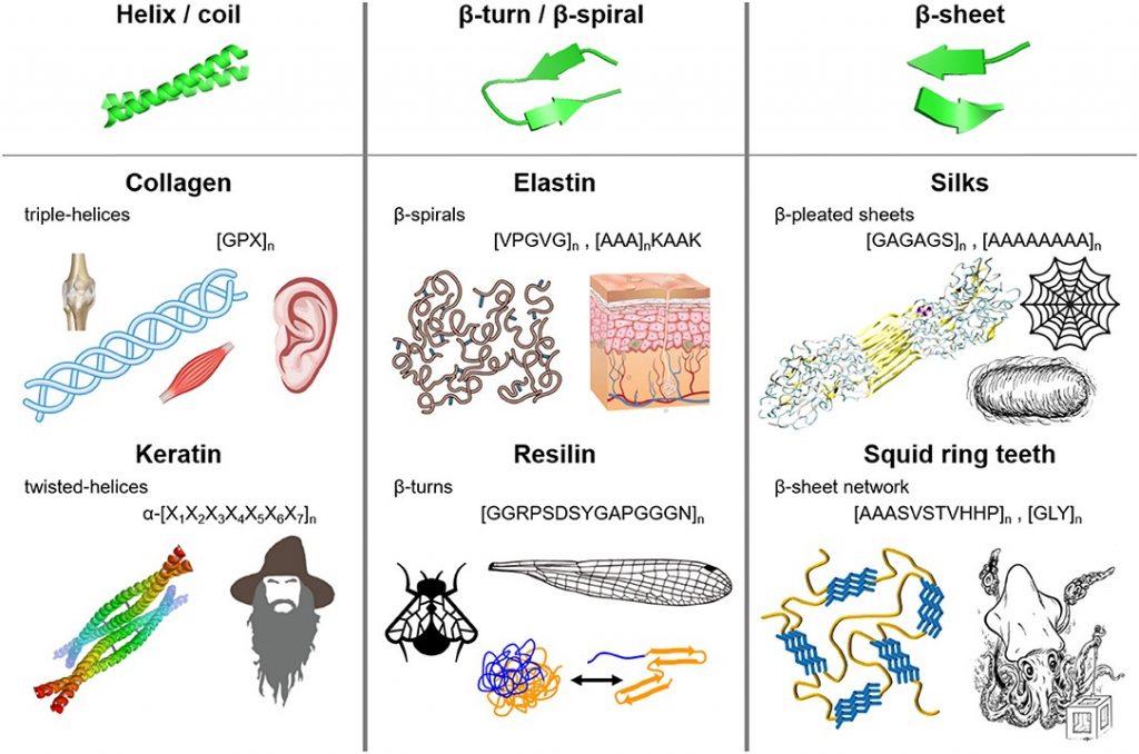
Figure 2. Fibrous protein polymers have molecular architecture that can include (i) helices and coils, (ii) β-turns and β-spirals, and (iii) β-sheets. Source

Video 1. SRT- coated fabrics that self-heal in water.

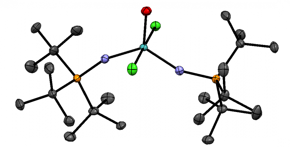
Squid ring teeth (SRT) are an especially promising candidate for making functional fibres and films due to its strength, conductivity and self-healing properties. The SRT are located inside the suction cups of the tentacles of squids and are composed of a naturally occurring protein complex. Fortunately, it is not necessary to harvest squid to obtain the SRT proteins as they can be biosynthetically produced since having their genome sequenced. The SRT-inspired monomer is repeated to create a polymeric chain and the resulting protein that is produced is named accordingly as tandem repeat (TR) proteins.

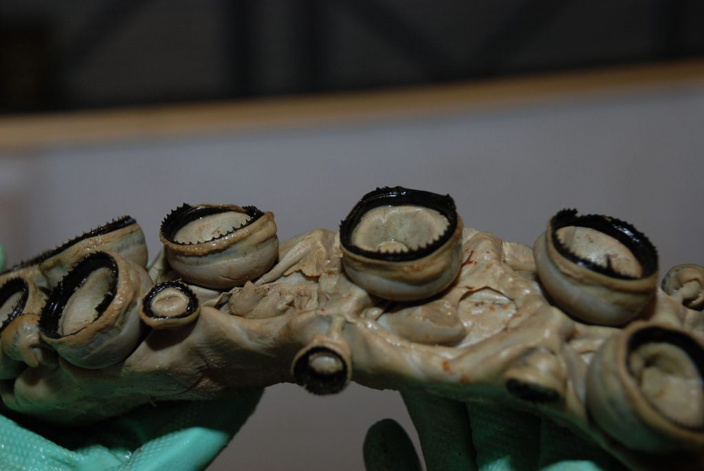
Figure 3. The Squid ring teeth of a giant squid. Wikipedia Commons

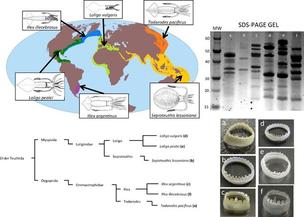
Figure 4. Optical images of squid ring teeth (SRT) and the six common squid species they originate from. Source

Films produced with SRT proteins consist of disordered domains that provide elasticity and flexibility to the materials in addition to ordered domains such as β-sheets that provide mechanical strength. One study designed four TR proteins denoted as TR-nX where X was the number of repeat units within the molecule. They measured the mechanical force of these four TR proteins and found that the ultimate strength of the protein’s scale linearly, with TR-n25 reaching an ultimate strength of 40 MPa. As seen in Figure 4 below, there is a limitation of the study due to sample size. The error bars of the TR-n12 and TR-n25 overlap, so it is not possible to say there is a statistically significant difference from each other.

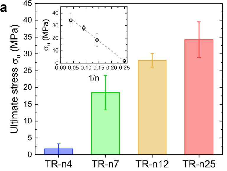
Figure 4. Mechanical testing of fully hydrated TR proteins (inset shows 1/n dependence) Source

Additionally, SRT protein films have been suggested as a potential solution to the issues related to the release of microfibers into the water from washing. A cloth made from polyester was coated with an SRT protein film and was found to dramatically increase the cloths resistance to abrasion (and microfiber release) compared to cloths that were not coated with SRT protein films.

While there is still a lot of further research to be done, SRT-based proteins are a promising avenue for making our world a little bit less plastic.

~Isla

Is Westernized Food Killing Our Gut?

Well not really killing your gut, but the vast and diverse biome of microbes that live in your gut. That’s right, you have around one hundred TRILLION helpful bacteria living inside of you! They have many jobs, ranging from helping break down food, helping with the production of vitamins, are a key component in our immune system and much more.

Studies have shown that when humans migrate from less westernized cultures to North America, their gut’s microbiome was significantly reduced in diversity and was predominated by two bacteria, Bacteroides and Prevotella. This decrease in diversity is shown to increase the inflammation in your gut, leading to gut-related diseases that are skyrocketing in modern society. Some of these are obesity, diabetes, Crohn’s disease, ulcerative colitis, allergies, and asthma, to name a few. This change in our microbiome was shown to be due to the Westernized high protein/fat/sugar diet whereas more developing countries have diets of very high fibre with fewer meats and fats.

source: Phys.org

Dr. Dan Knights, an assistant professor at the University of Minnesota, has done research on this change of microbiomes using wild monkeys and their captive counterparts to see if there were any differences. He found that the monkeys had much higher microbe diversity in the wild than when they were confined in a zoo.

Shows that the diversity of the primates microbiome decreases significantly when removed from the wild. Error bars indicate SD, and asterisks denote significance at **P<0.01, and ***P<0.001. Source: PNAS

Another exciting result he found was that two different wild monkey species with very different gut microbiomes converged to similar lower diverse microbiomes when captive, even though they did not live in the same zoo, never mind the same continent. They were converging towards the microbiome that modern humans have today.

As primates move from wild to captive, their microbiomes converge in the direction of modern humans. Non-western humans also have higher gut microbiome diversity than humans living in westernized areas. Source: TED

The data also showed that non-western humans followed this trend of having higher microbe diversity and subsequently losing this diversity when moving to the USA. This results in these migrants increasing their risk of obesity, diabetes and other gut-related diseases. These results beg the questions on what this ultimately means for our health? Further, this really makes you wonder, are captive monkeys becoming more like modern-day humans or are we just an example of super-captive primates??

~ Amanda Fogh

Image

New technology might allow mammals to have super-visual capabilities in the future

Radio waves, gamma rays, visible light, and all the other parts of the electromagnetic spectrum are electromagnetic radiation. However, a typical mammalian eye can only respond to visible light, which is a small portion (<1%) of the electromagnetic spectrum. But a recent study shows a new technology that may enable humans to sense near-infrared light.

The electromagnetic spectrum. Retrieved from Wikipedia Common.

The group of Professor Tian Xue from the University of Science and Technology of China and the group of Professor Gang Han from the University of Massachusetts State University have for the first time achieved the naked-eye infrared light perception in mice. Mice were able to see near-infrared light after being injected with special nanoparticles into their eyes. The special nanoparticles named pbUCNPs can anchor tightly on the retinal photoreceptors of mice and convert near-infrared light into visible green light. Additionally, these nanoparticles can stay in the eye for over two months without any obvious side effect.

The injection of the nanoparticles into the eyes of the mice. Image created by Ma et al.

Xue said: “This research breaks through the limitations of traditional near-infrared spectroscopy and develops a naked-eye passive infrared vision expansion technology, suggesting that humans have the potential for super-visual capabilities.”

To prove that the injected mice could see near-infrared light, the scientists did two experiments.

One experiment called pupillary light reflex. The pupillary light reflex gives the constriction of pupils in response to stimulation of eyes by light. The researchers shined near-infrared light into the eyes of injected and non-injected mice. The pupils of the injected mice constricted, while the non-injected mice showed no response.

pbUNCPs allow for detection of near-infrared (NIR) light. (A) Images show only the mouse injected with pbUCNPs gives a reflex when exposed to NIR light (980 nm), indicating that pbUCNP-injected mice are able to sense NIR light. (B) The curve shows the more intensive the NIR light is, the greater the pupil constriction response. Data are mean±SD. (Ma et al., 2019)

In the second experiment, as mice prefer to stay in the dark, the researches designed a box with two connected compartments. One compartment was completely dark, and the other was illuminated with near-infrared light. Scientists observed that the injected mice spent more time in the dark compartment, while the non-injected mice spent similar amounts of time in both compartments. 

The set-up of the Light-Dark Box. (Ma et al., 2019)

pbUCNP-injected mice recognize and respond to NIR light. Control mice and those injected with pbUCNPs responded to visible light (525 nm). However, when the light was in the NIR range (980 nm), only mice injected with pbUCNPs responded. Data are mean±SD. (Ma et al., 2019)

These two experiments proved that the injected mice perceived near-infrared light. Moreover, the scientists showed that the nanoparticles would not affect the normal vision of the injected mice.

This technique can potentially be applied to humans not only for generating super vision but also for repairing visible spectrum defects, such as colour blindness. 


Wenxin Zhao

Blood test: the future diagnostic method for Alzheimer’s Disease

Alzheimer’s disease (AD) is a type of brain disease that causes problems with thinking, memory, and behavior and leads to dementia. AD is frustratingly common among seniors over the age of 65. Approximately there are 5.8 million people in the United States are suffering; by 2050, this number will probably increase to 14 million.

How Alzheimer’s Changes the Brain. Source: https://www.youtube.com/watch?v=0GXv3mHs9AU

Although there is no current cure, an early and accurate diagnosis can help patients to access proper treatments which can slow the worsening of symptoms and improve quality of life for those suffering from AD. However, no single and efficient test can provide a reliable diagnosis. After doctors conduct interviews with patients with possible signs of symptoms, several blood tests and brain imaging are needed to rule out the other brain illnesses and confirm the diagnosis. This process may take several months, and the accuracy is only up to 75 to 85%. Researchers have been working on better and more efficient diagnostic methods. One advanced tool is Positron emission tomography (PET) scans which can detect the hallmark of abnormal protein clusters in brains and afford reliable results. However, these tests can cost thousands of dollars, and most people do not have access and never get tested.

PET scan showing glucose metabolism associated with decreased cognitive function.
Source: https://www.sciencedaily.com/releases/2009/07/090714085812.htm

For decades, researchers have been on the quest to develop a blood test for AD for blood testing is the most common and affordable medical diagnostics. The exciting news is that researchers have identified blood-based biomarkers of the disease that can provide fast and accurate measurements. A biomarker is a substance whose detection indicates a particular disease; in the case of AD, the particular substance is a protein called amyloids. One significant pathological signature of AD is the appearance of clumps of abnormal amyloid protein in brains. Those clumps are made of a mixture of peptides which form from the breakdown of the amyloid precursor protein (APP). In 2010, Bateman and his colleagues from Washington University School of Medicine found that the amount of a peptide known as amyloid-b 42 (Ab42) is significantly higher in the in a patient’s blood sample. However, several follow-up studies have suggested that the number of amyloid peptides, including Ab42, increases as people grow old, so the method of detecting Ab42 is proved unhelpful. In recent years, some research shows that the ratio of Ab42 to another peptide Ab40 indicates the significant difference in the diseased brain from a cognitively normal brain. Written in Nature last year, Yanagisawa and his team from the National Center for Geriatrics and Gerontology reported that using the ratio of these two peptides as the biomarker provides highly accurate results.

Figure 1: The clearance rate of amyloid- 40 and 42 peptides of 12 Alzheimer’s disease participants (red triangles) and 12 control (blue circles). The average clearance rate of amyloid-40 and 42 peptides is slower for AD individuals compared with cognitively normal control groups, suggesting the potential usage of these two peptides as biomarkers. Source: https://www.nature.com/articles/nature25456.

Although blood tests are not approved for commercially used yet, most researchers in the field believe that an affordable and accurate blood test for everyone will be commercially available in five years, especially when more proteins, such as neurofilament light polypeptide, are also found to be good candidates for biomarkers.

References:

National Institute on Aging. What Is Alzheimer’s Disease? https://www.nia.nih.gov/health/what-alzheimers-disease (accessed on March 21, 2019)

Alzheimer’s Association. Facts and Figures. https://www.alz.org/alzheimers-dementia/facts-figures

RadiologyInfo.org. Positron Emission Tomography-Computed Tomography. https://www.radiologyinfo.org/en/info.cfm?pg=pet (accessed on March 21, 2019)

Strimubu, K.; Tavel, J. A., Curr. Opin. HIV AIDS., 2010, 5, 463-466

O’Brien., R. J.; Wong, P. C., Annu. Rev. Neurosci. 2011, 34, 185-204

Amyloid precursor protein, Wikipedia.org, https://en.wikipedia.org/wiki/Amyloid_precursor_protein (accessed on March 21, 2019)

Mawuenyega, K. G.; Sigurdson, W.; Ovod, V.; Munsell, L.; Kasten, T.; Morris, J. C.; Yarasheski, K. E.; Bateman, R. J., Science, 2010, 330, 1774

Arnaud, C. H., Study tests plasma biomarkers for Alzheimer’s. https://cen.acs.org/articles/96/i6/Study-tests-plasma-biomarkers-Alzheimers.html (accessed on March 21, 2019)

Nakamura, A.; Kaneko, N.; Villemagne, V.L., Kato, T.; Doecke, J.; Dore, V.; Fowler, C.; Li, Q.; Martins, R.; Rowe, C.; Tomita, T.; Matsuzaki, K.; Ishii, K.; Ishii, K.; Arahata, Y.; Iwamoto, S.; Ito, K.; Tanaka, K.; Masters, C. L.; Yanagisawa, K., Nature, 2018, 554, 249-254

Lewczuk, P.; Ermann, N.; Andreasson, U.; Schultheis, C.; Podhorna, J.; Spitzer, P.; Maler, J. M.; Kornhuber, J.; Blennow, K.; Zetterberg, H., Alzheimer’s Research & Therapy. 2018, 10

Insomnia has a shared genetic risk with mental illness and metabolic disorders

Recently, Professor Danielle Posthuma from the Free University of Amsterdam and Professor Eus JW Van Someren from the Netherlands Institute of Neuroscience convened an international research team. This team has identified the cell types, regions, and biological processes that mediate the genetic risk of insomnia for the first time. Genetic risk is the contribution genes play in the chance of developing certain diseases. These findings, published in Nature Genetics, are a significant step in mastering the mechanisms that cause insomnia.

Image from Marygrace Taylor

Insomnia is a very common disorder in today’s society. Currently, one out of every 10 persons has a poor quality of long-term sleep and suffers from the serious consequences of insomnia during the day. Nearly 800 million people worldwide suffer from chronic insomnia.

Prevalence of Insomnia in the General Adult Population by Age. Data taken from DOI: 10.3988/jcn.2009.5.1.20

Although treatments relieve some symptoms, most people with insomnia still complain that they cannot sleep well. It has been found that susceptibility to insomnia often runs in families, and it seems that the problem is related to the brain. Before this study, researchers had only discovered several genes associated with insomnia susceptibility, and it was not clear which areas of the brain do these genes work in. To solve those problems, such a large study was conducted.

The findings were achieved by evaluating the DNA and sleep characteristics of more than 1.33 million people. The researchers identified a total of 956 genes that contribute to the risk of insomnia. They found that some of these genes have a major impact on the function of axons. Axons are a protrusion from a neuron that allows brain cells to communicate with each other. Another part of these genes is active in specific cell types in the frontal cortex and subcortical nucleus of the brain.

Neuron Structure. Retrieved from Wikipedia Common.

Brain Domain. Image Created by Dorling Kindersley

The researchers compared risk genes for insomnia with risk genes for other diseases. Surprisingly, Insomnia is genetically more related to psychiatric traits ( depression, anxiety, etc.) and metabolic traits (obesity, diabetes, etc.) than to other sleep traits. Here is a YouTube video about insomnia, depression, and anxiety.

Danielle Posthuma, a research author and professor of statistical genetics, said: “Our study shows that insomnia, like so many other neuropsychiatric disorders, is influenced by 100’s of genes, each of small effect. These genes by themselves are not that interesting to look at. What counts is their combined effect on the risk of insomnia. We investigated that with a new method, which enabled us to identify specific types of brain cells.” Guin Smit, a neurobiologist at the Free University of Amsterdam, said: ” These findings are a breakthrough since we can now for the first time start searching for underlying mechanisms in individual brain cells in the laboratory.”


Wenxin Zhao


Evolution of Enzymes is The New Trend

Last year, chemical engineer Frances H. Arnold from the California Institute of Technology earned the 2018 Nobel Prize in Chemistry for her pioneering and brilliant work with the directed evolution of enzymes. She became the second female to win the prize in Chemistry.

Figure 1: The flowchart for the directed evolution of enzymes.
Source: Advanced Information. NoblePrize.org. https://www.nobelprize.org/prizes/chemistry/2018/advanced-information/

Enzymes are biological catalysts to promote biochemical reactions in living organisms, and different enzymes specialize in different reactions. When the environment changes, genes mutate, and hence enzymes evolve to help an organism develop desired traits and adapt to the new environment. Although natural enzymes are excellent at doing their job, there are limitations: they only make chemicals that organisms need and only function in water at room temperature. These restrictions narrow the application of enzymes in the chemical and pharmaceutical industries. To tackle these problems, Dr. Arnoldhas used the same strategy as nature does-introduce mutations to existing enzymes-and obtained evolved enzymes which can quickly adapt to unusual environments, i.e., organic solvents, and speed up desired reactions. In the early 1990s, she reported the first case using subtilisin E, a digesting enzyme, to make an enzyme with much higher activity. This well-designed enzyme is 256 times more efficient to function the same reactions than the original enzyme in a polar organic solvent. This work has been seen as the benchmark achievement for the field of directed evolution of enzymes.

Figure 2: Reaction rate of hydrolysis of sAAPF-pna by subtilisin E variants in the solution containing 40% (vol/vol) DMF. Data source: Chen, K.; Arnold, F. H., Proc. Natl. Acad. Sci. USA, 1993, 90, 5681-5622


Dr. Arnold and her colleagues have been devoting to developing a variety of enzymes to deal with different synthetic challenges. For example, written in Nature this year, they describe a new iron-based enzymatic system to activate inert C-H bonds, replacing noble-metal catalysts. As the directed evolution proceeds, the system has a higher total turnover number (TTN) which represents how much product can be made until the catalyst is no longer active. Higher TTN means that the system becomes increasingly active as the enzyme evolves. The evolved enzyme CHF exhibits excellent stereoselectivity which is significant in pharmacology as human bodies react differently to enantiomers.

Figure 3: The bar chart represents the mean total turnover number (TTN) values averaged over four reactions; the grey dots show each TTN; green diamonds demonstrate enantioselectivity data.
Source: https://www.nature.com/articles/s41586-018-0808-5

Another recent pioneering work done by Arnold group is using a natural enzyme to form C-Si bond which is unknown in nature although Silicon is the most abundant element in Earth’s crust. Silicon has extensive applications in chemistry and material science, including pharmaceutical developments and productions of semiconductors, and preparations of silicon-containing molecules, especially organic compounds, usually require multi-step and unsustainable synthetic routes. This innovative and environmentally friendly method offers new avenues of producing organosilicon compounds and opens up more opportunities in pharmaceutical research. These findings also shine the lights on what silicon-based life might look like, which has long been a fantasy in science fictions!

Figure 4: The active environment of the enzymatic system for C-Si bond formation
Source: http://science.sciencemag.org/content/354/6315/1048

Figure 5: Artist rendering of Si-based life form
Source: https://media3.s-nbcnews.com/j/newscms/2017_16/1969741/organosilicon-based-life_c18e68cad6b3bf817a28e03558a7bfba.fit-2000w.jpg

As a winner of the Nobel Prize, Dr. Arnold will encourage more people, especially women, to do science, and inspire more research in biocatalysis. As she pointed out in her essay, using well-functionalized enzymes rather than transition metals as catalysts allows for the development of sustainable chemical and pharmaceutical industries, and hence producing many of chemicals with biocatalysts will be the trend in the near future.

Dr. Arnold’s Nobel Lecture: Innovation by Evolution. Source: YouTube

References

Directed evolution, Wikipedia.org, https://en.wikipedia.org/wiki/Directed_evolution (accessed on Feb. 28, 19)

Gibney, E.; Noorden, R.; Ledford, H.; Castelvecchi, D.; Warren, M., Nature, 2018, 562, 176

Enzyme, Wikipedia.org, https://en.wikipedia.org/wiki/Enzyme (accessed on Feb. 28, 19)

Chen, K.; Arnold, F. H., Proc. Natl. Acad. Sci. USA, 1993, 90, 5681-5622

Zhang, R. K.; Chen, K.; Huang, X.; Wohlschlager, L.; Renata, H.; Arnold, F. H., Nature, 2019, 56, 67-72

Williams, K.; Lee, E., Drugs, 1985, 30, 333-354

Kan, S. B. J.; Lewis, R. D.; Chen, K.; Arnold, F. H., Science, 2016, 354, 1048-1051

Silicon, Wikipedia.org, https://en.wikipedia.org/wiki/Silicon

Kincaid, P. Life, but not as we know it. https://www.newscientist.com/article/mg15821335-600-life-but-not-as-we-know-it/ (accessed on Feb. 28, 19)

Arnold, F. H., Angew. Chem. Int. Ed., 2018, 57, 4143-4148


Image

Forensic: How to Measure the Unknown Time of Death from Only Bone and Hair Remains

Typically, TV forensic shows, such as CSI and Bones, portray how forensic cases are fast and easy to solve within a couple of episodes. However, that is not true. Unlike in forensic shows, forensic cases take a long time to solve in real life, due to challenges that investigators have to face.

One of the challenges that criminal investigators have to solve is to determine the post-mortem interval (PMI) from decomposed skeletons. Post-mortem interval is the time after someone has died, which is important when the cause of death is unknown. Usually, medical assessment of corpses can determine the PMI after the body is dead for the first few days. However, when the victim’s bones and hairs are found after a few years, the accuracy of the PMI lowers. Therefore, new method is required to provide a more accurate estimation of the extended time of death.

Currently, there are numerous studies that use different methods for solving the PMI. Many methods include analyzing soil chemistry and insects at the crime scene. Since these methods could be complementary to each other, the scientists in Switzerland believe that combining these methods would improve the long-term PMI estimate, while illustrating how the crime scene could have occurred. Therefore, the Swiss scientists have attempted to use five different approaches simultaneously to estimate the PMI of the bone and hair remains. Once the soil, bone, and hair samples are collected from the Swiss forest, the scientists use five different approaches for analyses, which include using radiocarbon dating, analyzing pH and soil chemistry, counting and classifying nematodes and mites, and sequencing DNA of soil micro-eukaryotes.


After analyzing the findings, the scientists are able to propose a possible PMI, as well as the crime scene. First of all, the radiocarbon dating determines that the bones belong to a young adult male. Secondly, chemical, nematodes, and micro-eukaryotic analyses suggest that the remains have been partly decomposed in the forest for at least 8-9 months. Finally, the evidence from mites suggests that the corpse is partly decomposed in a separate confined place, because these mite species are only found in confined environment. Therefore, the suspect(s) could have allowed the corpse to decompose in a confined area before relocating it to the Swiss forest. As a result, the PMI of the dead victim appears to be at least 8-12 months before the body is discovered.

The skeleton that is found in a Swiss forest. Ildikó Szelecz, Sandra Lösch, Christophe V. W. Seppey, Enrique Lara, David Singer, Franziska Sorge1, Joelle Tschui, M. Alejandra Perotti & Edward A. D. Mitchell, Source, Creative Commons Attribution 4.0 International Licence

Overall, the study shows that using five approaches simultaneously in a forensic case study can estimate the PMI, while illustrating a possible crime scene of how the victim could have died. Moreover, all of the approaches can be complementary with each other, in order to provide more evidence from scarce remains. In conclusion, it is possible to further develop this technique in order to estimate PMI in other forensic cases.

Update: Blog post has been revised on 2019, Feb 15th.

Reference

Rasmussen College. How Accurate are Crime Shows on TV? Debunking 7 Common Myths. https://www.rasmussen.edu/degrees/justice-studies/blog/crime-show-myths-debunked-forensic-experts-tell-what-life-is-real/

Keele University. Chemical Ecology. PMI in Forensic Entomology. https://www.keele.ac.uk/chemicalecology/projects/pmi/

American Council on Science and Health. How Chemistry, Microbiology Could Help Catch A Killer. https://www.acsh.org/news/2018/01/18/how-chemistry-microbiology-could-help-catch-killer-12422 .

Szelecz, I.; Lösch, S.; Seppey, C. V. W.; Lara, E.; Singer, D.; Sorge, F.; . . . Mitchell, E. A. D. . Comparative analysis of bones, mites, soil chemistry, nematodes and soil micro-eukaryotes from a suspected homicide to estimate the post-mortem interval. Scientific Reports. [Online] 2018, 8(1), 25. doi:10.1038/s41598-017-18179-z.

Video

A Potential Memory Enhancing Drug

Do you easily forget things? Would you like to improve your memory? A recent study led by Professor Yuji Ikegaya and Dr. Hiroshi Nomura of the University of Tokyo suggests that your long-term memory might be improved with pro-histamine treatment.

The structural formula of histamine. Retrieved from Wikipedia Common.

Pro-histamine drugs increase the level of histamine, which in the central nervous system is associated with learning and memory. Scientists believe that elucidating the role of histamine in memory may help alleviate the symptoms of dementia.

Continue reading