
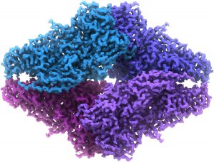
High Resolution of Detailed Structures (Credits to Nicolae Sfetcu)
About one month ago, Jacques Dubochet, Joachim Frank and Richard Henderson awarded the Nobel Price in Chemistry 2017 for developing the cryo-electron microscopy. The National Institutes of Health named cryo-electron microscopy ‘method of the year’. Cryo-electron microscopy can image frozen-hydrated specimens in native state without dyes at low temperatures through electron microscopy. Using this technology, scientists have produced three-dimensional images to target cancer drugs and demystify the Zika virus.
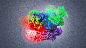
Cancer drug target visualized at atomic resolution (credits to NIH Image Gallery)
Actually, the development of cryo-electron microscopy has a long history. Previously, scientists used cumbersome dyes, stains or labels to visualize cell function, which would change the behaviour of the cell function and only provide a coarse two-dimensional image. This made scientists hard to understand molecular biology clearly since how the components in the cells looked like and what functions they performed remained unknown.
However, from 1975 to 1986, Joachim Frank stitched two-dimensional micrographs together to yield a sharp three-dimensional image. In 1990, Richard Henderson used this principle to visualize a protein in three-dimensional down to atoms with an electron microscope. In the early 1980s, Jacques Dubochet discovered that water would form a solid shell without freezing by rapidly cooling a specimen before putting it in an electron microscope, which could keep biological structures in original shape during scanning. They produced the desired atomic resolution in 2013. And researchers can now routinely produce three-dimensional structures of biomolecules.
Combining these theories, scientists could take biologically-accurate snapshots of the tiniest units of life. This technology helps scientists understand diseases better and develop better drugs. For instance, scientists found unique parts of the pathogen’s structure in the Zika virus and identified a potential target for a vaccine.
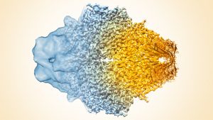
Improving resolution by cryo-EM (credits to NIH Image Gallery)
Engineers have developed better hardware to help improve cryo-electron microscopy by visualizing detailed structures instead of shapeless blobs. Scientists claim that the limited physical knowledge confines the resolution bt they will obtain better visualizations of biological structures in the coming year.