Learning more about Tuberculosis from the Fur Trade

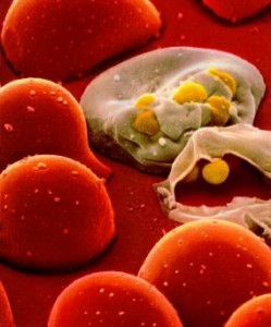
One of the most memorable things when learning about the 18th century Canadian fur trade in history class was how the Europeans spread diseases to the indigenous peoples by means of the trade. One of such diseases is tuberculosis, which infectious disease specialist Caitlin Pepperell and colleagues at Standford University in Palo Alto, California have been studying. In the new study published in the Proceedings of the National Academy of Sciences, they have puzzled out why TB didn’t become an epidemic until after the fur trade era even though the indigenous peoples of western Canada were repeatedly exposed to the strain by French Canadian voyageurs during 1710 – 1870.

Canadian Fur Trade / Source:Cartouche from William Faden

It wasn’t until the late 1800s that TB epidemics began to break out, which was about 150 years from the first introduction. Pepperell explains that the conditions most likely to trigger epidemics in Canada were the relocation of the native peoples  onto reservations, declining health conditions with relocation, and the biggest factor of them all, malnutrition. The key message from the results indicates that TB is infectious and remains dormant in host until stressful conditions – crowding, poor nutrition, weakening hosts – give the bacteria a leg up and turns on into active mode.
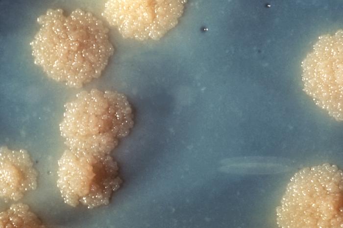
TB Culture / Source: CDC

Pepperell first discovered the same TB strain signature, or “fingerprint” in the native peoples of Quebec and a French Canadian population that didn’t live near the native communities when she received a DNA profile of the bacteria in the latter from a colleague. After using mathematical modeling and statistical analysis, they were able to trace the spread back to the 18th century, which was when waves of French traders carrying TB came to Canada and married indigenous women, resulting in the disease remaining latent in the native communities for several decades until the pressures of relocation and shortage of buffalo occurred.

Tuberculosis’s stealth nature has had it difficult to combat the disease without knowing how it spreads. The World Health Organization estimates that 1/3 of the people on Earth are infected with the disease. The new findings of the study can only point out how tenacious the disease actually is, bring us one step closer to understanding the tuberculosis bacterium and how we can control it.

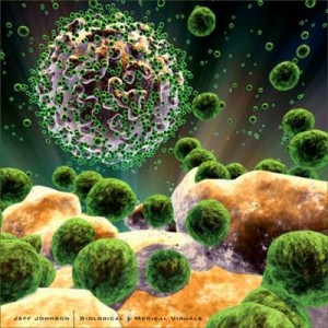
Hide and Go Seek: HIV vs Mathematical Model

Math may help overcome ‘sleeper cell’ hurdle in HIV treatment
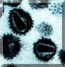
New research on HIV treatment is important for helping the 33 millionpeople who are affected in North America, Sub-Saharan Africa and the rest of the world.HIV is a Human immunodeficiency virus.  It is a condition in humans where the infection causes the immune system to fail, leading to life threatening infections and diseases.


The HIV virus can survive in two places in the body; inside cells or free floating in the blood.Inside a cell, the HIV virus has two options it could begin making copies of itself using the cells own DNA xerox machine, once enough copies are made the new viruses can break out of the cell and go on to infect other cells in the body. Other times the virus may simply hide out in the cell in a dormant phase. This dormant phase is what makes antiretroviral medication less than 100% effective. The medication can only target the free viruses and the ones that are making copies. The ones that are hiding could become active later on with out any warning.

See podcast for more on dormant cells.

Audio clip: Adobe Flash Player (version 9 or above) is required to play this audio clip. Download the latest version here. You also need to have JavaScript enabled in your browser.

The unpredictable activation of dormant cells led to some unique research by mathematicians at the University of British Columbia. They have used a mathematical model to track and predict how virus levels change in a patient when the dormant cells wake up. The research shows that these cells don’t wake up because of a trigger (like failing drug treatment) but are due to random activations.

See Video for more on the mathematical model.

https://www.youtube.com/watch?v=xMulji8omcA&hd=1

HIV can be detected through the screening of the blood. HIV infection occurs by the transfer of bodily fluids such as breast milk, blood, semen, vaginal fluid and pre-ejaculate. The most common routes of transmission of the infection is through unprotected sex, contaminated needles, breast milk and transmission from an infected mother to her child at birth.

The mechanism behind HIV is that it primarily infects the cells in the human immune system (T cells, white blood cells, macrophages and dendritic cells) which essentially protect the human body from infections. CD4+ T cells are a type of white blood cell that guides other white blood cells to fight infections. HIV infection leads to low levels of these CD4+ T cells, through three main mechanisms:
  1. direct viral killing of infected cells
  2. increased suicide rates in infected cells (most cells in our body are programmed to self destruct when they get old)
  3. killing of infected CD4+ T cells by CD8 cytotoxic lymphocytes that recognize infected cells.  These are like cops that hunt down the infected cells.

When the level of CD4+ T cells declines to a critical level, the protection provided by white blood cells is lost, and the body becomes more susceptible to infections. This is commonly known as AIDS.

Grabbing hold of Alzheimer’s disease: A new treatment

By: David Sawatzky, Paula Samper, Moh Mehrabi, and Daniel Passaseo

There is a new drug that can target and block harmful metal ions in the brains of Alzheimer’s patients. This drug developed by Dr. Chris Orvig from the University of British Columbia, is known as a chelating drug, which means it grabs hold of its target and makes it unable to do more damage. This is a massive breakthrough for treating Alzheimer’s disease, for which there is no cure.

This research is very important because you can only treat the symptoms of Alzheimer’s, and the leading treatments do a very poor job and targeting the affected brain cells. Dr. Orvig’s model drug has a sugar molecule to help deliver it to the brain. It is a very innovative idea, and there have been no previous Alzheimer’s drugs that are effective at getting the drug into the brain.

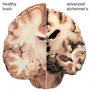
One of the effects of Alzheimer’s is neurodegeneration, which means the brain cells die. As these brain cells die, the patients lose their memory and motor skills, making it hard for them to live on their own. These patients then require family members and or caregivers to help with daily activities like preparing food and bathing. This takes a large emotional and financial toll on the patient’s family.

Photo from Google images

The actual causes of the disease are not known very well, because there may be many factors that contribute to this disease. But it is known that Alzheimer’s is not genetically inherited, although some genes may contribute to the risk of developing Alzheimer’s. For more information about what causes Alzheimer’s disease listen to our podcast!

Audio clip: Adobe Flash Player (version 9 or above) is required to play this audio clip. Download the latest version here. You also need to have JavaScript enabled in your browser.

The following video does a great job summarizing Dr. Orvig’s research and what Alzheimer’s disease is all about.

https://www.youtube.com/watch?v=VTrjcV_EyfI

There is hope that research like Dr. Orvig’s will eventually lead to a better understanding of this disease and eventually a cure.

A Breakthrough in Prostate Cancer Detection

UBC researchers have developed a new test that promises to overcome current issues and increase the reliability of screening.

By Amanda Au, Navi Dasanjh, Kushani Jayasundera and Martha Talbot

Dr. Eric Lagally, an assistant professor and researcher at the University of British Columbia, believes he has discovered a new method of detecting prostate cancer. Prostate cancer is the most common type of cancer in men, yet the two current tests for prostate cancer are inadequate and often give false positives and negatives. Being able to correctly identify prostate cancer cells could reduce the rates of misdiagnosis and over treatment.

Dr. Lagally’s new detection method looks at an enzyme called telomerase, which helps prevent premature cell death. He uses this molecule to help differentiate between cancerous cells and normal cells more precisely using a technology known as microfluidics.

Learn more about telomerase by listening to this UBC Mastermind Productions’ Podcast.

Audio clip: Adobe Flash Player (version 9 or above) is required to play this audio clip. Download the latest version here. You also need to have JavaScript enabled in your browser.

Microfluidics allows researchers to analyze small fluid samples using a chip the size of a postage stamp etched with miniscule channels and chambers. The width of these channels and chambers is similar to the width of a human hair. The small scale allows researchers to precisely test small samples.

Microfluidic chips like the ones that Dr. Lagally uses in his lab.

Currently, microfluidics is being used for many biomedical applications, such as DNA analysis. This new technology boasts many advantages, including the ability to analyze very small fluid samples at a minimal cost and with little power. This may also be quite beneficial to the new prostate cancer screening method Dr. Lagally hopes to implement. As he points out, the use of microfluidic chips will enable doctors, nurses, and other health practitioners to analyze test results at the bedside, eliminating the need to transfer samples to a testing facility.
As of today, Dr. Lagally’s microfluidic test is a long way from human applications. Alterations need to be made to the chip, which will then face gaining the approval of the Food and Drug Administration (FDA), a process that could take up to ten years.

As Dr. Lagally works on the process of transferring this technology from the lab to the clinic, he is also educating the public about microfluidics. He speaks about microfluidics and has developed a method of teaching students microfluidics by fabricating their own large-scale chip out of Jello. Try it yourself!

By educating the public, as well as other researchers, the hope is that this technology will be able to make a smooth transfer so that microfluidics can begin to positively affect people who are in desperate need of this technology.