Author Archives: Jenny Zhong

New skin from transgenic stem cells

Gene therapy is a promising medical development that can save people suffering in perilous diseases. One outstanding case of gene therapy is transplanting transgenic skin cells to replace over 80% (0.85 m2) of a young boy’s outer layer of skin (called the epidermis), effectively treating a severe skin disease called junctional epidermolysis bullosa (JEB) without long-term adverse effects. This accomplishment was published by Professor Michele de Luca and the collaborating medical team from Ruhr-Universität Bochum’s burn unit and the Center for Regenerative Medicine at the University of Modena (Italy), on November 8, 2017, in the journal Nature.

JEB is a disease caused by mutations in any of three genes that encodes a protein called laminin, which anchors the epidermis to the dermis, the inner layer of skin. Failure to do so results in fragile skin with low mechanical resistance and elasticity, manifesting as blisters and wounds in many areas and increased vulnerablility to infections. The patient in the study had extremely severe JEB caused by mutation in one gene and lost an amount of skin equal 80% of his total body surface area, including arms, legs, chest and back.

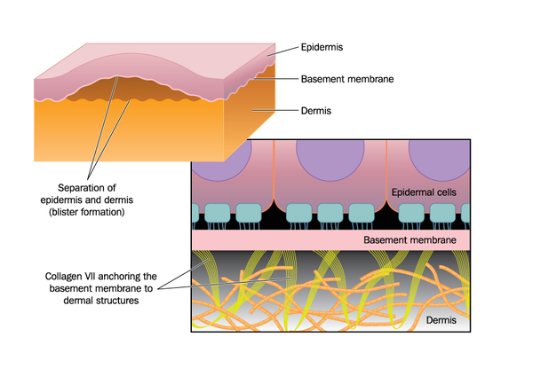
mutation in the laminin protein (not shown) causes detachment of the epidermis (outer skin) from the basemebt membrane and inner skin (dermis) in junctional epidermolysis bullosa. source: https://ghr.nlm.nih.gov/condition/junctional-epidermolysis-bullosa

The treatment started two years ago in 2015, with doctors removing a small area of normal skin to establish skin cultures, which were artificially infected (transduced) with a virus (called a retroviral vector) carrying the normal laminin-encoding gene. After growing the cells to 0.85 m2 , the new skin was sequentially grafted on sites of exposed inner skin, using either plastic or fibrin as the adhesive base. Both graft types were equally effective: the regenerated skin did not blister or damage after pinching; furthermore, after 21 months followup, the grafted skin did not produce antibodies by the body, indicating it was safe and the body recognizes it as belonging to itself. The new skin regenerates monthly by a small number (about 5% of the skin after 8 months) of long-lived stem cells called holoclones, which could regenerate themselves and develop into half-differentiated cells (meroclones) and almost fully differentiated cells (paraclones), which cannot divide further but replace old cells and gradually disappear. 

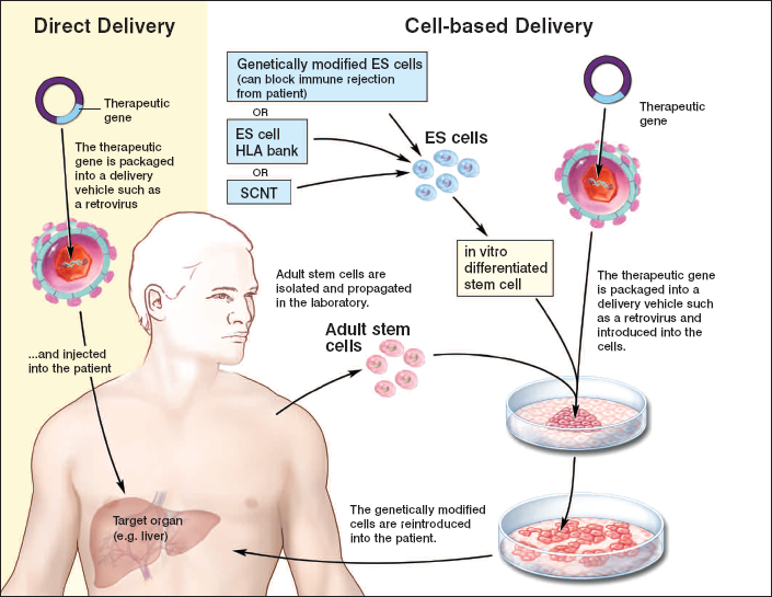
general stem cell therapy scheme. Skin cells from the patient are harvested and a virus introduces the desired gene into the skin culture to create genetically modified cells. Source: https://stemcells.nih.gov/info/Regenerative_Medicine/2006Chapter4.htm

However successful it was, the treatment was actually pretty risky. The retroviral vector could insert, or integrate, the normal laminin gene anywhere in the patient’s genetic blueprint and disrupt other normal genes and cause unregulated tissue growth, resulting in tumours or cancers. Fortunately, genetic sequencing of the transplanted skin revealed that the normal gene was inserted mostly in regions not coding for proteins (introns and intergenic regions) with only 5% inserted in protein-encoding regions, but these genes were not involved in cancer. In addition, the transplant did not cause specific cells to survive better than others and cause tumour formation. However, long term monitoring is still required.

While this technology is currently limited to injuries with an intact dermis, it is less invasive and more effective than surgery as it does not entail infections, and it could be applied to early diagnosis stages to prevent skin diseases as well as restoring large areas of damaged skin. This is a new treatment for epidermolysis bullosa, a condition affecting 500 000 people worldwide, and a major stepping stone to developing stem-cell therapies for many debilitating diseases.

Written by Jenny Zhong

The Invisible Blue Halo

How do flowers attract bees? Well, you might have thought of their bright, colourful petals or their delightful scent, but there’s something else that is not that obvious. If you have looked carefully at certain flowers, like the Queen of the Night tulip, and noticed a certain blue hue when switching to different angles, you’ve got it – but what is it?

Queen of the Night Tulipa. source: Fine Art America

another beautiful tulip, source: Gardener’s Dream

This blue results from diffraction of light by microscopic ridges found in the cuticle (outer filmy layer covering the outer skin layer) of petals, researchers at the University of Cambridge found. Blue pigments, for unknown reasons, are hard to make naturally. The researchers, led by Professor Beverley Glover, found that distantly related flower species with different anatomies had surprisingly similar ridges of similar height, width, and spacing – a phenomenon of convergent evolution. The ridges run across the petal cuticles, parallel to each other, but were not perfectly shaped like artificial diffraction gratings. They scatter light and generate a blue halo only visible at a plane perpendicular to the direction of ridges, which is easily missed by humans.

To confirm what the blue halo does, researchers made artificial “flowers” with the ridges of either with perfect, artificial dimensions or simulating a natural messy-ness, and flowers with smooth surfaces, on underlying colour pigments – yellow, blue, or black – placed next to a sugar solution. The flowers with smooth surfaces appeared the same no matter the viewing angle, but those with microscopic ridges, perfect or messy, were iridescent – changing colour as viewing angle changes; but only those with disordered ridges have the blue halo. They found that bees found the iridescent flowers faster than the smooth-surfaced flowers with the same underlying pigment. The effect is reduced in blue-coloured flowers, likely because the blue halo and blue pigment appear quite the same.

Artificial “flowers” made on underlying black background. Sm= smooth surface, Di= surface with disordered diffraction ridges, and Or= ordered, regularly repeating ridges. Adapted from Figure 4 in “Disorder in convergent floral nanostructures enhances signalling to bees”.

So, why do bees like blue? Previous research has shown that bees, with more sensitive receptors to the blue-ultraviolet region of light, are more attracted to the shorter wavelengths of visible light, and blue flowers were found to produce more nectar. Interestingly, the appearance of neat ridges in two major classes of flowers, monocots and eudicots, coincided with the appearance of nectar-gathering insects, increasing the evidence of the blue halo attracting bees to flowers. Knowing how colour affect bees could help beekeeping and possibly increase pollination effects of a flower, if these ridged structures can be coated onto flower petal surfaces without harming them.

What about the flowers that do not have the microscopic ridges? Although blue is difficult to generate, it turns out flowers have a few ways to make blue: increasing the pH of vacuoles, the big cellular compartment that stores nutrients, ions, and pigments, or by making “metal-pigment complexes that blue-shift” the colour of anthocyanin, a pigment that is purple in neutral pH solutions, as the original research article states.

All in all, what I take away from this is: don’t judge a flower by its colour.

Designing new organelles

Many people may have heard about cutting and pasting genes from a genome, the complete blueprint of an organism, into microorganisms to make them magically produce compounds that they could never produce naturally. However, few experiments have endeavored to make a novel organelle, an artificial factory within a living cell. Genetic evidence shows that the mitochondrion, the cell’s power plant, was actually a bacterium that got swallowed by an ancestral cell, and it revolutionized how life works. Wouldn’t it be extremely cool if scientists could do something like that and change how cells behave?


Organelles are compartments performing specific functions, found within the cells of many organisms. They concentrate reactants for chemical reactions while not disturbing the cell’s other activities. For example, the organelle called peroxisome is a processing plant and protector of plant and animal cells. It degrades very long chain fatty acids from dietary fats and produces toxic hydrogen peroxide, which is safely reduced to water within. Zellweger syndrome is due to dysfunctional peroxisomes, where a person accumulates of toxic metabolites and cannot make myelin for nerve cells and bile for fat digestion, which may be fatal with no available treatment. Therefore, it is important to maintain healthy peroxisomes and their protein machineries.

Structure of Peroxisome in fixed cells, labelled with SelectFX® Alexa Fluor® 488 Peroxisome Labeling Kit. Adapted from Thermo Fisher Scientific: Peroxisome Structure

Interestingly, peroxisome proteins are imported directly from the cytosol, the fluid-filled space surrounding organelles within the cell, without going through a central protein-sorting system in another organelle, the web-like endoplasmic reticulum.

In September 2017, chemical biologist Stuart Warriner and his team at the University of Leeds, England, published their work on tweaking the natural peroxisomal protein import system by modifying two proteins involved in the pathway. They found that their new pathway overrides the natural protein carrying pathway with no adverse side effects for the cell. Theoretically, scientists could design it in whichever way they want through this simple, subtle change.

Figure 1. Scheme of research. Adapted from Figure 1 of “Towards designer organelles by subverting the peroxisomal import pathway” by Warriner et. al, 2017.

A big family of proteins called PEX (peroxins) serve as porters and receptors that recognize and dock cytosolic proteins with the five-letter amino acid peroxisomal targeting sequence (PTS; like a password) to import them into peroxisomes. However, the researchers only changed the PTS and a protein called PEX5, which wanders in the cytosol looking for homeless, PTS-carrying proteins.


Using known crystal structures of PEX5 protein, the researchers generated several versions of PEX5 by mutating (changing) one or two amino acids at one end that binds the original PTS. They modified the PTS by changing its last two amino acids, and identified a new PTS that rcognized the new PEX5’ but not the original.

Adapted from Figure 3 of “Towards designer organelles by subverting the peroxisomal import pathway” by Warriner et al., 2017, showing how the new PEX5 protein selectively imports new PTS-containing proteins into peroxisomes.

Unexpectedly, viewing cells with fluorescence microscope showed the natural PTS was imported by the new porter protein PEX5’, meaning PEX5’ is more lenient on PTS. However, when fluorescent PTS and PTS’ were inserted in PEX5′-containing cells, only the new PTS’ ended up in peroxisomes, leaving the PTS in the cytosol. Evidently, the new and original PTS compete for import, with the original PTS outcompeted – how they interact with other PEX proteins and by what mechanism are still mysteries. However, this means scientists can manipulate peroxisomes by simply switching to the new PEX5′ protein.


Custom organelles has many potential applications, such as more efficient and sustainable production of therapeutics and other synthetic compounds without compromising the cell’s other activities and health. Scientists could also design a novel waste management system for the cell by designing PTS-containing proteins that scavenges certain chemical wastes from the cytosol and import them for processing in peroxisomes. Without knowing when that day will come, meanwhile, we should appreciate what our wonderful organelles do to keep us alive!

Written by Jenny Zhong