Hey Team! I have chosen the term ‘Dental Radiographs’. As you guys know I am in the dental field and work with dental radiographs everyday. Though there are many crazy terms in the dental realm I chose this because I’m sure you’ve all had some taken! For this assignment we were to write three definitions: parenthetical, sentence and expanded and be able to understand and value the different types of definitions to offer the audience at hand.
Parenthetical Definition
Dental radiographs also known as X-Rays are images of the teeth and surrounding structures which help the dentist to assess the condition of the patient’s oral health.
Sentence Definition
| Term | Class | Distinguishing Features |
| Dental Radiograph | Diagnostic Tool | Small film or sensor placed in one’s mouth to capture a diagnostic image with the use of radiation |
A dental radiograph is a diagnostic tool in the form of a small film or sensor that is placed in one’s mouth and is emitted by radiation to capture an image of one’s oral cavity including bone and teeth.
Expanded Definitions
The history of radiography began as early as 1895 by a German University professor Wilhelm Conrad Roentgen. He made a breakthrough discovery experimenting in his lab with a cathode-ray tube that consisted of both positive and negative electrodes. When the air and the tube was evacuated, and a high voltage applied- the tube itself produce a fluorescent glow consequently creating the beginning of radiation imaging! The ray of radiation from the x-ray tube head passes through the mouth, takes an image and stores it on the film or sensor in the patient’s mouth. Most radiation is absorbed by the teeth and bone structure, making the teeth appear lighter – which helps the Dentist to diagnose decay or infection- which appears darker on the image. Dental Radiographs are one of the best diagnostic tools in assisting the dentist and oral health team to evaluate the periodontium and surrounding structures. The main uses of dental radiographs are: assessing for decay and periodontal health, detection and diagnoses of oral pathologic lesions and calcified arteries as well as evaluation of growth and development and jaw position for orthodontic patients.
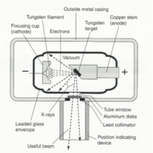
Figure 1 X-Ray Tube. Housing of Radiation.
Source: Wilkins, Esther M. “Clinical Practice of the Dental Hygienist.” 11th edition. 2013; 159.
The visual above represents the x-ray tube where radiation is produced and exits the tube to hit a target – in this case the film or sensor in the patients mouth which captures the image.
References
Cherney, B. K. and K. (2018, September 29). Dental X-Rays: Purpose, Procedure, and Risks. Retrieved from https://www.healthline.com/health/dental-x-rays#when-to-get-one
Darby, M. L., & Walsh, M. M. (201AD). Dental Hygiene Theory and Practice (3rd ed.). St. Louis, MO: Elsevier.
History of Radiography. (n.d.). Retrieved from https://www.nde-ed.org/EducationResources/CommunityCollege/Radiography/Introduction/history.htm
How do Dental X-rays work? (2019, July 9). Retrieved from https://www.foundationeducation.edu.au/articles/2018/05/how-do-dental-x-rays-work
Wilkins, E. M. (2013). Clinical Practice of the Dental Hygienist (11th ed.). Philadelphia: Wolters Kluwer.
Hello everyone! I am currently a Registered Dental Hygienist practicing in Southern Ontario. I am enrolled to complete my ' Dental Sciences' degree!
Leave a Reply