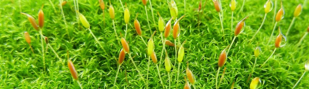
 Scanning Electron Microscopy (SEM) enables users to observe structures on specimens that may be difficult to distinguish using light microscopes or the naked eye. Thus, SEM is particularly useful for viewing the ornate morphology of tiny bryophyte structures. The SEM images acquired below were taken at the University of British Columbia’s Electron Microscopy lab of the Biology department. Please click on a link below to view various SEM images from different bryophyte classes.
PHYLUM BRYOPHYTA
PHYLUM MARCHANTIOPHYTA
PHYLUM ANTHOCEROTOPHYTA