
According to the Canadian Cancer Society, about 2 in 5 Canadians will develop some form of cancer during their lifetime, and a shockingly 1 in 4 Canadians will die from cancer. An estimated 187 600 Canadians will be diagnosed with cancer in 2013. Although there are various therapies designed to beat cancer, a definite treatment plan that will be suitable for all types of cancers has yet to be made. With cancer having a larger toll on the medical industry, it is not surprising to find that the quest to finding a cure has become one frequently travelled on by scientists and researchers with various backgrounds.
A group of scientists from Stanford University may have discovered a possible solution. Researchers have spent a considerable amount of time focusing on understanding the mechanisms malignant tumor cells utilize to cheat death. This may be the key to the cells’ undoing.
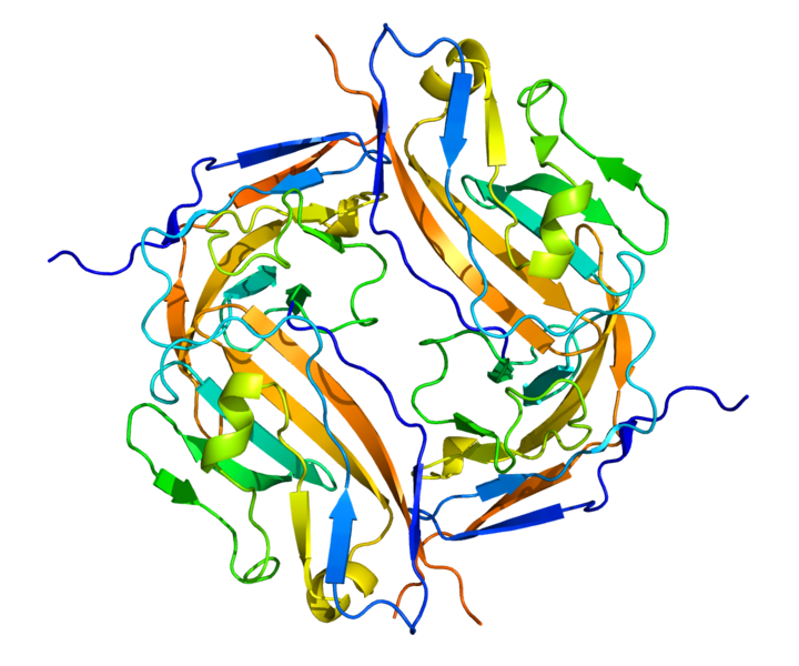
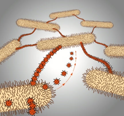
Photo Courtesy of Pleitrope
The 3D rendering image in the left provides a visualization of the CD47 protein, which is produced in large quantities by malignant cells. When released into the surrounding environment, CD47 proteins become a “do not attack” signal to phagocytic cells, and help malignant cells escape their inevitable death. These cells are able to continue replicating as the proteins cloak the fact that they have already reached their expiration date.
CD47 concentration has been correlated with patient survival rate in the past. Patients with a high concentration and expression of CD47 proteins and mRNA tend to have a decreased probability of survival, and struggle with severe symptoms in comparison to patients with reduced CD47 expression and concentration. Knowing this trend, researchers have developed an anti-CD47 antibody that will literally eat up the CD47 proteins, and prevent them from affecting the performance and capabilities of phagocytic cells. Similar to the concept of a lock and a key, the anti-CD37 antibodies detect CD47 proteins, and render them useless. These antibodies are also able to disrupt cancer by preventing metastasis – the spread of cancerous cells throughout the body.
Benign cells also express CD47 proteins and mRNA. In fact, CD47 proteins play a critical role in overall cell function. Due to this reason, researchers are not aiming to completely wipe out expression and concentration. The key to this solution is moderation, and researchers aim to lower concentration and expression to a reasonable range.
Coupled with other therapies, using these anti-CD47 antibodies have led to amazing results. Once the antibodies were introduced into immune systems surrounding malignant tumor cells, researchers found that the antibodies would activate macrophages, which would proceed to consume the malignant cells. Using this technique and technology, researchers will be able to customize cancer treatment plans for each patient, and tailor the dosages their needs. This treatment may be more sensitive to the needs of the patients, have a higher success rate, and be associated with fewer negative side effects.
Literature Referenced:
Cohen, J.D., Contreras-Trujillo, H., Dalerba, P., Gentles, A.J., Lovelace, P., Martin, R., Mitra, S.S., Sahoo, D., Volkmer, J-P., Wang, J., Willingham, S.B., et al. 2012. The CD47-signal regulatory protein alpha (SIRPa) interaction is a therapeutic target for human solid tumours. Proceedings of the National Academy of Science of the United States of America. 109(17): 6662-6667.


