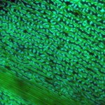
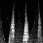
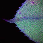
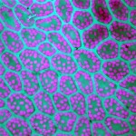
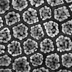
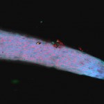
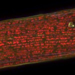
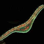
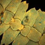
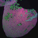
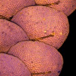
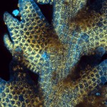
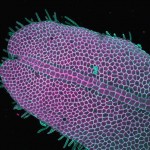
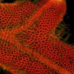
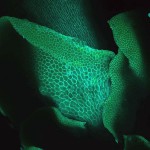
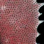
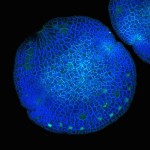
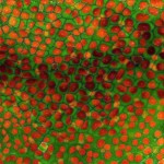
The Olympus FV1000 MPE confocal images showcase various structures of the following Bryophytes. Lasers were used to fluoresce structures within specimens. The 405 wavelength laser was utilized to emit light upon these specimens. Commonly fluorescing structures were the chloroplasts, cell walls, and cytoplasm. Different structures fluoresced at different wavelengths. Chloroplasts generally fluoresced at higher wavelengths than any other fluorescing structures. Dyes particular to certain wavelengths, were used to differentiate between these structures.
-Photos by: Francesca Salas
Phylum Bryophyta
Class Bryopsida
Dicranum scoparium leaf whole mount, showing the cells and chloroplasts of the costa and lamina.
Objective lens: 40x
Hypnum circinale peristome teeth, showing the cells of the exostome and endostome.
Objective lens: 40x
Plagiomnium insigne leaf whole mount, showing the cells and chloroplasts of the costa, lamina, and margin.
Objective lens: 10x
Plagiomnium insigne leaf whole mount, showing the cells and chloroplasts of the lamina.
Objective lens: 40x
Rhizomnium glabrescens leaf whole mount, showing the cells and chloroplasts of the costa, lamina, and margin.
Objective lens: 10x
Rhizomnium glabrescens leaf whole mount, showing the cells and chloroplasts of the lamina.
Objective lens: 40x
Rhizomnium glabrescens calyptra whole mount, showing the cells and chloroplasts of an immature calyptra.
Objective lens: 10x
Class Tetraphidopsida
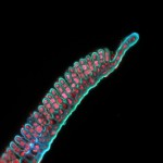
Tetraphis pellucida seta whole mount, showing the cells and chloroplasts of an immature seta.
Objective lens: 40x
Class Polytrichopsida
Polytrichum formosum leaf cross-section, showing the cells and chloroplasts of the costa, lamina and lamellae.
Objective lens: 10x
Polytrichum formosum leaf cross-section, showing the cells and chloroplasts of the costa, lamina, and lamellae.
Objective lens: 40x
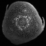
Polytrichum formosum stem cross-section, showing the conducting cells, cortex, leaf traces, and epidermal cells.
Objective lens: 10x
Polytrichum juniperum leaf cross-section, showing the cells and chloroplasts of the costa, lamina, and lamellae.
Objective lens: 20x
Class Sphagnopsida
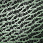
Sphagnum sp. branch leaf whole-mount, showing the hyaline cells, pores, and chlorophyllose cells.
Objective lens: 40x
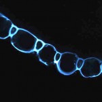
Sphagnum sp. branch leaf cross-section, showing the hyaline cells.
Objective lens: 20x
Phylum Marchantiophyta
Class Jungermanniopsida
Bazzania denudata ventral whole mount, showing the cells and chloroplasts of the leaves and stem.
Objective lens: 10x
Calypogea muelleriana lateral leaf whole mount, showing the laminar cells and chloroplasts.
Objective lens: 10x
Calypogea muelleriana lateral leaf whole mount, showing the laminar cells, chloroplasts, and oil bodies.
Objective lens: 40x
Frullania sp. dorsal side whole mount, showing the laminar cells and chloroplasts.
Objective lens: 20x
Lepidozia reptans dorsal whole mount, showing the laminar cells and chloroplasts.
Objective lens: 25x
Metzgeria conjugata dorsal thallus whole mount, showing the cells, rhizoids, and chloroplasts.
Objective lens: 10x
Metzgeria conjugata ventral thallus whole mount, showing the cells, rhizoids, and chloroplasts.
Objective lens: 10x
Porella sp. ventral whole mount, showing the cells and chloroplasts of the underleaves, stem, and lateral leaves.
Objective lens: 10x
Scapania bolanderi lateral leaf whole mount, showing the cells and chloroplasts of the lamina.
Objective lens: 25x
Class Marchantiopsida
Lunularia cruciata gemma whole mount, showing gemma cells and chloroplasts.
Objective lens: 10x, zoomed in with Olympus software to 20x
Phylum Anthocerotophyta
Hornwort dorsal thallus whole mount, showing the cells and chloroplasts.
Objective lens: 20x
Thank you,
Skylight, collection contributions from Steve Joya, those at the UBC BioImaging Facility, and Shona Ellis for collecting specimens and the opportunity to work on this project.

