Author Archives: harnoor shoker

The Changing Field of Stroke Medication

Strokes are the fifth leading cause of deaths in North America. If one is fortunate enough to survive a stroke, the rehabilitation process is long and painful depending on the amount of damage done to the brain. There are two types of strokes – ischemic and hemorrhagic. Ischemic strokes are the result of a clot forming in an artery and preventing blood flow, whereas hemorrhagic strokes are the result of an artery bursting and and the brain literally bleeding out.

Many researchers have worked towards improving and developing treatments to reduce the amount of brain damage a patient suffers during a stroke. One of the events that takes place during a stroke is called excitotoxicity, where brain cells literally excite themselves to death.

Receptors like NMDA as well as calcium are key culprits in causing damage to brain tissue. NMDA is a protein that is present on nerve cells and binds to the neurotransmitter glutamate. When a stroke occurs, nerve cells release large amounts of glutamate which bind to these NMDA receptors. The binding of glutamate to an NMDA receptor causes it to open. Calcium which is present in excess on the outside of the nerve cell, enters the cell. The calcium alongside with glutamate go on to wreck havoc in the nerve cell ultimately leading to its death. 

Courtesy of Khashayar.

Dr. Nicolas Weilinger investigated what happens at a cellular level during a stroke and the mechanism which works to damage brain cells. While researching, Dr. Weilinger discovered a new signalling pathway that had broad reaching implications for brain physiology and pathology.

YouTube Preview Image Courtesy of Harnoor Shoker

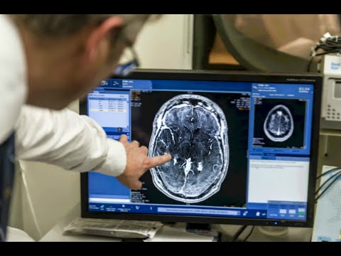
The findings of this study are important because current treatments in place to protect the brain during and after a stroke are not as effective as they should be. One of the main findings of Dr. Weilinger’s paper was that another channel much bigger than NMDA called pannexin gets activated during a stroke. Pannexin is physically connected to the NMDA receptor so when the NMDA receptor opens it signals pannexin to open as well. The opening of another channel therefore allows more calcium and glutamate to enter at an even faster pace. Using this information, a new drug was designed that would prevent the NMDA receptor from communicating with pannexin – in other words it would block the physical connection between the two proteins.

The wider implications of Weilinger’s paper is to hopefully improve stroke treatment. Future research into Dr. Weilinger’s findings could potentially be the first step in discovering a new drug type that can be used to reduce brain damage suffered during a stroke.

**We would like to thank Dr. Nicholas Weillinger for his time and the SCIE 300 team for guiding us and providing feedback.**

Harnoor, Khashayar, Matthew.

Special K to treat the Blues

Ketamine is a drug that was developed in 1962 and is commonly used as an anesthetic during surgical procedures in humans as well as in animals. However, like most drugs, ketamine made its way into the party scene during the 90s due to it’s hallucinogenic as well as dissociative effects (alter a person’s perception of reality). Furthermore, it also became notorious for being a date rape drug and was eventually classified as a Schedule III drug.

A vial containing ketamine.

A vial containing ketamine. Courtesy of Wikimedia Commons

In an interesting turn of events, ketamine is now undergoing clinical trials for its potential use in the treatment of depression. Depression itself is a debilitating condition with approximately 121 million people globally affected by it. The same drug that was being used as a date rape drug is now being used to treat depression?!

Yes! Ketamine therapy is being used to treat patients with severe major depression who don’t respond to traditional antidepressant medication. The treatment consists of giving a low dose of ketamine to the patient. The most common way to administer ketamine is by injecting it or by intranasal (smelling it) use. The positive effects in mood can be seen within 24 hours and can last up to ten days. This is one of the biggest advantages of using ketamine because the effects are noticeable immediately compared to traditional antidepressants which can take up to several months to work.

A typical neuron. Courtesy of Wikimedia Commons

A typical neuron. Courtesy of Wikimedia Commons

Depression is a multi-faceted disorder with several causes, one of them being a reduction in synaptic connections – the area between two neurons. For example, picture a neuron being a tree during spring, with many branches and leaves. When depression comes along, that tree now looks shrivelled up with bare branches and no leaves. Introduce a bit of Special K at low doses, and it converts our sad looking tree into a healthy tree once again. In other words, the synaptic connections are restored. The exact mechanism behind this is still unknown and being investigated.

Like with any other treatment, ketamine therapy also has a few side effects. When patients are first given a dose of ketamine they experience dissociative effects which are only temporary. Another down side of ketamine treatment is cost. The effects of ketamine treatment are short lived so patients often have to get regular infusions and since insurance does not cover the cost of treatment, it can get expensive; a single dose can cost anywhere from $525 to $800.

YouTube Preview Image

Courtesy of The Doctors

Furthermore, many opponents are concerned about the possibility of patients developing an addiction to ketamine. The dose used in clinical trials is well below the dose used by recreational users so it is very unlikely for the patient to develop an addiction. The future of ketamine therapy in treating depression looks promising and further studies should explore the long term effects of it before it becomes a standard in treating depression cases that don’t respond to traditional therapy.

Harnoor Shoker

You Shall Not Pass – Breaking the Blood-Brain Barrier

The blood-brain barrier is a protective covering that surrounds the brain and prevents contaminants, pathogens and other toxins from entering the brain. Sounds amazing, right? Yes! It’s the brain’s own defence mechanism to protect itself, however it can come in the way when trying to treat brain disorders like Parkinson’s, Alzheimer’s or even brain tumours.

Blood Brain Barrier. Courtesy of Wikimedia Commons

Blood Brain Barrier. Courtesy of Wikimedia Commons

Well, the problem is that the barrier only allows certain molecules such as water, glucose, lipid soluble molecules and some gases to pass and enter the brain. When designing drugs to combat brain diseases, researchers must find ways to bypass the blood-brain barrier which isn’t as easy as it seems. The cells that make up the barrier are tightly clustered together making it nearly impossible for drugs to enter into the brain.

In order to overcome this problem, researchers in Toronto designed a technique that was a breakthrough in breaking the blood-brain barrier. The technique, which has not yet been named, is currently being tested on patients with brain tumours. The first step in this technique is to administer a dose of a chemotherapy drug and also inject the patient with microbubbles which are smaller than red blood cells. The microbubbles and drug travel to the brain and come face to face with the blood-brain barrier. The patient is then placed in an MRI machine and the exact location of the tumour is identified.  Once the tumour has been located, ultrasound waves are emitted to that specific region causing the microbubbles to vibrate really fast.

Courtesy of Newswise

These vibrations cause the tight junctions of the cells that make up the blood-brain barrier to loosen up, creating a small passage for drugs to pass through. Once the ultrasound waves stop, the microbubbles are reabsorbed by the lungs and the passage which forms closes within a six to twelve-hour window.

Current techniques being used to penetrate the blood-brain barrier are invasive and not as targeted as this technique. If researchers can find a way to bypass the blood-brain barrier in a non-invasive manner, it will change therapeutic approaches to treating brain disorders. Like any other procedure which involves opening the blood-brain barrier, there are risks associated with this technique as well. The blood-brain barrier remains open for several hours after the initial procedure is done and during this time toxins and other contaminants can enter the brain through that passage. This technique is currently undergoing clinical trials but if it proves to be effective it can revolutionize the way we treat brain diseases.

YouTube Preview Image Courtesy of Sunnybrook Hospital

Harnoor Shoker