Category Archives: Biological Sciences

Borrowing from Mosquitos – A Potential Broad-Spectrum Antiviral

A team of researchers at the National Institute of Environmental Health Sciences (NIEHS) have found a promising antiviral in the strangest place – a protein called AEG12 found in the gut of mosquitos. This recent study lead by Dr. Alexander Foo found that AEG12 strongly inhibits the family of viruses that cause Yellow Fever, Dengue, West Nile, and Zika, and that the protein may also inhibit their distant relative: coronaviruses. Although the virus-killing AEG12 protein sounds promising, it may still be a long time before its safe to use in humans.
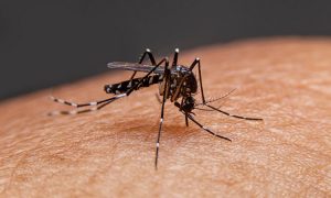
The Aedes aegypti mosquito, which can carry viruses such as Yellow Fever, Dengue, and Zika (Image source)

Foo and His Findings

Scientists have long been on the hunt for effective antivirals, and researchers like Dr. Foo have been making great progress in the field. Foo’s recent study showed that AEG12 was effective at killing flaviviruses (such as Dengue and Zika) but was less effective at killing coronaviruses. Using methods and software tools for understanding how proteins work and specialized laboratory techniques to study viruses, the team was able to piece together how AEG12 is such a successful virus-destroyer.

Flaviviruses are enveloped: meaning that each virus particle is surrounded by a membrane. The infection process, in the video below, shows how the virus must first attach to the surface a human cell (0:28) and then fuse its membrane with ours to get inside (1:12).

Essentially, the AEG12 protein carries little membrane pieces of its own, which are different from the type found in the virus’ membrane. When it comes across a virus, AEG12 trades its pieces for the virus’. Once enough chunks are swapped out, the virus’ membrane becomes unstable and can’t fuse with our cells: effectively “killing” the virus.
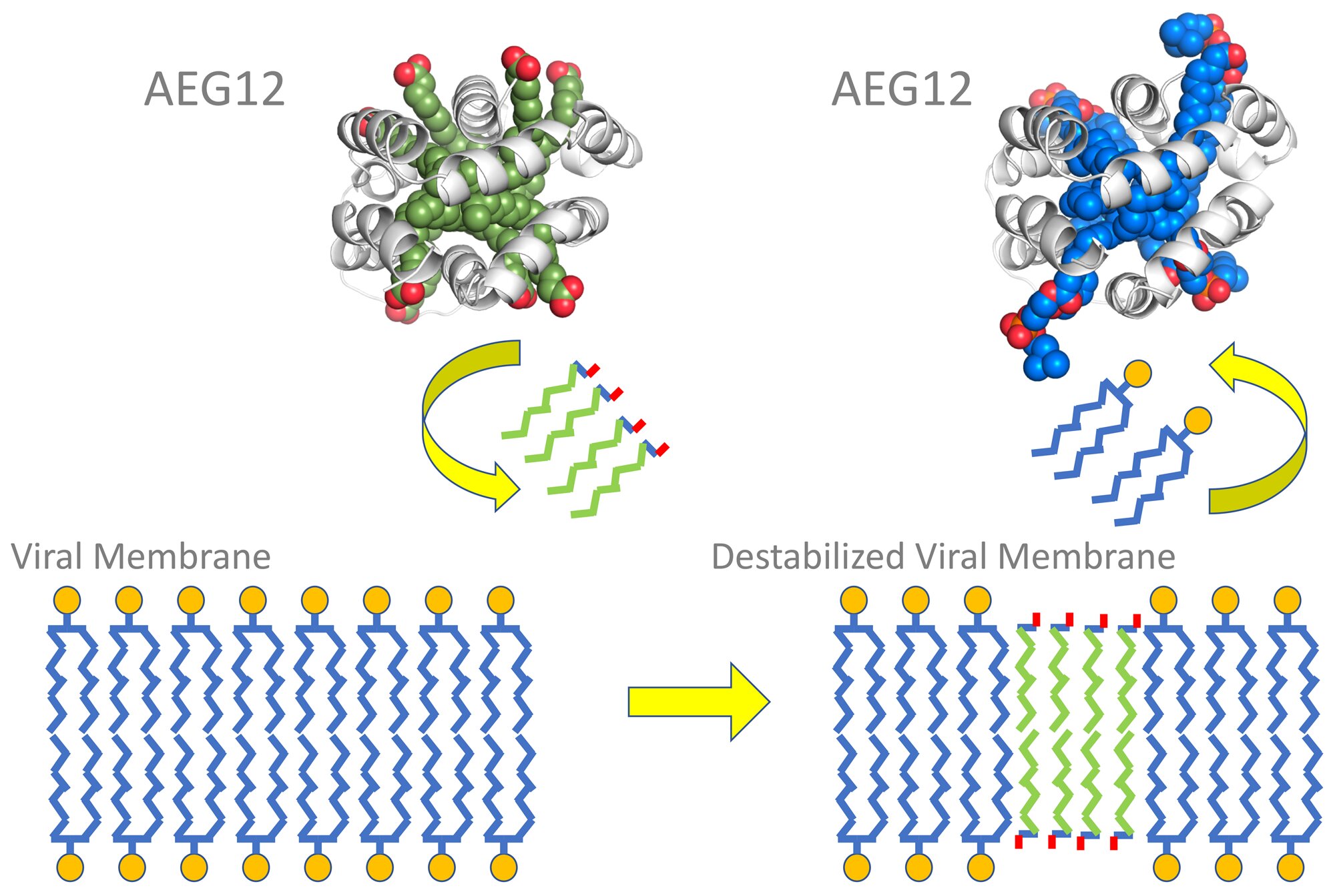
The AEG12 protein swaps out viral membrane pieces for unstable pieces (Image source)

Even though AEG12’s virus-killing abilities sound promising, Foo’s team found that AEG12 breaks apart red blood cells. Although useful to a mosquito that needs to digest a blood meal, use of AEG12 in humans would lead to a very serious blood disorder that could be fatal if left untreated.

There is Still Hope

So, AEG12 isn’t safe for use in humans… Yet. With some clever solutions and careful bioengineering, further research could find a way to modify AEG12 so that it targets viruses and ignores human cells. The development of a drug or molecule effective against enveloped viruses could save lives across the globe – addressing not only the headlining COVID-19 pandemic, but the ongoing epidemics as well.

-Maya Bird

Mice Grown In Vials: Are Humans Next?

For at least a hundred years, researchers have been struggling to answer one question: how does a single cell become a full grown human.

One major barrier to fully understanding this process, was that we could never see it happening before our own eyes. Luckily, a team of biologists led by Dr. Jacob Hanna at the Weizmann Institute of Science had a major breakthrough: mice grown in vials. 

The Problem

In an interview with the New York Times, biologist Dr. Paul Tesar said:

“The holy grail of developmental biology is to understand how a single cell, a fertilized egg, can make all of the specific cell types in the human body and grow into 40 trillion cells. Since the beginning of time, researchers have been trying to develop ways to answer this question.”

Each one of us started the same, as just one cell. In our mothers, one cell became two, then four, which eventually led to us. During these beginning stages of development, we were what researchers call an embryo, or an early-stage animal. Embryos are located in a mother’s uterus, which acts like a house that provides everything needed to grow. 

To see what is going on inside this house, researchers have tried many different tricks including taking pictures, or even removing the uterus fully from animals such as mice to get a better look. What researchers have not been able to do thus far, is watch the embryo grow continuously outside of the mother. This has not only made research in the field difficult, but has restricted the work being done.

The Solution

Although the researchers at Weizmann were not the first to come up with the idea, up until now, mice grown outside of a mother have either not been able to survive, or did not grow correctly. In fact, it took Dr. Hanna and his colleagues 7 years to perfect their technique. 

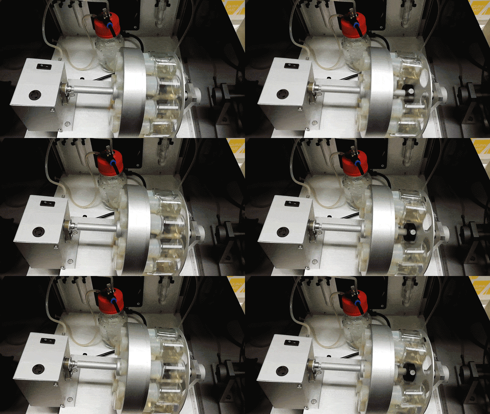
The entire process consists of two steps. First, the uterus of a recently pregnant mouse is removed. Second, the uterus is transferred to a vial filled with liquid containing all the food it will need. As seen in the video below, the embryo is slowly spun to make sure it does not attach itself to the wall of the vial as that could result in death. 

Mice embryos growing inside spinning vials (Video from MIT Technology Review)

The mice were under constant observation and images, as seen below, were taken and compared to mice developing inside a mother. The two were identical. 

Mice developing over a 5-day period (Image from The New York Times)

Implications

As a result of this breakthrough, researchers will be able to better understand events including birth defects and miscarriages. Additionally, researchers can now easily change the environment that the embryo grows in to see what conditions can affect development. Although it may still be some time until this research is transferred to humans, this breakthrough certainly marks a big step in the right direction.

-Jessica Petrochuk

Combating Antibiotic Resistance with “Nanoparticles”

The Centers for Disease Control and Prevention (CDC) calls antibiotic resistance “one of the biggest public health challenges of our time.” But what is antibiotic resistance? How is it affecting our lives? and How can we use nanoparticles to fight it?

Antibiotic Resistance Crisis:

Antibiotics are powerful medications that are widely used for the treatment of infections caused by bacteria. However, taking antibiotics too often or for the wrong purpose caused bacteria to evolve various antibiotic resistance mechanisms.
Some bacteria have developed resistance to nearly all the antibiotic treatments available and can cause serious fatal diseases that were once easily treatable with antibiotics.

Without the invention of new strategies to counteract drug-resistant infections, they are likely to kill more than 10 million people each year by 2050. This is more than the number of  people currently dying from cancer.

Ongoing studies are analyzing the ways nanoparticles (small particles ranging between 1 to 100 nanometres in size) can be used to defeat antibiotic-resistant bacteria. The size of nanoparticles and their flexible antibacterial properties make them a favorable solution to this problem since they can be used to not only deliver antibiotics but also to fight bacteria themselves.

The following video explains what nanoparticles are, how they are produced, and how they can enter and kill the bacterial cells:

YouTube Preview Image

Source: TCTTPC YouTube

Nanoparticles as Antibiotic Carriers:

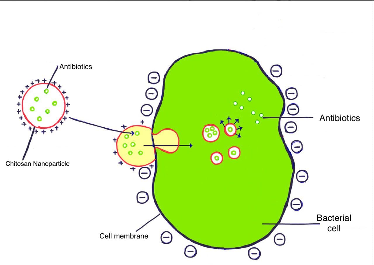
According to this study conducted by Zhang and his colleague in late 2020, some nanoparticles can penetrate into the bacterial cells while carrying and protecting the antibiotic agents. These nanoparticles —developed using materials such as metals and chitosan (a type of fiber)— can save the antibiotic from chemicals released by bacteria that can otherwise destroy them.

 Chitosan nanoparticle possesses a positive charge making it able to attach to bacterial cells that have a negative charge on their membrane (outer layer of the cell). Source: ResearchGate

Nanoparticles as Antibiotic Drugs: 

Nanoparticles can also defeat bacteria directly using mechanisms such as the generation of reactive oxygen species (ROS). ROS are unstable molecules that can easily react with other biomolecules (DNA, protein, etc.) in a cell, disrupt them, and cause cell death.

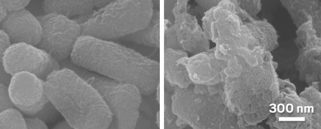
Silver nanoparticles (SNPs), for instance, can destroy the bacterial membrane and interact with interior components of the bacterium by releasing silver ions that can generate ROS inside the cell. Indeed, severe cellular damages in 5 different types of bacteria were reported when treated with SNPs.

Effect of Nanoparticles on Bacteria

E.coli (a type of bacteria) (left) is severely damaged when treated with a  silver nanoparticle (right). Source: pubs.acs.org

Nanoparticles appear to be a promising solution to address the problem
of antibiotic resistance; however, the main factor that limits their application in treatments is that researchers often face side-effects related to nanoparticle toxicity when interacted with biological systems like human cells. For instance, the ROS generated by a high dose of SNPs can damage the human cell components.

New strategies are being investigated to direct the target of nanoparticles to bacterial cells only and reduce their toxicity in order to develop safe and efficient antibacterial nanoparticles.

– Samin Shadravan

Need to Sober Up? Just Breathe Out the Booze!

With regards to alcohol, many of us have previously reached the so-called point of no return: a moment where the pleasant buzz is replaced by a throbbing headache (and massive amounts of regret). If only there was a simple way to quickly sober up…

Alcohol! Source: awee_19, Flickr

A simple overview of ethanol breakdown

First, let’s dive into how our bodies break down alcohol. Once ethanol arrives at the stomach and intestines, it is absorbed into the bloodstream. From there, most of the alcohol ends up in the liver. The liver is responsible for detoxifying 90% of the ethanol that we consume; the remaining 10% is eliminated through sweat, urine, and breath.

However, the rate at which the liver breaks down ethanol is zeroth-order: meaning that the breakdown rate is always constant, no matter how much ethanol is in your system. This explains why we haven’t been able to develop techniques to speed up the rate of ethanol breakdown in our livers.

Naturally, the next step would be to see whether we can speed up the elimination of the remaining 10% of ethanol in our bloodstreams. Turns out, we can! Remember how we said that some ethanol is breathed out? This works the same way that we exhale carbon dioxide: diffusion! Since the ethanol concentration in our bloodstream is higher than in the air that we breathe in, some ethanol diffuses into our lungs and we breathe it out!

Diffusion Explained.
Source: Free Animated Education, YouTube

A breathalyzer uses the fact that we breathe out ethanol to determine our blood alcohol concentration (BAC). Source: Dave Shea, Flickr

So can I just hyperventilate until I start to feel sober?

In theory, you could… but you really shouldn’t. Hyperventilating will reduce your ethanol levels, sure, but it will also decrease your CO2 levels: causing your brain’s blood vessels to narrow, and ultimately depriving your brain of oxygen. Thankfully, a recent study has found a simple and effective solution, utilizing isocapnic hyperpnea.

Isocapnic hyperpnea: what is it?

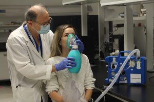
To put it simply, isocapnic hyperpnea (IH) is when you deeply (sometimes rapidly) breathe in air that has an equal concentration of carbon dioxide as in your bloodstream. This lets you breathe out all the nasty ethanol, while your CO2 levels stay steady. In the study, participants drank vodka, then were connected to a device which supplied air which had a CO2 concentration similar to what would be found in normal blood vessels. The results of the study showed that the participants who underwent IH were able to get ethanol out of their system more than three times faster than participants who breathed regular air!

A demonstration of the IH apparatus. Source: UHN

This technology could be widely available in the near future, since IH has already been approved as a treatment for clearing our bodies of other chemicals. IH could help paramedics in clearing the alcohol out of a patient’s system in a timely manner, which could ultimately save their lives. Remember to always drink responsibly!


– Sam Jung

Treating Depression: Personalized Deep Brain Stimulation

How would you feel if the treatment or medication you were taking had little to no effect in suppressing your symptoms? Unfortunately, this is the case for 1 in 3 patients diagnosed with depression. These patients fall under a category known as treatment-resistant depression. Personalized deep brain stimulation, a promising alternative to conventional treatments, has the potential to treat various forms of depression by allowing physicians to tailor treatment to an individual.

THE PROBLEM

Depression, which is characterized by low mood, is linked to an imbalance of serotonin, norepinephrine, and dopamine neurotransmitters in the brain. It is a common mental illness that affects the way someone feels, thinks, and acts. However, it is important to note that depression varies significantly among individuals and many other factors play a role.

Source: flickr.com

As stated by Ben Paul from USC Viterbi School of Engineering,

“Mental disorders can manifest differently in each patient’s brain.”

There is no single treatment that can effectively treat the symptoms of depression among all diagnosed individuals. This makes it hard for physicians to provide the best treatment for their patients.

WHAT IS DEEP BRAIN STIMULATION?

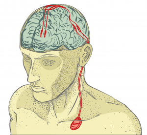
Deep brain stimulation (DBS) is a surgical procedure where electrodes are implanted within specific areas of the brain. By electrically stimulating these parts of the brain, physicians can reduce the symptoms associated with depression. The amount of stimulation is controlled by a pacemaker that is placed under the skin on the chest.

The video below explains this procedure further and contains an interview with Edi Guyton, a patient who had this surgery: 

Source: CNN | Youtube

PROMISING ALTERNATIVE: PERSONALIZED DEEP BRAIN STIMULATION 

Even with deep brain stimulation, each patient’s response to treatment will be different. However, one of the pros of deep brain stimulation is that it results in immediate changes. This is the key component that allows physicians to personalize treatment. A 2021 research study led by Dr. Maryam Shanechi and her team at the USC Viterbi School of Engineering explains an approach that can be used to predict and see how an individual’s brain responds to stimulation. Her research will allow physicians to monitor brain regions in real-time.

How is this done? 

       Two tools have been designed: 

  1. Electrical stimulation wave to map brain activity
  2. Machine-learning techniques that can learn the mapped brain activity which is collected during stimulation 

The stimulation wave tool randomly changes the characteristics (amplitude and frequency) of the electrical impulse over time. A change in these characteristics is the equivalent of changing the dosage of a medication. Analysis of brain activity during these changes will help physicians determine the correct stimulation doses. 

THE FUTURE: TREATING DEPRESSION

Dr. Maryam Shanechi’s research will allow physicians to personalize deep brain stimulation for all patients diagnosed with depression. This can help physicians overcome the difficulty of assisting individuals with treatment-resistant depression. Success within this field of personalized deep brain stimulation not only holds great potential for treating depression but can also lead to improved treatments for other psychiatric disorders. 

Source: flickr.com


– Samantha Nalliah

Forgot Something? Suspect Your Dopamine

You open up an internet browser, but you forget what you were going to search for or why you even started your computer. Have you ever came across these situations?  The recent research on temporary memory loss (2021) has discovered the betrayer within our body: the dopamine.

Dopamine has a reputation as the “happy hormone.” It’s a neurotransmitter, a substance that conveys signals between the neurons that mediate pleasure in our brain and make you crave for the things you love. For instance, you constantly refresh your Instagram feed or click on the next recommended video on YouTube because your brain remembers and relate those activities to pleasure. Then every time you perform those activities your brain release dopamine, which makes you feel good.

Problems with Dopamine Addiction

In a nutshell, dopamine acts very similar to recreational drugs because it constantly tempts and controls you to do things that provide instant gratification and make you crave for stronger pleasure. The problem of dopamine addiction recently arose because of the rapid technological advancement, which allowed for easy access to activities that release dopamine such as social media, video games, and pornography. People have hard time focusing on their work and managing their time because they crave that dopamine shots.

Here’s a video that talks about the effect of dopamine on human and possible solution to overcome addiction:

YouTube Preview Image

Source: TopThink (youtube.com)

In addition, the researchers from Scripps Research Institute revealed dopamine’s another dark secret. They discovered the mechanism in which the stimulation of dopamine circuit is responsible for transient forgetting, also known as temporary memory loss. In particular, the biochemical team specified a single pair of dopamine-releasing neuron, named PPL1- α2α’2, that causes the universal nuisance in our brain.

In summary, the research team trained Drosophila, commonly known as the fruit fly, to associate a certain scent with an unpleasant shock. However, when the flies were introduced to stimuli such as a puff of air or blue light, the type of light emitted from the screens of your electronic devices, the dopamine released due to the stimuli interfered with the flies’ memory signal and the flies temporarily forgot the negative association with the scent.

Furthermore, the research found that the increased strength or intensity of the stimulus increases the duration of the temporary memory loss . In other words, exposure to stronger stimulus, such as illicit drug or concentrated alcohol, requires more time to recover from the transient memory loss.

During the unprecedented period of pandemic, without social interactions, you probably consume more social media feeds or YouTube videos for your daily dose of dopamine. However, think of dopamine as sugar for now. Your body needs it to continue the happy life, but addiction could bring potential complications with regards to your success and health. Experts recommend activities such as the dopamine detox or meditation to overcome the dopamine addiction. Control your dopamine; don’t let the YouTube recommendations control you!

-Matthew Lim

Microalgae Used to Create Biodiesel: An Organism That Can Save Our Planet?

As time continues to move forward, global climate change is becoming an ever-growing issue. In order to mitigate the effects of global warming, we as a society need to change our ways of living. Due to rapid industrialization and urbanization, there is an increased amount of pollutants emitted into the atmosphere that are slowly damaging the earth. In today’s world, most of the energy production is coming from fossil fuel burning, which is the key source of carbon dioxide (CO2) emissions to the atmosphere. Energy demand will continue to increase and as of right now, fossil fuels still contribute to 82% of the global demand. The figure below displays gas emissions from various modes of transportation.

Source: bbc.com

An article written in 2020, by Ashokkumar Veeramuthu and team, describes the potential use of microalgae to produce biodiesel. You may be asking, what are microalgae and what the heck is biodiesel? Let’s jump straight into it.

What is Biodiesel?

Biodiesel is made from materials such as plant oils. It’s an alternative to petroleum diesel and emits much less harmful substances into the atmosphere. Since we are slowly killing our planet, replacing our non-renewable energy sources with green alternative sources doesn’t sound like a bad idea.

Why use Microalgae?

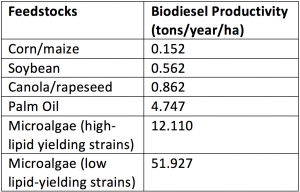
You may be wondering, what is so special about microalgae? Why can’t we use some type of terrestrial plant like corn to produce biodiesel? Studies show that the use of microalgae is the best option for the production of a renewable and sustainable source of energy. Microalgae are photosynthetic organisms living in wet environments that can convert sunlight, water and COinto biofuel. There has been a shift of attention towards microalgae to produce biodiesel because microalgae provide many advantages over terrestrial plants. The benefits of microalgae include high lipid concentrations (which can easily be converted to biodiesel through a process called transesterification), rapid growth and minimal nutrient requirements. The table below compares values of the biodiesel productivity of microalgae and other plants.

Biodiesel productivity of different feedstocks. Source: intechopen.com

Microalgae also tend to grow 10x more rapidly than terrestrial plants and less than 10% of the land is required to produce the same amount of biomass. Additionally, microalgae don’t require large amounts of fertilizers to grow, unlike terrestrial plants. The cultivation of microalgae can be carried out by using wastewater, since it is rich in key nutrients. Furthermore, the use of wastewater decreases costs greatly and makes biodiesel production commercially viable.

This video showcases the general process of biodiesel production in a nutshell:

Source: David T. Kearns (YouTube)

In today’s world, there’s a shift of attention to deal with the issue of climate change. From Elon Musk creating fully electric vehicles to Joe Biden rejoining the Paris climate accord within hours into presidency, we as humans are finally taking initiative to save our planet. The future of creating fuels from microalgae sounds promising and having a range of renewable sources of green energy will be beneficial to us in the coming time.

– Parwaz Gill